Case Report Intraoperative Endoscopy in Transient Adult Jejunojejunal Intussusception
←
→
Page content transcription
If your browser does not render page correctly, please read the page content below
Hindawi
Case Reports in Gastrointestinal Medicine
Volume 2021, Article ID 3718089, 6 pages
https://doi.org/10.1155/2021/3718089

Case Report
Intraoperative Endoscopy in Transient Adult
Jejunojejunal Intussusception

           Takeshi Okamoto ,1,2 Hidekazu Suzuki ,2 and Katsuyuki Fukuda                                       1

           1
               Department of Gastroenterology, St. Luke’s International Hospital, 9-1 Akashicho, Chuo-ku, Tokyo 104-8560, Japan
           2
               Department of Gastroenterology and Hepatology, Department of Internal Medicine, Tokai University School of Medicine,
               143 Shimokasuya, Isehara, Kanagawa 259-1143, Japan

           Correspondence should be addressed to Takeshi Okamoto; tak@afia.jp

           Received 19 April 2021; Accepted 8 July 2021; Published 12 July 2021

           Academic Editor: Yoshihiro Moriwaki

           Copyright © 2021 Takeshi Okamoto et al. This is an open access article distributed under the Creative Commons Attribution
           License, which permits unrestricted use, distribution, and reproduction in any medium, provided the original work is
           properly cited.
           Despite improvements in imaging modalities, causative lead points in adult intussusception may be difficult to diagnose. Such lead
           points can be malignant, causing recurrence or metastases if left unresected. We describe a case of transient adult jejunojejunal
           intussusception, in which intraoperative endoscopy was used to confirm the absence of a lead point. A 39-year-old woman with a
           history of laparoscopic oophorectomy presented with epigastric pain, nausea, and vomiting. Contrast computed tomography
           revealed jejunojejunal intussusception, with no visible lead point. Spontaneous reduction was confirmed during exploratory
           laparoscopy. After lysis of adhesions, intraoperative peroral jejunoscopy was performed with the surgeons’ assistance. Endoscopy
           confirmed the absence of tumor, and bowel resection was avoided. No recurrence has been observed during 24 months of follow-
           up. Intraoperative endoscopy may provide additional reassurance for the absence of a lead point in cases where preoperative
           enteroscopy cannot be performed and no lead points can be identified on imaging.

1. Introduction                                                          exploratory laparoscopy with bowel resection remains the
                                                                         most widely accepted treatment for AI, the need for such
While intussusceptions in children are commonly idio-                    interventions has recently been brought into question [6, 7].
pathic, adult intussusceptions (AI) generally have a causative           Almost 5% of AI patients were treated conservatively in a
lead point. Enteric intussusceptions, the most common type               meta-analysis [1]. On the other hand, avoiding surgery may
accounting for about 50% of AIs, have been reported to                   occasionally come at the cost of missing a malignant small
result from malignant tumors in 22.5% of cases, with                     bowel tumor, which may be undetected despite recent ad-
metastatic carcinoma being the most common malignant                     vances in imaging modalities [8–12]. Such an event may
lead point [1]. On the other hand, about 15% of AIs are                  have various devastating results, including recurrence and
idiopathic, with no apparent lead point.                                 metastatic disease.
    One type of AI, classified as idiopathic by some authors,                 Although not always possible preoperatively, entero-
is caused by adhesions resulting from prior surgery [1–3].               scopy offers an alternative to confirm the absence of a lead
Truly idiopathic AI may be caused by mechanisms such as                  point without bowel resection. Herein, we report a case of
bowel hyperactivity and be transient, causing chronic, in-               intraoperative endoscopy performed on a patient with
termittent, or nonspecific symptoms [4, 5]. While                         jejunojejunal AI which demonstrated the absence of tumor
2                                                                                   Case Reports in Gastrointestinal Medicine

in the jejunum, providing additional reassurance that bowel           Exploratory laparoscopy was performed the next day.
resection could be avoided.                                       Severe jejunal hyperactivity was observed intermittently
                                                                  throughout the laparoscopy. However, no intussusception
2. Case Presentation                                              was observed, suggesting spontaneous reduction. Tumors
                                                                  and segmental edema in the proximal jejunum were also
A 39-year-old woman presented with epigastric pain, in-           notably absent. Adhesions from previous laparoscopic oo-
termittent nausea, frequent vomiting, and loss of appetite        phorectomy were observed near a port placed in the right
over the last seven days. She denied weight loss. She could       lower quadrant (Figure 3(a)). While adhesiotomy was
pass gas but had not passed stool over the last four days. She    performed, the adhesions were distant from the proximal
was able to tolerate a full meal the night before presenting to   jejunum and appeared as an unlikely cause of
the hospital, but nausea and vomiting had resumed within          intussusception.
several hours.                                                        Intraoperative peroral jejunoscopy was performed with a
    Her medical history was significant only for laparoscopic      long colonoscope (PCF-H290L, Olympus Corp., Tokyo,
oophorectomy for a left ovarian cyst almost 20 years prior.       Japan) and carbon dioxide insufflation. A thorough lapa-
She was taking no medications, herbal remedies, or nutri-         roscopic exploration was completed prior to commencing
tional supplements. She admitted to chronic alcohol abuse         intraoperative endoscopy, as endoscopic insufflation would
with frequent visits to the emergency department due to           hinder the laparoscopic view. The surgeons used laparo-
alcohol intoxication, but had never smoked cigarettes. She        scopic grasping forceps to apply gentle pressure to the
denied any recent sexual contact, overseas travel, raw food       stomach to facilitate scope insertion (Figure 3(b)). When the
ingestion, or sick contacts.                                      endoscope reached the jejunum, the laparoscopic camera
    Upon presentation, the patient appeared to be in              was pointed to a location in the jejunum believed to be distal
moderate distress. Her respiratory rate was 24 breaths per        to the reduced intussusception. Forceps were also gently
minute, but vital signs were otherwise stable. She com-           placed at this location to facilitate insufflation (Figure 3(c)).
plained of discomfort on palpation of the epigastric region.      The endoscope was inserted until light from the laparoscopic
The upper abdomen was soft but mildly distended. No mass          camera was visualized, confirming the absence of tumors or
was palpated. Small scars from previous laparoscopy were          other lesions which may serve as a lead point for intus-
noted.                                                            susception (Figure 4). Short bowel resection was therefore
    Laboratory results were only remarkable for a mildly          not performed. The patient was diagnosed with transient
elevated C-reactive protein of 0.44 mg/dL. Esophagogas-           jejunojejunal intussusception, more likely associated with
troduodenoscopy (EGD) performed 20 hours after the pa-            chronic alcoholism rather than adhesions from previous
tient’s last meal revealed a mildly distended stomach with        surgery.
significant food residue in the esophagus and stomach                  The patient experienced complete resolution of her
(Figure 1). The patient vomited copious food residue during       symptoms after the surgery. The postsurgical course was
the procedure, precluding a thorough examination for fear         uneventful and the patient was discharged two days later,
of aspiration. No gross abnormalities were found up to the        with instructions to stop drinking alcohol. No recurrence
third part of the duodenum. Food residue and intraluminal         was observed during 24 months of follow-up.
air were suctioned to the extent possible at the end of the
examination.                                                      3. Discussion
    Despite the patient’s ability to pass gas, an emergency
computed tomography (CT) scan was conducted to rule out           AI presents two problems for the patient: symptoms relating
small bowel obstruction. CT without contract was largely          to bowel obstruction and a potentially malignant lead point.
unremarkable, with no visible signs of tumor or bowel                 AI can be difficult to diagnose due to its rarity and the
obstruction (Figure 2(a)). However, CT with contrast taken        chronic, nonspecific nature of its symptoms [1, 4]. In
several minutes later revealed the “target sign,” a bowel-in-     general, CT has a sensitivity of 83% in diagnosing the eti-
bowel configuration measuring 7 cm in the proximal jeju-           ology of small bowel obstruction and of 82% in diagnosing
num with invaginated mesentery (Figure 2(b)). No lead             small bowel tumors as the cause [13]. Helical CT-enter-
point was identified. No proximal distension was observed,         oclysis had particularly high pooled sensitivity (92.8%) and
most likely as a result of vomiting and suction during EGD.       specificity (99.2%) for small bowel tumors in a meta-analysis
The patient was diagnosed with jejunojejunal intussuscep-         [9]. On the other hand, only 52% of enteric AIs were rec-
tion, most likely of a transient nature.                          ognized preoperatively in a study of 44 patients [7]. Another
    While the possibility of spontaneous reduction was            study of 318 patients found that AI patients presented with
explained, the patient wished to undergo exploratory lap-         symptoms of complete and partial bowel obstruction in only
aroscopy due to the severity of her symptoms. The surgeons        27% and 15% of cases, respectively [8]. CT failed to identify
also agreed to exploratory laparoscopy in light of the severe     lead points in reports of jejunal AI caused by heterotopic
obstructive symptoms, surgical history with possible ad-          gastric mucosa, laterally spreading tumor, and gastrointes-
hesions, and possible recurrence if left untreated. Consent       tinal stromal tumor [10–12].
for intraoperative endoscopy was also obtained to evaluate            While symptoms can be relieved by reduction or bowel
the jejunum for a possible lead point, as the patient was not     resection, recurrence has been reported in about 6.5% of all
in a condition to undergo preoperative enteroscopy.               AI cases [1]. This figure may be higher in enteric AI, as one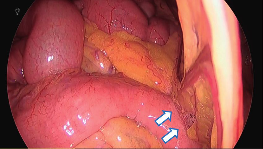
Case Reports in Gastrointestinal Medicine                                                                                            3

Figure 1: Esophagogastroduodenoscopy performed 20 hours after the patient’s last meal revealed significant food residue in the esophagus
and stomach, suggesting possible bowel obstruction.

                                         (a)                                              (b)

Figure 2: (a) Computed tomography (CT) without contract was largely unremarkable, with no visible signs of tumor or bowel obstruction
in the jejunum (arrow). (b) CT with contract taken several minutes later revealed a bowel-in-bowel configuration with invaginated
mesentery, consistent with jejunojejunal intussusception (arrow). No mass was visualized.

report found recurrences in 21 of 230 enteric AI patients            bowel hyperactivity due to chronic alcoholism played a role
(9.1%) [8]. All recurrences occurred at the site of the initial      in the pathogenesis of AI in our case. The effects of alcohol
AI and 63% required surgery. Recurrence occurs frequently            on small bowel motility depend on the alcohol concentration
in celiac disease, Crohn’s disease, and polyposis syndromes          of consumed beverages and chronicity of alcohol use, with
such as Peutz–Jeghers syndrome due to their multifocal               chronic consumption of large doses of alcohol accelerating
involvement [14–16]. Recurrence has also been reported in            small bowel transit [21, 22]. There is only one report of small
idiopathic enteric AI [17].                                          bowel intussusception in a patient with chronic alcoholism,
    Transient intussusceptions with spontaneous reduction            although various other factors such as malnutrition and
are very common in children as they are generally idiopathic         brown bowel syndrome were also present in that case [23].
in nature, particularly when the length of the intussuscep-          The role of alcohol use in transient AI has not been studied;
tion is less than 3 cm [18, 19]. Similarly, intussusception          the history of alcohol use is generally missing from case
length of less than 3.5 cm is an independent predictor for           reports on AI. While alcohol is also associated with diarrhea
self-limiting AI [20]. The diagnosis of AI on CT may be more         due to inhibited water and sodium absorption as well as
common than once believed, in part due to improvements in            mucosal injury in the duodenum and upper jejunum, none
imaging technology. In a study of 37 CT diagnoses of AI, 31          of these findings were observed in our case [24]. The use of
were cared conservatively and none required surgery during           scopolamine during preoperative EGD may have tempo-
a mean follow-up of 5.2 months [20]. Although we suspected           rarily reduced bowel hyperactivity, contributing to tempo-
transient AI based on the discrepancy between the plain and          rary resolution of AI before the CT scan.
contrast CT scans in our case, exploratory laparoscopy was               Both antegrade enteroscopy and retrograde enteroscopy
believed to be indicated due to the length of the intussus-          have been used to diagnose various types of lead points
ception (7 cm), severity of symptoms, possibility of recur-          including gastrointestinal stromal tumor, inverted Meckel’s
rence, and the wishes of the patient.                                diverticulum, inflammatory fibroid polyp, Peutz–Jeghers
    As the adhesions observed during laparoscopy were                syndrome, heterotopic pancreatic mass, malignant mela-
distant from the site of intussusception, we suspect that            noma, and mass-forming fibrogranulation from a healed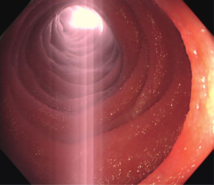
4                                                                                         Case Reports in Gastrointestinal Medicine

                                  (a)                                                              (b)

                                                                   (c)

Figure 3: (a) Adhesions from previous laparoscopic oophorectomy observed near a port placed in the right lower quadrant (arrows). (b) The
surgeons used laparoscopic grasping forceps to apply gentle pressure to the stomach to facilitate scope insertion during intraoperative
peroral jejunoscopy. (c) When the endoscope reached the jejunum, the laparoscopic camera was pointed distal to the suspected location of
the reduced intussusception to signal the desired destination for endoscopic viewing. Forceps were also gently placed at this location to
facilitate insufflation.

Figure 4: Light from the laparoscopic camera showing through the jejunal wall confirmed passage of the endoscope beyond the site of
intussusception.

ulcer [25–31]. Preoperative enteroscopy can also identify                bleeding [32–34]. Intraoperative endoscopy can be per-
signs of bowel ischemia, which occurs in about 15% of cases              formed via the peroral route, the transanal route, and
[1]. Furthermore, the balloon used in double-balloon                     enterotomies, achieving total small bowel visualization in
enteroscopy has also been shown to be useful in achieving                57–100% of cases [34]. Advantages over single-balloon or
reduction of the intussusception [25, 28].                               double-balloon enteroscopy include one-stage intervention
    Endoscopy has been used in various surgical procedures               when a tumorous lead point is discovered, the surgeon;s
such as laparoscopic endoscopic cooperative surgery, con-                manual assistance during scope insertion, the use of
firmation of anastomoses during gastrointestinal surgery,                 enterotomies, and performance under general anesthesia in
and in exploratory laparotomy for obscure gastrointestinal               the operating theater [35, 36]. However, single-balloon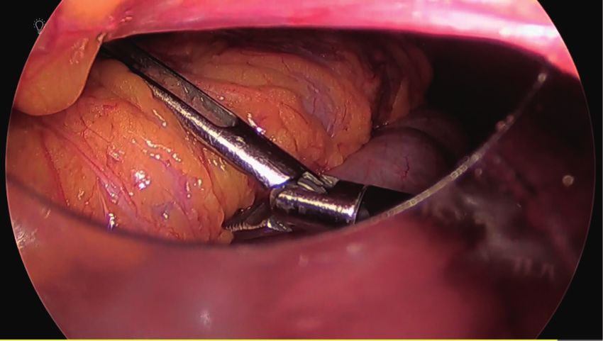
Case Reports in Gastrointestinal Medicine                                                                                              5

enteroscopy and double-balloon enteroscopy have reduced              [2] M. G. Sarr, D. M. Nagorney, and D. C. McIlrath, “Postop-
the need for intraoperative endoscopy for diagnostic pur-                erative intussusception in the adult,” Archives of Surgery,
poses, which is now only used when preoperative entero-                  vol. 116, no. 2, pp. 144–148, 1981.
scopy cannot be performed or when the diagnosis remains in           [3] F. Agha, “Intussusception in adults,” American Journal of
question [37]. In our case, preoperative enteroscopy was not             Roentgenology, vol. 146, no. 3, pp. 527–531, 1986.
                                                                     [4] O. Catalano, “Transient small bowel intussusception: CT
realistic without deep sedation and intubation, as copious
                                                                         findings in adults,” The British Journal of Radiology, vol. 70,
vomiting, strong gag reflexes, and body movement were                     no. 836, pp. 805–808, 1997.
observed throughout the preoperative EGD. As the site of             [5] T. E. Napora, K. E. Henry, T. J. Lovett, and M. S. Beeson,
intussusception could be reached with a long colonoscope,                “Transient adult jejunal intussusception,” The Journal of
we avoided the use of intraoperative double-balloon                      Emergency Medicine, vol. 24, no. 4, pp. 395–400, 2003.
enteroscopy which would require additional time, cost, and           [6] N. Aydin, A. Roth, and S. Misra, “Surgical versus conservative
preparation.                                                             management of adult intussusception: case series and review,”
    Reports of intraoperative endoscopy in the setting of AI             International Journal of Surgery Case Reports, vol. 20,
are mainly limited to Peutz–Jeghers syndrome [38, 39].                   pp. 142–146, 2016.
There are isolated reports of intraoperative endoscopy for AI        [7] M. Barussaud, N. Regenet, X. Briennon et al., “Clinical
due to Meckel’s diverticuli, cavernous hemangiomas, and                  spectrum and surgical approach of adult intussusceptions: a
duodenal pseudopolyps [40–42]. To the extent of our search,              multicentric study,” International Journal of Colorectal Dis-
                                                                         ease, vol. 21, no. 8, pp. 834–839, 2006.
there are no reports in the literature on intraoperative en-
                                                                     [8] M. A. Amr, S. F. Polites, M. Alzghari, E. O. Onkendi,
doscopy in suspected transient AI. However, it is difficult to             T. E. Grotz, and M. D. Zielinski, “Intussusception in adults
be confident that there is no lead point, as CT scans can give            and the role of evolving computed tomography technology,”
false-negative results. Patients are often unable to tolerate            The American Journal of Surgery, vol. 209, no. 3, pp. 580–583,
preoperative enteroscopy, and capsule endoscopy is con-                  2015.
traindicated in AI presenting with small bowel obstruction.          [9] P. Soyer, M. Aout, C. Hoeffel, E. Vicaut, V. Placé, and
    Insertion of the long colonoscope could be achieved                  M. Boudiaf, “Helical CT-enteroclysis in the detection of small-
without fluoroscopy with the surgeons’ assistance. Although               bowel tumours: a meta-analysis,” European Radiology, vol. 23,
surgeons’ hands in a laparotomy would be ideal, grasping                 no. 2, pp. 388–399, 2013.
forceps during laparoscopy provided helpful resistance              [10] K. R. Ahn, J. S. Koo, H. I. Kim et al., “Endoscopic treatment of
during scope insertion in our case. The camera light clearly             jejunal heterotopic gastric mucosa that caused recurrent in-
                                                                         tussusception,” Clinical Endoscopy, vol. 50, no. 6, pp. 605–608,
showed the planned destination for the enteroscopy in an
                                                                         2017.
otherwise uniform bowel. Intubation under general anes-
                                                                    [11] A. Ponte, R. Pinho, A. Rodrigues et al., “Sporadic jejunal
thesia allowed for a painless procedure with no risk of                  lateral spreading tumor: a rare cause of recurrent jejunoje-
aspiration.                                                              junal intussusception,” GE-Portuguese Journal of Gastroen-
    In conclusion, we report a case of transient jejunojejunal           terology, vol. 24, no. 3, pp. 154-155, 2017.
AI in which the absence of tumor was confirmed by                    [12] J. J. Sam, R. Mustard, G. Kandel et al., “Colonoscopy leads to a
intraoperative endoscopy. Intraoperative endoscopy may be                diagnosis of a jejunal gastrointestinal stromal tumour
helpful to assess the need for small bowel resection when no             (GIST),” Gastroenterology Research, vol. 4, pp. 277–282, 2011.
lead point can be identified on preoperative imaging and             [13] Z. Li, L. Zhang, X. Liu, F. Yuan, and B. Song, “Diagnostic
preoperative enteroscopy cannot be performed.                            utility of CT for small bowel obstruction: systematic review
                                                                         and meta-analysis,” PLoS One, vol. 14, no. 12, Article ID
                                                                         e0226740, 2019.
Data Availability                                                   [14] A. D. Beggs, A. R. Latchford, H. F. A. Vasen et al., “Peutz-
                                                                         Jeghers syndrome: a systematic review and recommendations
The data used to support the findings of this study are                   for management,” Gut, vol. 59, no. 7, pp. 975–986, 2010.
available from the corresponding author upon request.               [15] T. A. Gonda, S.-U.-Z. Khan, J. Cheng, S. K. Lewis, M. Rubin,
                                                                         and P. H. R. Green, “Association of intussusception and celiac
Conflicts of Interest                                                    disease in adults,” Digestive Diseases and Sciences, vol. 55,
                                                                         no. 10, pp. 2899–2903, 2010.
The authors declare that they have no conflicts of interest.         [16] S. M. Sainaba, A. S. Ganapath, A. Sivakumar, A. V. Gayathri,
                                                                         and I. P. Yadev, “Adult intussusception at a tertiary care
                                                                         center: a retrospective study,” Nigerian Journal of Surgery:
Acknowledgments                                                          Official Publication of the Nigerian Surgical Research Society,
                                                                         vol. 26, pp. 63–65, 2020.
The authors would like to thank Dr. Shuntaro Hirose and Dr.         [17] N. Nkwam, A. Desai, and S. Radley, “Adult idiopathic jejuno-
Akihiro Suzuki, Department of Surgery, St. Luke’s Inter-                 ileal intussusception,” Case Reports, vol. 2010, Article ID
national Hospital for performing the laparoscopy.                        bcr0520103050, 2010.
                                                                    [18] A. Kornecki, A. Daneman, O. Navarro, B. Connolly,
References                                                               D. Manson, and D. J. Alton, “Spontaneous reduction of in-
                                                                         tussusception: clinical spectrum, management and outcome,”
 [1] K. D. Hong, J. Kim, W. Ji, and S. D. Wexner, “Adult intus-          Pediatric Radiology, vol. 30, no. 1, pp. 58–63, 2000.
     susception: a systematic review and meta-analysis,” Tech-      [19] Q. Wang, M. Luo, X. Xie, Y. Wu, and B. Xiang, “Can in-
     niques in Coloproctology, vol. 23, no. 4, pp. 315–324, 2019.        tussusceptions of small bowel and colon be transient? A
6 Case Reports in Gastrointestinal Medicine
prospective study,” European Journal of Pediatrics, vol. 178, gastrointestinal bleeding,” Digestive and Liver Disease, vol. 45,
no. 10, pp. 1537–1544, 2019. no. 4, pp. 277–284, 2013.
[20] N. Lvoff, R. S. Breiman, F. V. Coakley, Y. Lu, and R. S. Warren, [36] V. Wadhwa, S. Sethi, S. Tewani et al., “A meta-analysis on
“Distinguishing features of self-limiting adult small-bowel efficacy and safety: single-balloon vs. double-balloon
intussusception identified at CT,” Radiology, vol. 227, no. 1, enteroscopy,” Gastroenterology Report, vol. 3, no. 2,
pp. 68–72, 2003. pp. 148–155, 2015.
[21] S. Grad, L. Abenavoli, and D. Dumitrascu, “The effect of [37] T. Voron, G. Rahmi, S. Bonnet et al., “Intraoperative
alcohol on gastrointestinal motility,” Reviews on Recent enteroscopy,” Gastrointestinal Endoscopy Clinics of North
Clinical Trials, vol. 11, no. 3, pp. 191–195, 2016. America, vol. 27, no. 1, pp. 153–170, 2017.
[22] F. Izbéki, T. Wittmann, S. Csáti, E. Jeszenszky, and [38] R. G. Panos, F. G. Opelka, and J. J. Nogueras, “Peutz-Jeghers
J. Lonovics, “Opposite effects of acute and chronic admin- syndrome. A call for intraoperative enteroscopy,” The
istration of alcohol on gastric emptying and small bowel American Surgeon, vol. 56, pp. 331–333, 1990.
transit in rat,” Alcohol and Alcoholism, vol. 36, no. 4, [39] E. M. H. Mathus-Vliegen and G. N. J. Tytgat, “Intraoperative
pp. 304–308, 2001. endoscopy: technique, indications, and results,” Gastroin-
[23] W. M. Drake, T. A. Winter, S. K. Price, and S. J. O’Keefe, testinal Endoscopy, vol. 32, no. 6, pp. 381–384, 1986.
“Small bowel intussusception and brown bowel syndrome in [40] M. Kopácová, J. Bures, L. Vykouril et al., “Intraoperative
association with severe malnutrition,” The American Journal enteroscopy: ten years’ experience at a single tertiary center,”
of Gastroenterology, vol. 91, pp. 1450–1452, 1996. Surgical Endoscopy, vol. 21, pp. 1111–1116, 2007.
[24] C. Bode and J. Christian Bode, “Effect of alcohol consumption [41] Y. Wang, X. Zhao, and X. You, “Blue rubber bleb nevus
on the gut,” Best Practice & Research Clinical Gastroenter- syndrome coexisted with intestinal intussusception: a case
ology, vol. 17, no. 4, pp. 575–592, 2003. report,” The Pan African Medical Journal, vol. 17, p. 212, 2014.
[25] E. Rahimi, S. Guha, O. Chughtai, A. Ertan, and N. Thosani, [42] B. S. de Bakker, S. S. K. S. Phoa, M. Kara et al., “The vanishing
“Role of enteroscopy in the diagnosis and management of duodenal polyp: mesenteric invagination presenting as du-
adult small-bowel intussusception,” Gastrointestinal Endos- odenal pseudopolyp,” BMJ Case Reports, vol. 2017, Article ID
copy, vol. 84, no. 5, pp. 863-864, 2016. bcr2016214998, 2017.
[26] Y. Kawasaki, S. Shinozaki, T. Yano et al., “Intussusception due
to an inverted Meckel’s diverticulum diagnosed by double-
balloon enteroscopy,” Case Reports in Gastroenterology,
vol. 11, pp. 632–636, 2017.
[27] T. Miyata, H. Yamamoto, H. Kita et al., “A case of inflam-
matory fibroid polyp causing small-bowel intussusception in
which retrograde double-balloon enteroscopy was useful for
the preoperative diagnosis,” Endoscopy, vol. 36, no. 4,
pp. 344–347, 2004.
[28] Y. Miura, H. Yamamoto, K. Sunada et al., “Reduction of
ileoileal intussusception by using double-balloon endoscopy
in Peutz-Jeghers syndrome (with video),” Gastrointestinal
Endoscopy, vol. 72, no. 3, pp. 658-659, 2010.
[29] T. Peeraphatdit, T. C. Smyrk, and G. L. Alexander, “Recurrent
jejunal intussusception caused by heterotopic pancreas mass,”
Clinical Gastroenterology and Hepatology, vol. 15, no. 12,
pp. e175–e176, 2017.
[30] K. Kouladouros, D. Gärtner, S. Münch, M. Paul, and
M. R. Schön, “Recurrent intussusception as initial manifes-
tation of primary intestinal melanoma: case report and lit-
erature review,” World Journal of Gastroenterology, vol. 21,
no. 10, pp. 3114–3120, 2015.
[31] T. Okamoto and K. Fukuda, “Ileocolic intussusception caused
by mass-forming fibro-granulation from healed ulcer
masquerading as small bowel lipoma,” Clinical Journal of
Gastroenterology, vol. 14, no. 2, pp. 522–530, 2021.
[32] M. Sakon, M. Takata, H. Seki, K. Hayashi, Y. Munakata, and
N. Tateiwa, “A novel combined laparoscopic-endoscopic
cooperative approach for duodenal lesions,” Journal of Lap-
aroendoscopic & Advanced Surgical Techniques, vol. 20, no. 6,
pp. 555–558, 2010.
[33] Y. Sakanoue, K. Nakao, Y. Shoji, H. Yanagi, M. Kusunoki, and
J. Utsunomiya, “Intraoperative colonoscopy,” Surgical En-
doscopy, vol. 7, no. 2, pp. 84–87, 1993.
[34] T. A. Bowden Jr., V. H. Hooks, and A. R. Mansberger Jr.,
“Intraoperative gastrointestinal endoscopy,” Annals of Sur-
gery, vol. 191, no. 6, pp. 680–687, 1980.
[35] S. Bonnet, R. Douard, G. Malamut, C. Cellier, and P. Wind,
“Intraoperative enteroscopy in the management of obscureYou can also read