Case Report Right Atrioventricular Valvular Dysplasia in a New Zealand White Rabbit
←
→
Page content transcription
If your browser does not render page correctly, please read the page content below
Hindawi
Case Reports in Veterinary Medicine
Volume 2021, Article ID 6674024, 4 pages
https://doi.org/10.1155/2021/6674024

Case Report
Right Atrioventricular Valvular Dysplasia in a New Zealand
White Rabbit

          Scott D. Reed            and Melanie E. Blaisdell
          Biocompatibility Department, NAMSA, Northwood, Ohio, USA

          Correspondence should be addressed to Scott D. Reed; dzdreed@gmail.com

          Received 27 October 2020; Revised 25 January 2021; Accepted 29 January 2021; Published 4 February 2021

          Academic Editor: Giuseppe Mazzullo

          Copyright © 2021 Scott D. Reed and Melanie E. Blaisdell. This is an open access article distributed under the Creative Commons
          Attribution License, which permits unrestricted use, distribution, and reproduction in any medium, provided the original work is
          properly cited.

          A sixteen-week-old, male New Zealand White rabbit was euthanized following an acute onset of respiratory distress and cyanosis.
          On necropsy, the rabbit had marked right atrioventricular eccentric hypertrophy, absence or rudimentary presence of the septal
          leaflet of the right atrioventricular valve, focally extensive left ventricular infarction, diffuse hepatic chronic passive congestion,
          and diffuse pulmonary edema. To our knowledge, right atrioventricular valvular hypoplasia, dysplasia, or aplasia has not been
          previously described in rabbits.

1. Introduction                                                          drome, and both humans and dogs have had the anomaly
                                                                         mapped to a specific genetic defect [4]. Similarly, it is likely
Congenital heart defects are seldom reported in rabbits. The             that most RAV anomalies are genetic in other species; how-
few cases described in the literature include ventricular septal         ever, developmental anomalies of the RAV have also been
defect with aortic valve insufficiency [1], atrial septal defect           associated with prenatal exposure to cyclooxygenase inhibi-
[2], and partial atrioventricular septal defect [3]. No publica-         tors [6] and RAV insufficiency secondary to trauma in a kit-
tions referring to right atrioventricular valvular (RAV) hypo-           ten has been described [7].
plasia, dysplasia, or aplasia in rabbits have been published. In             It should be noted that rabbits are unique in that they
contrast, congenital lesions in dogs, cats, and humans are               normally have a bicuspid right atrioventricular valve; there-
well documented and RAV insufficiency and related congen-                  fore, RAV is the anatomic term used throughout this manu-
ital defects have been well documented in these species [4–              script to include both the bicuspid valve in rabbits and the
12]. In humans, the most commonly reported defect of the                 tricuspid valve in other species. In this report, the gross
RAV is Ebstein’s anomaly (EA) [5]. EA is a malformation                  pathology of the first known reported case of right atrioven-
of the RAV and right ventricle characterized by (1) adher-               tricular valvular dysplasia in rabbits is described and
ence of the septal and posterior leaflets to the underlying               depicted.
myocardium; (2) apical displacement of the functional annu-
lus; (3) dilation of the “atrialized” portion of the right ventri-       2. Materials and Methods
cle, with various degrees of hypertrophy and thinning of the
wall; (4) redundancy, fenestrations, and tethering of the ante-          A twelve-week-old, male New Zealand White rabbit was
rior leaflet; and (5) dilation of the right atrioventricular junc-        received from Robinson Services Incorporated (RSI), 158
tion. Although EA includes all aforementioned criteria,                  Kayla Trail, Post Office Box 1057, Mocksville, NC 27028,
various RAV anomalies that do not include all criteria have              and underwent quarantine and acclimation. After five days
also been described [5, 6, 9–11]. Most of the nonhuman spe-              of quarantine and acclimation, the animal was maintained
cies with described RAV anomalies do not fit all criteria for             in a vivarium with other stock rabbits prior to being placed
EA, but the dog has been suggested as a model of the syn-                onto the study. Four weeks following receipt, the animal
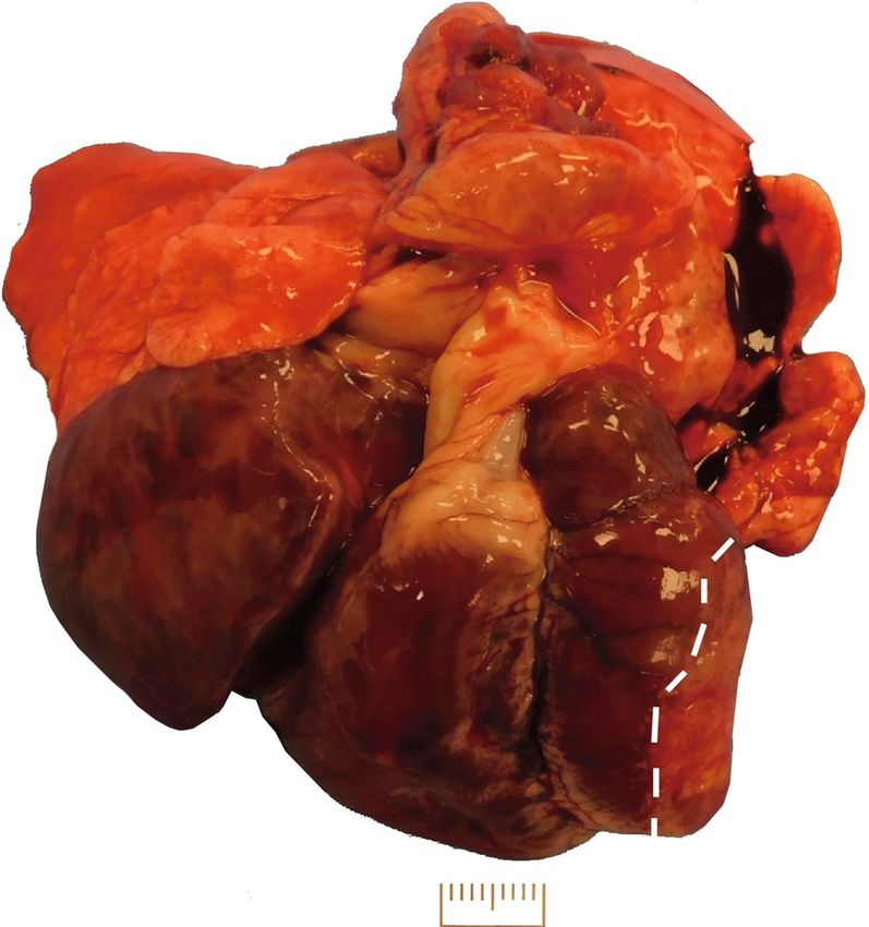
2                                                                                                 Case Reports in Veterinary Medicine

Figure 1: Gross image of the heart and lungs—caudal view of the heart illustrating a markedly enlarged right atrium (RA) and an enlarged
right ventricle (RV). The left ventricle (LV) was slightly smaller than the right ventricle. Lungs were partially compressed by the enlarged
heart, were moist and exuded fluid on sectioning, and had a rubbery texture. The pulmonary artery and entire pulmonary outflow tract
(POT) were also enlarged and dilated. Inset: an image of an age-matched rabbit is provided for comparison. Scale bar = 1 cm.

Figure 2: Gross image of the heart and lungs—caudal view of the heart. The left ventricle had a focally extensive depressed firm white area
encompassing approximately half of the ventricular free wall (outlined in white dashes). Scale bar = 1 cm.
Case Reports in Veterinary Medicine                                                                                                          3

Figure 3: Gross image of the right atrial and ventricular lumina. The lumina of the right atrium and ventricle were markedly dilated as was the
atrioventricular annulus. A fibrous ridge characterizes the annulus between the atrium and ventricle, and a single valve leaflet (free wall) is
seen with a hypoplastic cusp (right arrow). The location of where the septal leaflet was expected (left arrow) has no leaflet (cusp aplasia)
and no associated chordae tendineae. The chordae tendineae associated with the free wall leaflet were more characteristic of a thickened
fenestrated membrane than individual well-developed chordae tendineae. The right wall thickness was within normal limits, but overall
right heart mass was increased (eccentric hypertrophy). Inset: an image of an age-matched rabbit is provided for comparison. Scale bar = 1
 cm.

started exhibiting clinical signs of acute respiratory distress           the only other notable finding was a liver with minimal
and cyanosis. Based on the animal’s deteriorating condition               enlargement, an accentuated lobular pattern, and rounded
and distress, euthanasia was performed by intravenous injec-              margins.
tion of a pentobarbital-based euthanasia solution. A full nec-
ropsy was performed, and findings are the subject of this                  4. Discussion
manuscript.
    Animal husbandry, euthanasia, and all procedures were                 We describe a rabbit with decompensated congestive heart
in compliance with all applicable principles set forth in the             failure resulting from developmental anomalies in the RAV.
National Institutes of Health Guide for the Care and Use of               On gross necropsy, the rabbit had moist, rubbery, partially
Laboratory Animals (8th Edition, revised 2011).                           compressed but not collapsed lungs, a slightly enlarged liver
                                                                          with an accentuated lobular pattern and rounded margins,
3. Results                                                                and a markedly enlarged heart dominated by right atrial
                                                                          and ventricular enlargement. The heart had malformation
On necropsy, diffusely wet firm rubbery lungs were partially                of the RAV characterized by a hypoplastic free wall valve leaf-
compressed by a markedly enlarged heart. Heart enlarge-                   let, malformed chordae tendineae associated with the free
ment was characterized by marked right-sided heart enlarge-               wall leaflet, aplasia of the septal wall leaflet, and associated
ment composed of a right atrium that was more than doubled                chordae tendineae, and a markedly dilated annulus. Associ-
in size and a right ventricle with eccentric hypertrophy and              ated with the RAV anomaly was a markedly dilated right
enlargement by approximately 80-100% (Figure 1). The left                 atrium and right-sided eccentric myocardial hypertrophy.
ventricular free wall was characterized by partially thinned,             Additionally, an incidental finding of left ventricular free wall
white, fibrous tissue comprising approximately 30% of the                  infarction was seen.
ventricle consistent with a chronic infarct (Figure 2).                        This is the first case of RAV malformation published in
    Upon opening the heart, the right atrioventricular annu-              the rabbit. Clinical as well as pathologic cardiac and systemic
lus was markedly dilated and no definitive septal cusp was                 changes were consistent with RAV insufficiency. Liver find-
identified (chordae tendineae truncated at the fibrous ridge                ings were consistent with chronic passive congestion which
forming the right atrioventricular annulus); the free wall cusp           is an expected finding with decompensated right heart fail-
appeared smaller than normal and had a decreased number                   ure. Pulmonary changes were consistent with pulmonary
of chordae tendineae attaching to it (Figure 3). Systemically,            edema which is an expected finding with decompensated
4 Case Reports in Veterinary Medicine
congestive left heart failure. Both the hepatic and pulmonary Medical and Biological Research, vol. 39, no. 7, pp. 925–934,
findings were characteristic of biventricular heart failure as 2006.
were the antemortem clinical findings. [7] J. M. Closa and A. Font, “Traumatic tricuspid insufficiency in a
Non-RAV anomaly cardiac changes in the right heart kitten,” Journal of the American Animal Hospital Association,
were consistent with RAV insufficiency as well. Right atrial vol. 35, no. 1, pp. 21–24, 1999.
dilation is an expected outcome of chronic RAV regurgita- [8] T. R. Famula, L. M. Siemens, A. P. Davidson, and M. Packard,
tion. Right-sided myocardial eccentric hypertrophy is an “Evaluation of the genetic basis of tricuspid valve dysplasia in
expected compensatory mechanism associated with RAV Labrador Retrievers,” American Journal of Veterinary
insufficiency. The infarct of the left ventricular free wall is Research, vol. 63, no. 6, pp. 816–820, 2002.
not completely unexpected given the propensity of animals [9] R. Formigari, P. Francalanci, P. Gallo et al., “Pathology of
with valvular incompetence to have reflux rheostatic alter- atrioventricular valve dysplasia,” Cardiovascular Pathology,
ations resulting in turbulence and increased susceptibility to vol. 2, no. 2, pp. 137–144, 1993.
thromboemboli. It is uncertain if the left heart failure alter- [10] R. Kobza, D. J. Kurz, E. N. Oechslin et al., “Aberrant tendinous
ations were a consequence of infarction of the left ventricle chords with tethering of the tricuspid leaflets: a congenital
or of preload decreases secondary to right heart failure. anomaly causing severe tricuspid regurgitation,” Heart,
Given the unique structure of the rabbit RAV (bicuspid), vol. 90, no. 3, pp. 319–323, 2004.
it is not surprising that many of the RAV anomaly changes in [11] D. Lang, R. Oberhoffer, A. Cook et al., “Pathologic spectrum of
this species are different than those described in other species malformations of the tricuspid valve in prenatal and neonatal
and the consequences of malformation of a single leaflet are life,” Journal of the American College of Cardiology, vol. 17,
no. 5, pp. 1161–1167, 1991.
much more consequential. Criteria associate with human
EA were not met, but some of the changes seen in that anom- [12] N. A. Robinson and A. G. Armíen, “Tubular hypoplasia of the
aly were present in this case. Given the lack of any history of aorta and right atrioventricular valve dysplasia in a bulldog,”
Journal of Veterinary Diagnostic Investigation, vol. 22, no. 4,
any gestational or postgestational xenobiotic use and the lack
pp. 667–670, 2010.
of a history of early trauma, this rabbit’s RAV anomalies were
thought to be genetic in this rabbit and other changes were a
consequence of RAV insufficiency.
Abbreviations
EA: Ebstein’s anomaly
RAV: Right atrioventricular valve.
Conflicts of Interest
The authors declare that they have no conflicts of interest.
References
[1] K. Voros, F. Seehusen, S. Hungerbuhler, A. Meyer-Lindenberg,
and N. von der Hoeh, “Ventricular septal defect with aortic
valve insufficiency in a New Zealand White rabbit,” Journal
of the American Animal Hospital Association, vol. 47, no. 4,
pp. e42–e49, 2011.
[2] M. Nakata, Y. Miwa, J. K. Chambers, T. Saito, and K. Uchida,
“Ostium secundum type of atrial septal defect in a rabbit,”
Journal of Veterinary Medical Science, vol. 80, no. 8,
pp. 1325–1328, 2018.
[3] N. di Girolamo, C. Palmieri, M. Baron Toaldo et al., “First
description of partial atrioventricular septal defect in a rabbit,”
Journal of Exotic Pet Medicine, vol. 27, no. 4, pp. 5–9, 2018.
[4] G. Andelfinger, K. N. Wright, H. S. Lee, L. M. Siemens, and
D. W. Benson, “Canine tricuspid valve malformation, a model
of human Ebstein anomaly, maps to dog chromosome 9,”
Journal of Medical Genetics, vol. 40, no. 5, pp. 320–324, 2003.
[5] C. H. Attenhofer Jost, H. M. Connolly, J. A. Dearani, W. D.
Edwards, and G. K. Danielson, “Ebstein’s anomaly,” Circula-
tion, vol. 115, no. 2, pp. 277–285, 2007.
[6] F. Burdan, J. Szumilo, J. Dudka, A. Korobowicz, and
R. Klepacz, “Congenital ventricular septal defects and prenatal
exposure to cyclooxygenase inhibitors,” Brazilian Journal ofYou can also read