The somatic and autonomic innervation of the clitoris; preliminary evidence of sexual dysfunction after minimal invasive slings
←
→
Page content transcription
If your browser does not render page correctly, please read the page content below
Chapter 2
                   The somatic and autonomic innervation of the
                   clitoris; preliminary evidence of sexual dysfunction
                   after minimal invasive slings

                   (Submitted)

                   Milou D. Bekker1, Cornelis R.C. Hogewoning1, Chris Wallner2, Henk W. Elzevier1,
                   Marco C. De Ruiter3

                   1. Department of Urology, Leiden University Medical Center
                   2. Department of Anatomy en Embryology, Academic Medical Center
                   3. Department of Anatomy en Embryology, Leiden University Medical Center
                   The Netherlands

Milou BW.indd 21                                                                                     22-Feb-11 11:24:34 AM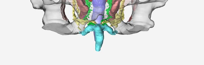
Milou BW.indd 22 22-Feb-11 11:24:34 AM
The somatic and autonomic innervation of the clitoris   23

                   Introduction

                   Within a decade, the midurethral (vaginal) slings became by far the most popular surgical
                   treatments for stress urinary incontinence (SUI), with more than one million women treated (1).
                   Despite the numerous studies on objective and subjective outcomes of this minimal invasive
                   procedure, very few studies have addressed the impact of vaginal sling procedures on sexual-
                   ity. Small series evaluating the sexual well-being before and after the tension-free vaginal tape
                   (TVT), the transobturator in-out (TVT-O) and/or the transobturator out-in (TOT) procedures
                   show conflicting results. Of these studies, some suggest deterioration (2-7) of sexual function,
                   some improvement (2-4;8-11), whereas others were equivocal (12-16). A prognostic factor in
                   the improvement of the sexual functioning of these patients is the cure of incontinence during
                   intercourse (17;18). Negative effect on sexuality is hypothesized to be related to the implanted
                   material because of damage to important vascular and/or neural genital structures (2-7).

                   The clitoris plays an important role in achieving female orgasm by sexual stimuli (19). It is
                   innervated by the dorsal nerves of the clitoris (DNC). These peripheral sensory afferents of the
                   clitoris originate from the pudendal nerve. The clitoris is also innervated with fibres coming
                   from the autonomic pelvic plexus (also known as the inferior hypogastric plexus): the cavern-
                   ous nerves of the clitoris. Clitoral and labial swelling during sexual arousal, is associated with
                   parasympathetic vasodilator mechanisms, among which nitric oxide (NO) appears to be a
                   primary neurotransmitter contributing to the mediation of this function (20;21). NO control of
                   vasodilatation and neuronal signaling between the cavernous nerves and the dorsal nerve of
                   the clitoris contribute to the engorgement and subsidence of clitoral tissue. This supports the
                   initiation of sexual arousal by tactile stimuli of the clitoris (22). Therefore, in theory, injury to the
                   clitoris and/or its innervating nerves, both somatic and autonomic, could lead to altered sexual
                   function.

                   To investigate the anatomical relation of vaginal slings to important neural genital structures,
                   basic knowledge about and detailed descriptions of the neuro-anatomy of the clitoris are
                   needed. The clitoris is a structure of which few diagrams and minimal descriptions are provided,
                   potentially impacting its preservation during surgery. Research has demonstrated the integral
                   relationship between the clitoris, and the distal urethra and vagina (23-25). O’Connell et al.
                   provided a major contribution to the research on the anatomy of the clitoris (23;26). They found
                   the clitoris to be intimately related to the distal urethra, which lead them to suggest that the
                   role of the urethra in sexual function is related to the position of the surrounding erectile tissue
                   rather than the urethral sphincter (23). The DNC was described in detail, the autonomic nerves,
                   however, were poorly addressed.

Milou BW.indd 23                                                                                                                22-Feb-11 11:24:34 AM
Disruption of the somatic innervation of the clitoris can lead to a diminished sensibility of the
clitoris, thereby affecting sexual arousal due to the absence of tactile stimuli. The DNC is located
along the medial aspect of the ischiopubic ramus (IPR) where it runs along the pubic bone in a
sulcus described as the sulcus nervi dorsalis clitoridis (27). Risk of injury to the DNC along the
IPR had been suggested by Delorme with the medial to lateral passage of the needle which
is used for placing vaginal slings and may alter postoperative sexual function such as arousal,
orgasmic function or pain (28). This possible risk has been illustrated by Lowenstein, showing
the topographic relation of mid-urethral sling for stress urinary incontinence to critical female
genital structures (29). In a cadaveric study, performed by Achtari et al., the potential risks
of three vaginal slings to the DNC were evaluated. Distances of a TVT, transobturator in-out
(TVT-O) and transobturator out-in (Monarc) to the DNC were similar (11-12 mm) (30). Given
the outside-in course, the Monarc was claimed to (theoretically) be the safest device. Another
cadaveric study found similar results, although they only documented the course of the DNC
from the piercing of the perineal membrane to its terminal branching and not its course along
the IPR (31).
The cavernous nerves of the clitoris are involved in the neural control of vasocongestion
and, consequently, the lubrication-swelling response. Disruption of these nerves could lead
to altered vascular function during sexual arousal and possibly disordered orgasm. However,
although important for normal sexual function, in afore mentioned studies no attention was
Chapter 2
paid to possible disruption of the cavernous nerves of the clitoris (29-31).
24
The aim of this study was to reinvestigate the neuro-anatomy of the clitoris by performing dis-
section in an adult female pelvis and by using (immuno)histochemical and three-dimensional
(3D) reconstruction techniques on a female fetus. In this study we focus on 1) the autonomic
innervation of the clitoris, 2) the course of the DNC, 3) to investigate the anatomical sites of
potential nerve damage during vaginal sling surgery for SUI.
During dissection, it is difficult to recognize and dissect small nerve fibers such as those from
the pelvic plexus. Therefore, there is a high risk of artifacts because tissue strings may be mis-
taken for small nerves. The study of serially sectioned human fetuses has an advantage over
conventional cadaver dissections, because of immunohistochemical staining; structures of
interest are easily recognized (32-35).
Dimensions may change but the topographic relationships of tissues persist throughout
fetal development which makes the study of fetuses excellent for describing anatomy (32;36).
Furthermore, 3D reconstructions of the innervation of the clitoris can be prepared to deduce its
anatomical relationship to other pelvic structures and provide an insightful illustration which
can be used by pelvic surgeons.
Milou BW.indd 24 22-Feb-11 11:24:34 AMThe somatic and autonomic innervation of the clitoris 25
Methods
Fetal 3D-reconstruction
Fetal pelves from the collections in the departments of Anatomy and Embryology at the Leiden
University Medical Centre and at the Amsterdam Medical Centre were studied. Eleven paraffin
embedded fetuses (all female), ranging from 10 to 27 weeks of gestation; 6-26 mm crown-rump
length (CRL) were serially sectioned. Six were stained with haematoxylin and eosin, three with
haematoxylin-azophloxine and two with both haematoxylin and neurofilament (35). The fetal
tissue was fixed in 4% paraformaldehyde, embedded in paraffin and transversely sectioned into
serial sections of 10 μm. Analysis of the transverse sections was performed from superior part
of the pubic arch to just below Alcock’s canal. Digital images were taken of the serial sections,
photographing every second section. These images were used to prepare three dimensional
reconstructions with the Amira software package (v3.0, Visage Imaging GmbH, Fürth, Germany).
Unfortunately, in the transversed sections, the perineal membrane (or urogenital diaphragm)
was not recognized and thus not reconstructed.
Cadaver study
The pelvis of a female cadaver without signs of any pelvic surgery was used for this study. Usu-
ally, the age of death of these women is older than 70 years of age. Preservation of the cadaver
was performed by injection of the embalming fluid AnubiFiX™ into the femoral artery. Due
to this fixation process, the cadaver remains flexible which enables a natural dissection and
allows surgical procedures. The cadaver was donated to the university for medical research and
hence does not require separate ethics approval to be dissected. A trained urologist (H.W.E.)
performed parts of both procedures (TVT and TVT-O) on one cadaver, one sling placement on
each side. Both procedures were performed exactly similar as they are normally performed on
patients.
The pelvis was sectioned through the midline from the pubic symphysis anteriorly to the
sacrum posteriorly. The urethra was sectioned along its full length in the midline. The clitoris
and its somatic and autonomic nerves were dissected and the shortest distance between the
needle/sling and the nerves were measured and various stages of the dissection were recorded
photographically.
An interactive three-dimensional reconstruction is attached to this article, available as a
supplementary file showing the pelvis, the pelvic organs, the clitoris, its autonomic (cavernous)
nerves and the branches of the pudendal nerve, the obturator nerve and the levator ani muscle
of a female foetus (12 wk of gestation; same foetus as shown in Figures 1A-H).
Milou BW.indd 25 22-Feb-11 11:24:34 AMResults
Three-dimensional reconstruction
Anatomy of the clitoris
The clitoris is a multiplanar structure positioned deep to the labia minora, labial fat and vascula-
ture, bulbospongiosus and ischiocavernosus muscles, inferior to the pubic arch and symphysis.
It has a broad attachment to the pubic arch, and via extensive supporting tissue to the mons
pubis (the adipose tissue lying above the pubic bone of adult females, anterior to the symphysis
pubis) and labia. The clitoris consists of a tip, also known as the glans clitoridis, the erectile body
and the crura (or corpora caverosa) (Fig. 1A). The clitoris has a close relationship to the distal
urethra and vagina (Fig. 1B). Because this study involved human fetuses, aged 10-27 weeks of
gestation, these two structures are still partly merged.
Dorsal nerve of the clitoris
The three-dimensional reconstruction illustrates the course of the dorsal clitoral nerves. They
originate from the pudendal nerve in the Alcock’s canal immediate medial to the pubic bone
and lateral to the rectum, forming a bundle that fannes out laterally, passing the levator ani
muscle and ascending to the clitoral bodies (Fig. 1C, D, E and F). The 3-D images also revealed
Chapter 2
that both dorsal nerves of the clitoris run medial and close to the puboischial ramus (Fig. 1E).
Furthermore, the close relationship of the clitoral crura to the clitoral dorsal nerve was notable.
26
They further traverse distally along the clitoral crura and run posterior to the body of the cli-
toris before hooking over it in anterolateral direction. There, they medially cross the cavernous
nerves and pass over the clitoral body (Fig. 1E and G). As they pass over the body, they merge
with the autonomic nerves. After passing over the body, the merged somatic and autonomic
nerves pass further caudal and anterior over the clitoral body to the glans clitoridis (Fig. 1E
and H).
Autonomic nerves of the pelvic plexus
From the superior hypogastric plexuses, two nerves (the hypogastric nerves) run bilaterally into
the small pelvis, to be joined by the pelvic splanchnic nerves coming from sacral roots S2 to S4,
to form the pelvic plexus, also known as the inferior hypogastric plexus (IHP) on both sides of
pelvic organs (Fig. 1A). The IHP showed to be a triangularly shaped plexus in a sagitally plane.
It is in close contact with its target organs as a flat meshed plaque of nerve tissue, stretching
from anterolateral to the rectum, passing the cervix and vagina laterally and extending from
the lateral vaginal wall to the base of the bladder and lateral to the urethra with branches to
the clitoris.
Its nerves extend onto the lateral walls of the proximal and mid-vagina, where they form a
dense network. These nerves travel superior to cover the proximal anterior vaginal wall, where
Milou BW.indd 26 22-Feb-11 11:24:34 AMCHAPTER 2
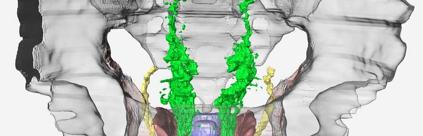
Figure 1
The(grey),
A. Anterior view (3D) of the clitoris (blue), the pelvis somaticthe
and autonomic innervation
urethra/vagina (U, of the clitoris 27
purple), the pudendal nerves (yellow), the hypogastric (autonomic) nerves
Figure 1
A. Anterior view(green)
(3D) oftravelling into (blue),
the clitoris the pelvis
theand forming
pelvis the the
(grey), inferior hypogastric plexus
urethra/vagina (IHP), the pudendal
(U, purple),
nerves (yellow), the hypogastric (autonomic) nerves (green)
the merged autonomic and dorsal nerves (light green).
travelling into the pelvis and forming the
inferior hypogastric plexus (IHP), the merged autonomic and dorsal nerves (light green).
Hypogastric nerves
IHP
crus
glans
B. Lateral view (3D) of the midsaggital cut pelvis with the symphysis (Sy) centered
(grey), the levator ani muscle (red) the clitoris (blue), the autonomic (green) and
somatic (yellow) nerves and the urethra/vagina (U, purple). IHP: inferior
B. Lateral view (3D) of the midsaggital cut pelvis with the symphysis (Sy) centered (grey), the levator ani
muscle (red)hypogastric
the clitorisplexus,
(blue), PN:
the pudendal
autonomic (green)
nerve, CN: and somatic
cavernous (yellow)
nerve, DNC:nerves
dorsaland the urethra/
vagina (U, purple). IHP: inferior hypogastric plexus, PN: pudendal nerve, CN: cavernous nerve, DNC:
nerve of the clitoris, Sa: sacrum.
dorsal nerve of the clitoris, Sa: sacrum.
Sa
IHP
Sy PN
CN
U
DNC
Milou BW.indd 27 22-Feb-11 11:24:34 AMC. Lateral view (3D) of the pelvis (grey) on the obturator foramen, the clitoris (blue), the autonomic
(green) and somatic (yellow) nerves and the urethra/vagina (purple). IHP; inferior hypogastric plexus,
C. Lateral view (3D) of the pelvis (grey) on the obturator foramen, the clitoris (blue), the autonomic
(green) and
PN; pudendal somatic
nerve, CN;(yellow) nerves
cavernous and the
nerve, urethra/vagina
DNC; (purple).
dorsal nerve of theIHP; inferior hypogastric plexus,
clitoris.
PN; pudendal nerve, CN; cavernous nerve, DNC; dorsal nerve of the clitoris.
IHP
ON
CN
PN
DNC
D. Posterior view (3D) of the pelvis with the pubic bone centered (grey), the clitoris
(blue), the autonomic (green) and somatic (yellow) nerves and the urethra/vagina
Chapter 2
(purple). IHP; inferior hypogastric plexus, ON; obturator nerve, PN; pudendal
D. Posterior view (3D) of the pelvis with the pubic bone centered (grey), the clitoris (blue), the autonomic
nerve, Obt. F; obturator foramen, CN; cavernous nerve, DNC; dorsal nerve of the
(green) and somatic (yellow) nerves and the urethra/vagina (purple). IHP; inferior hypogastric plexus,
28 ON; obturator
clitoris nerve, PN; pudendal nerve, Obt. F; obturator foramen, CN; cavernous nerve, DNC; dorsal
nerve of the clitoris
ON
IHP IHP
PN
Obt. F
CN
DNC
Milou BW.indd 28 22-Feb-11 11:24:34 AME. Anterior and slightly lateral view (3D), of the pelvis (grey) the obturator foramen
(Obt. F), the obturator nerve (ON), the clitoris (purple), the dorsal nerve of the
clitoris (DNC, yellow) and the cavernous nerves
The (green, CN)
somatic and cominginnervation
autonomic from the of the clitoris 29
inferior hypogastric plexus (IHP). ON; obturator nerve,
E. Anterior and slightly lateral view (3D), of the pelvis (grey) the obturator foramen (Obt. F), the obturator
nerve (ON), the clitoris (purple), the dorsal nerve of the clitoris (DNC, yellow) and the cavernous nerves
(green, CN) coming from the inferior hypogastric plexus (IHP). ON; obturator nerve,
ON
IHP IHP
Obt. F
DNC
CN
F. Stained section, showing the body of the clitoris (B) with its crura (Cr) close to the
ramus inferior of the pubic bone (IPR), and the dorsal clitoral nerves (yellow
F. Stained section, showing the body of the clitoris (B) with its crura (Cr) close to the ramus inferior of the
pubicarrows)
bone (IPR), and the
passing dorsal
along theclitoral nerves
obturator (yellow (Obt.
foramen arrows) passing along the obturator foramen
F.).
(Obt. F.).
Urethra/vagina
B
Cr Cr
IPR IPR
Obt. F Obt. F
rectum
Levator ani muscle
Milou BW.indd 29 22-Feb-11 11:24:34 AMG. Close-up of a stained section. The close relationship of the ramus inferior of the
pubic bone (IPR) to the clitoral dorsal nerve (yellow arrows) is notable and it
shows that the branches of the cavernous nerves of the clitoris pass medial to
G. Close-up of athe dorsalsection.
stained nerves The
(green arrows).
close B: clitoral
relationship of body, U: urethra/vagina.
the ramus inferior of the pubic bone (IPR) to the
clitoral dorsal nerve (yellow arrows) is notable and it shows that the branches of the cavernous nerves
of the clitoris pass medial to the dorsal nerves (green arrows). B: clitoral body, U: urethra/vagina.
B
IPR IPR
U
H. Stained section, showing both cavernous nerves (green arrows) and the dorsal
nerves of the clitoris (yellow arrows) hooking over the clitoral body and travelling
H. Stained section, showing both cavernous
further caudally nerves
alongside and (green
into the clitoralarrows)
body andand
glansthe
(reddorsal nerves of the clitoris
arrows).
(yellow arrows) hooking over the clitoral body and travelling further caudally alongside and into the
clitoral body and glans (red arrows).
Chapter 2
30 IPR
IPR
I. Stained section, the autonomic nerves merge with the branches of the dorsal
I. Stained section, the autonomic nerves merge with the branches of the dorsal nerves as they pass over
nerves as they pass over the clitoral body (light green arrows).
the clitoral body (light green arrows).
IPR
IPR
Milou BW.indd 30 22-Feb-11 11:24:34 AMThe somatic and autonomic innervation of the clitoris 31
they form the cavernous nerves at the 2 and 10 o’clock positions along the urethra. (Fig. 1B
and F)
There, they travel further caudal to the clitoral bodies crossing the dorsal clitoral nerve medi-
ally (Fig. 1C). The nerve bundles then travel alongside the branches of the dorsal nerve passing
over the clitoral body. After passing over the clitoral body, these autonomic nerves merge with
the branches of the dorsal nerve and travel further caudally alongside and into the clitoral body
and glans (Fig. 1E, H and I).
Dissection
Anatomy of the clitoris
The initially almost straight clitoral crura commence proximally running along the puboischial
ramus and join distally under the pubic symphysis as a single clitoral body that projects ante-
riorly into the glans. There it projects into the fat of the mons pubis. They are situated between
the clitoral crura and form a midline core in the triangular shaped clitoral structure. Dissection
shows that the apex of this triangular structure is the most superior point of the clitoral body,
where it attaches to the under surface of the pubic symphysis by the deep suspensory liga-
ment (Fig. 2). As the clitoral body projects from the bone into the mons pubic fat, it descends
and folds back on itself in a boomerang-like shape in a dorsalcaudalward direction forming
the glans clitoridis.
Figure 2.The glansview
Anterior of the clitoris
of the is a clitoris.
dissection relativelyThe small nodular
mons pubis has structure that
been opened to becomes
partially covered
show by
the the
deepglando-preputial lamella
suspensory ligament. and prepuce
Furthermore, (orcrus,
the clitoral clitoral
bodyhood).
and glans
are shown with the dorsal nerve of the clitoris (DNC) ascending along the inferior
Figure 2. Anterior view of the dissection clitoris. The mons pubis has been opened to show the deep
ramus of the pubic bone (IPR), hooking over the clitoral body whilst passing through
suspensory ligament. Furthermore, the clitoral crus, body and glans are shown with the dorsal nerve of
the ascending
the clitoris (DNC) suspensoryalong
ligament
theand branching
inferior ramusinto thepubic
of the clitoris.bone
(LMi:(IPR),
labiahooking
minora) over the clitoral
body whilst passing through the suspensory ligament and branching into the clitoris. (LMi: labia minora)
Milou BW.indd 31 22-Feb-11 11:24:34 AMDorsal nerve of the clitoris
The course of the pudendal nerve (PN) around the ischial spine was approached posteriorly by
removal of the skin and superficial fascia between the anterior inferior iliac spine, the ischial
tuberosity and the posterior superior iliac spine. The gluteus maximus muscle was dissected
from its origin to expose underlying structures. The sacrotuberous ligament was identified and
transected to identify the PN subjacent to the sacrotuberous ligament and around the ischial
spine of the pelvis. The entrance of PN into Alcock’s canal was identified. Alcock’s canal was
then unroofed which revealed the three main branches of the PN, namely, the inferior rectal
nerve, the perineal nerve and the dorsal nerve of the clitoris (DNC). The DNC was then followed
until its termination in the clitoris. The DNC travels along the perineal membrane (or urogenital
diaphragm) and runs inferior to the inferior pubic ramus. By opening the perineal membrane
the TVT-O tape was exposed. (Figure 3 A-B) The distance of the TVT-O to the DNC was 2 mm and
they were separated by the perineal membrane. (Figure 3)
Figure 4 is a schematic lateral view on a midsagittal sectioned right pelvis showing the
course of the DNC from the PN to the clitoris lateral from the levator ani muscle.
Figure 3 A. Frontal view of the right female genital and perineal area. In order to expose
the DNC the skin was opened between the right labia majora and minora. To show the
route of the TVT-O sling, the perineal membrane was opened subsequently. DNC;
Chapter 2
Figure
dorsal3nerve
A. Frontal viewclitoris,
of the of the right femalethe
TVT-O; genital and perineal
tensionfree area. In
vaginal order
tape to expose the
obturator; IPR;DNC the
inferior
skin was opened between the right labia majora and minora. To show the route of the TVT-O sling, the
32 perineal membrane
pubic ramus, LMa; waslabia
opened subsequently.
majora, DNC;minora.
LMi; labia dorsal nerve of the clitoris, TVT-O; the tensionfree
vaginal tape obturator; IPR; inferior pubic ramus, LMa; labia majora, LMi; labia minora.
ventral
lateral medial
dorsal
Milou BW.indd 32 22-Feb-11 11:24:34 AMFigure 3 B. Close-up of figure 3A. The bulbospongiosus muscle and clitoral crus were
moved medially to show the course of the DNC. To show the route of the TVT-O sling,
the perineal membrane was opened. DNC; dorsal nerve of the clitoris, TVT-O; the
The somatic and autonomic innervation of the clitoris 33
tensionfree vaginal tape obturator; IPR; inferior pubic ramus.
Figure 3 B. Close-up of figure 3A. The bulbospongiosus muscle and clitoral crus were moved medially to
show the course of the DNC. To show the route of the TVT-O sling, the perineal membrane was opened.
DNC; dorsal nerve of the clitoris, TVT-O; the tensionfree vaginal tape obturator; IPR; inferior pubic ramus.
ventral
lateral medial
dorsal
Milou BW.indd 33 22-Feb-11 11:24:34 AMFigure 4 A schematic lateral view on a midsaggital sectioned right pelvis showing
the course of the autonomic nerves from the hypogastric nerve to the target
organs and the course of the DNC from the pudendal nerve, lateral from the
levator ani muscle to the clitoris.
Figure 4 A schematic lateral view on a midsaggital sectioned right pelvis showing the course of the
autonomic nerves from the hypogastric nerve to the target organs and the course of the DNC from the
pudendal nerve, lateral from the levator ani muscle to the clitoris.
Chapter 2
Autonomic nerves
The superior hypogastric plexus was identified inferior to the bifurcation of the aorta. The proxi-
34
mal hypogastric nerves were identified in the subperitoneal layer (between the peritoneum
and the endopelvic fascia) and followed alongside the ureter into the small pelvis to the inferior
hypogastric plexus (IHP). Figure 4 is a schematic lateral view on a midsagittal sectioned right
pelvis showing the course of the autonomic nerves from the hypogastric nerve to the target
organs.
The IHP, a flat meshed plaque of nerves was dissected. Its branches, which follow the con-
nective tissue plane within the small pelvis which supports the uterine cervix, vagina and blad-
der, were identified and dissected into their target organs. Special attention was paid to the
branches passing along the urethra and innervating the clitoris. The autonomic nerves, running
from the IHP, were found to be pierced by the TVT-needle. (Figure 5)
Discussion
This study describes the neuro-anatomy of the clitoris; its somatic and autonomic pathways.
Previous studies on the innervation of the clitoris were mainly focused on the dorsal nerve of
the clitoris, paying no attention to the cavernous nerves coming from the pelvic plexus, which
play an important role in female sexual function (23;25-27;31;37-39). The cavernous nerves are
Milou BW.indd 34 22-Feb-11 11:24:34 AMFigure 5: View from above (abdominal view) into left female half pelvis. In order to expose the area lateral
to the vagina, the uterus was removed from the level of the cervix and the bladder and vagina had been
The somatic and autonomic innervation of the clitoris 35
retracted medially and anteriorly. The peritoneum and part of the fascia have been removed. IHP; inferior
Figure 5: View from above (abdominal view) into left female half pelvis. In order to expose the area lateral
to the vagina, the
hypogastric uterusB;
plexus, was removedV;
bladder, from the level
vagina, O;ofOvarian,
the cervixTVT;
and the
thebladder and vagina
TVT tape, had been
NF; nerve fibres from the IHP
retracted medially and anteriorly. The peritoneum and part of the fascia have been removed. IHP; inferior
to the vagina
hypogastric andB;clitoris,
plexus, bladder, Obt C; obturator
V; vagina, O; Ovarian,canal.
TVT; the TVT tape, NF; nerve fibres from the IHP to
the vagina and clitoris, Obt C; obturator canal.
involved in the neural control of vasocongestion and, consequently, the lubrication-swelling
response. Disruption of these nerves could lead to altered vascular function during sexual
arousal and possibly disordered orgasm.
In 1982, Walsh and Donker described the anatomic location of the pelvic plexus (or IHP) in
men and the nerves innervating the corpora cavernosa (40). These pioneering observations
and descriptions of the anatomical basis for radical prostatectomy fostered resurgence in the
use of surgery as treatment for localized prostate cancer and led urologic surgeons to refine a
nerve-sparing technique within the following two decades. Only in the recent years, attention
is paid to the IHP in females and nerve-sparing techniques are being developed in surgery for
cervical cancer (41-43).
Although the IHP has been described in females, little attention has been paid to the cav-
ernous nerves coming from this IHP and their anatomical relation to other pelvic structures.
Yucel et al reported that the cavernous nerve supply the female urethral sphincter complex and
clitoris (25). The branches of the cavernous nerve were described and, as in our study, noted to
join the clitoral dorsal nerves. The cavernous nerves have also been described in mice, using
immunostaining to show communicating nerves between the cavernous nerve and the dorsal
Milou BW.indd 35 22-Feb-11 11:24:34 AMnerve of the clitoris which supports the initiation of sexual arousal by tactile stimuli and follow-
ing clitoral swelling (22). This study underlines the importance of both somatic and autonomic
innervation of the clitoris in normal female sexual function.
In the study performed by O’Connell et al. the course of the dorsal nerve was described
but the cavernous nerves were poorly addressed (26). Other important studies on the neuro-
anatomy of the clitoris focused mainly on the DNC (26;27;39;44). Similar to our findings, the
DNC is described to originate from the pudendal nerve and to ascend along the ischiopubic
rami.
Vaginal sling procedures for stress urinary incontinence have been developed in the nine-
ties by Ulmsten (45). After research showed the procedure to be safe and effective, the TVT
and derived procedures became a well established surgical procedure for the treatment of
female urinary stress incontinence. Especially in these early years, no attention was paid to the
topographic relation to important genital structures. Only in recent years, the possible risk of
nerve damage during vaginal sling procedures, especially the obturator procedures, has been
suggested (29).
The aim of this study was not only to describe the neuro-anatomy of the clitoris, but also its
relation to surrounding structures which are anatomical landmarks in vaginal tape procedures
for SUI. When performing vaginal sling surgeries, a sagittal incision is made within the anterior
vaginal wall mucosa about 1 centimeter from the urethral meatus and the vaginal mucosa is
Chapter 2
dissected of the mid-urethra. When performing the TVT procedure, a tape is placed (blindly)
behind the pubic symphysis using trocars attached to the tape when performing the TVT
36
procedure (46).
When performing the TOT, the ‘outside-in’, procedure, a similar midline incision is made in
the anterior vaginal wall between the mid-urethra and bladder neck, enough to introduce the
index finger. Dissection is carried out laterally to the level of the vaginal sulcus without penetra-
tion of endopelvic fascia. The IPR of the pubic bone and the obturator foramen are located
manually, and the medial edge of the ramus is pinched between thumb and index finger. The
skin puncture is made at the level of the clitoris right above the pinching thumb. The curved
sling passer is guided from the thumb to the index finger and then rotated and delivered to the
vaginal incision with the tip on the index finger. The arm of the tape is hooked to the tip of the
passer and brought out to the skin (46).
When performing the TVT-O, the ‘outside-in’, a similar midline incision is made and peri-
urethral tunnels are developed bilaterally. Unlike the TOT, where the dissection stops at the
IPR, with the TVT-O the obturator membrane is perforated with the tip of the scissors. A winged
metal trocar, which is designed to help guide the tape around the IPR, is inserted into the peri-
urethral tunnel and the tip is pushed just beyond the perforated membrane. The trocar is then
rotated around the IPR to exit out the skin through stab incisions located 2 cm above a horizon-
tal line at the level of the urethral meatus and 2 cm outside the thigh folds. The same procedure
is performed on the other side and the sling is tensioned and the procedure completed (46).
Milou BW.indd 36 22-Feb-11 11:24:34 AMThe somatic and autonomic innervation of the clitoris 37
As described, during these procedures, the part of the mid-urethra along the anterior vagi-
nal wall is an important surgical site; here the first incision is made. This study illustrates that the
urethra is surrounded by autonomic nerves coming from the IHP. Not only do they travel to and
innervate the urethra, the cavernous nerves travel from the vaginal nervous plexus occupying
the 2 and 10 o’clock positions on the anterolateral vagina and they travel at the 5 and 7 o’clock
positions along the urethra. It is therefore, possible that during the mid-urethral incision in
all vaginal procedures and the para-urethral tunneling during the obturator procedures, the
cavernous nerves are disrupted. The results of the cadaveric part of this study showed that
indeed the cavernous nerves are being pierced during the TVT procedure.
The IPR plays an important role in the obturator procedures; the tapes are placed around
this bony structure. Furthermore, dissection is performed paraurethral to the IPR, where during
the inside-out technique the obturator membrane is perforated with scissors. Because the DNC
travels along the medial side of the IPR, especially at the level of the urethral meatus, it is at risk
to be damaged during obturator procedures, both outside-in and inside-out. The results of the
dissections confirmed that the DNC is at risk for nerve damage during the TVT-O procedure.
Achtari et al. have also dissected female cadavers and measured the distance of the DNC
to the TVT, TVT-O and Monarc. The results showed a distance varying from 19-40 mm, the
TVT-O being the closest to the DNC (30). A similar study in fresh cadavers measured a distance
between the DNC and the TOT of 3-14 mm. Despite this small distance, they concluded the TOT
to be a safe procedure. This study was however biased because straight needles were used to
mimic the course of the TOT, instead of curving trocars (31). As the results of this study shows,
it is possible that during dissection to the foramen and during placement of the tape, the DNC
is injured.
This study is the first to illustrate, in detail, both somatic and autonomic pathways of the
clitoris and thereby, significant progress has been made in the field of female sexual anatomy
and its representation. This may facilitate further research in the related fields of female sexual
health and education and can be used by surgeons in the field of urogynecology. Furthermore,
the topographic relation of vaginal slings to the important critical female genital structure, the
clitoris, has been illustrated and described for the first time. Future (clinical) research should be
performed to confirm these results and to investigate the consequences of injury to the clitoral
nerves on the clitoral sexual response and female sexual functioning.
Conclusion
This study shows and described the somatic and autonomic innervation of the clitoris in detail
including their relation to important surrounding structures.
Furthermore, the relation of vaginal sling procedures for SUI to the clitoris and its innervation
has been evaluated. Given the course of the dorsal clitoral nerve; inferior to the inferior pubic
Milou BW.indd 37 22-Feb-11 11:24:34 AMramus, it is at risk for iatrogenic injury after placement of transobturator tape. Furthermore,
the autonomic innervation of the vaginal wall is disrupted by the tensionfree vaginal tape
procedure, which could lead to altered lubrication-swelling response. When the “inside–out”
technique is used, the introducer can come into contact with the dorsal nerve of the clitoris
because the introducer to passes through the obturator foramen close to the ischio-pubic
ramus.
Acknowledgements
We wish to thank from the department of Anatomy and Embryology, Leiden University Medical
Center, M.J. Aarents, A.H. Van Immerseel and J. Den Boeft for their assistance with the anatomi-
cal dissections and L.J. Wisse for his assistance with the three-dimensional reconstruction.
Furthermore, we wish to thank the NutsOhra Fund for their financial support of this study.
Chapter 2
38
Milou BW.indd 38 22-Feb-11 11:24:34 AMThe somatic and autonomic innervation of the clitoris 39
Reference list
(1) Hermieu JF. [Suburethral bands in women urinary stress incontinence: a review of the various tech-
niques]. Ann Urol (Paris) 2005 June;39(3-4):124‑36.
(2) Berthier A, Sentilhes L, Taibi S, Loisel C, Grise P, Marpeau L. Sexual function in women following the
transvaginal tension-free tape procedure for incontinence. Int J Gynaecol Obstet 2008 August;102(2):
105‑9.
(3) Elzevier HW, Venema PL, Lycklama à Nijeholt AAB. Sexual function after tension-free vaginal tape
(TVT) for stress incontinence: results of a mailed questionnaire. Int Urogynecol J Pelvic Floor Dysfunct
2004 September;15(5):313‑8.
(4) Ghezzi F, Serati M, Cromi A, Uccella S, Triacca P, Bolis P. Impact of tension-free vaginal tape on sexual
function: results of a prospective study. Int Urogynecol J Pelvic Floor Dysfunct 2006 January;17(1):
54‑9.
(5) Mazouni C, Karsenty G, Bretelle F, Bladou F, Gamerre M, Serment G. Urinary complications and sexual
function after the tension-free vaginal tape procedure. Acta Obstet Gynecol Scand 2004 October;
83(10):955‑61.
(6) Rogers RG, Kammerer-Doak D, Darrow A, Murray K, Olsen A, Barber M, Qualls C. Sexual function after
surgery for stress urinary incontinence and/or pelvic organ prolapse: a multicenter prospective study.
Am J Obstet Gynecol 2004 July;191(1):206‑10.
(7) Yeni E, Unal D, Verit A, Kafali H, Ciftci H, Gulum M. The effect of tension-free vaginal tape (TVT) pro-
cedure on sexual function in women with stress urinary incontinence. Int Urogynecol J Pelvic Floor
Dysfunct 2003 December;14(6):390‑4.
(8) Jha S, Moran P, Greenham H, Ford C. Sexual function following surgery for urodynamic stress inconti-
nence. Int Urogynecol J Pelvic Floor Dysfunct 2007 August;18(8):845‑50.
(9) Jha S, Radley S, Farkas A, Jones G. The impact of TVT on sexual function. Int Urogynecol J Pelvic Floor
Dysfunct 2008 October 21;20(2):165‑9.
(10) Murphy M, van RH, Mercurio E, Haff R, Wiseman B, Lucente VR. Incontinence-related quality of life and
sexual function following the tension-free vaginal tape versus the “inside-out” tension-free vaginal
tape obturator. Int Urogynecol J Pelvic Floor Dysfunct 2008 April;19(4):481‑7.
(11) Pace G, Vicentini C. Female sexual function evaluation of the tension-free vaginal tape (TVT) and
transobturator suburethral tape (TOT) incontinence surgery: results of a prospective study. J Sex Med
2008 February;5(2):387‑93.
(12) Glavind K, Tetsche MS. Sexual function in women before and after suburethral sling operation for
stress urinary incontinence: a retrospective questionnaire study. Acta Obstet Gynecol Scand 2004
October;83(10):965‑8.
(13) Maaita M, Bhaumik J, Davies AE. Sexual function after using tension-free vaginal tape for the surgical
treatment of genuine stress incontinence. BJU Int 2002 October;90(6):540‑3.
(14) Marszalek M, Roehlich M, Racz U, Metzenbauer M, Ponholzer A, Rauchenwald M, Madersbacher S.
Sexual function after tension-free vaginal tape procedure. Urol Int 2007;78(2):126‑9.
(15) Sentilhes L, Berthier A, Caremel R, Loisel C, Marpeau L, Grise P. Sexual function after transobturator
tape procedure for stress urinary incontinence. Urology 2008 June;71(6):1074‑9.
(16) Shah SM, Bukkapatnam R, Rodriguez LV. Impact of vaginal surgery for stress urinary incontinence on
female sexual function: is the use of polypropylene mesh detrimental? Urology 2005 February;65(2):
270‑4.
(17) Serati M, Cattoni E, Salvatore S. Coital Incontinence: The Tip of the Iceberg? J Sex Med 2010 April 1.
Milou BW.indd 39 22-Feb-11 11:24:34 AM(18) Bekker M, Beck J, Putter H, Venema P, Lycklama à Nijeholt A, Pelger R, Elzevier H. Sexual Function
Improvement Following Surgery for Stress Incontinence: The Relevance of Coital Incontinence. J Sex
Med 2009 July 21;6(11):3208‑13.
(19) Leff JJ, Israel M. The relationship between mode of female masturbation and achievement of orgasm
in coitus. Arch Sex Behav 1983 June;12(3):227‑36.
(20) Burnett AL, Calvin DC, Silver RI, Peppas DS, Docimo SG. Immunohistochemical description of nitric
oxide synthase isoforms in human clitoris. J Urol 1997 July;158(1):75‑8.
(21) Hoyle CH, Stones RW, Robson T, Whitley K, Burnstock G. Innervation of vasculature and microvascula-
ture of the human vagina by NOS and neuropeptide-containing nerves. J Anat 1996 June;188 ( Pt 3):
633‑44.
(22) Martin-Alguacil N, Pfaff DW, Shelley DN, Schober JM. Clitoral sexual arousal: an immunocytochemical
and innervation study of the clitoris. BJU Int 2008 June;101(11):1407‑13.
(23) O’Connell HE, Hutson JM, Anderson CR, Plenter RJ. Anatomical relationship between urethra and
clitoris. J Urol 1998 June;159(6):1892‑7.
(24) Suh DD, Yang CC, Cao Y, Garland PA, Maravilla KR. Magnetic resonance imaging anatomy of the female
genitalia in premenopausal and postmenopausal women. J Urol 2003 July;170(1):138‑44.
(25) Yucel S, De SA, Jr., Baskin LS. Neuroanatomy of the human female lower urogenital tract. J Urol 2004
July;172(1):191‑5.
(26) O’Connell HE, Sanjeevan KV, Hutson JM. Anatomy of the clitoris. J Urol 2005 October;174(4 Pt 1):
1189‑95.
(27) Sedy J, Nanka O, Belisova M, Walro JM, Jarolim L. Sulcus nervi dorsalis penis/clitoridis: anatomic
structure and clinical significance. Eur Urol 2006 November;50(5):1079‑85.
Chapter 2
(28) Delorme E, Droupy S, de TR, Delmas V. Transobturator tape (Uratape): a new minimally-invasive
procedure to treat female urinary incontinence. Eur Urol 2004 February;45(2):203‑7.
40 (29) Lowenstein L. Topographic relation to mid-urethral sling for stress incontinence to critical female
genital structures. J Sex Med 2009;6(11):2954‑7.
(30) Achtari C, McKenzie BJ, Hiscock R, Rosamilia A, Schierlitz L, Briggs CA, Dwyer PL. Anatomical study
of the obturator foramen and dorsal nerve of the clitoris and their relationship to minimally invasive
slings. Int Urogynecol J Pelvic Floor Dysfunct 2006 June;17(4):330‑4.
(31) Tate SB, Culligan PJ, Acland RD. Outside-in transobturator midurethral sling and the dorsal nerve of
the clitoris. Int Urogynecol J Pelvic Floor Dysfunct 2009 November;20(11):1335‑8.
(32) Fritsch H. Topography of the pelvic autonomic nerves in human fetuses between 21-29 weeks of
gestation. Anat Embryol (Berl) 1989;180(1):57‑64.
(33) Wallner C, Maas CP, Dabhoiwala NF, Lamers WH, DeRuiter MC. Innervation of the pelvic floor muscles:
a reappraisal for the levator ani nerve. Obstet Gynecol 2006 September;108(3 Pt 1):529‑34.
(34) Wallner C, van WJ, Maas CP, Dabhoiwala NF, DeRuiter MC, Lamers WH. The contribution of the levator
ani nerve and the pudendal nerve to the innervation of the levator ani muscles; a study in human
fetuses. Eur Urol 2008 November;54(5):1136‑42.
(35) Wallner C, Dabhoiwala NF, DeRuiter MC, Lamers WH. The anatomical components of urinary conti-
nence. Eur Urol 2009 April;55(4):932‑43.
(36) Yucel S, Baskin LS. An anatomical description of the male and female urethral sphincter complex. J
Urol 2004 May;171(5):1890‑7.
(37) Baskin LS, Erol A, Li YW, Liu WH, Kurzrock E, Cunha GR. Anatomical studies of the human clitoris. J Urol
1999 September;162(3 Pt 2):1015‑20.
Milou BW.indd 40 22-Feb-11 11:24:34 AMThe somatic and autonomic innervation of the clitoris 41
(38) O’Connell HE, DeLancey JO. Clitoral anatomy in nulliparous, healthy, premenopausal volunteers
using unenhanced magnetic resonance imaging. J Urol 2005 June;173(6):2060‑3.
(39) Vaze A, Goldman H, Jones JS, Rackley R, Vasavada S, Gustafson KJ. Determining the course of the
dorsal nerve of the clitoris. Urology 2008 November;72(5):1040‑3.
(40) Walsh PC, Donker PJ. Impotence following radical prostatectomy: insight into etiology and preven-
tion. J Urol 1982 September;128(3):492‑7.
(41) Maas CP, Kenter GG, Trimbos JB, DeRuiter MC. Anatomical basis for nerve-sparing radical hysterec-
tomy: immunohistochemical study of the pelvic autonomic nerves. Acta Obstet Gynecol Scand 2005
September;84(9):868‑74.
(42) Maas CP, Trimbos JB, DeRuiter MC, van d, V, Kenter GG. Nerve sparing radical hysterectomy: latest
developments and historical perspective. Crit Rev Oncol Hematol 2003 December;48(3):271‑9.
(43) Pieterse QD, Ter Kuile MM, DeRuiter MC, Trimbos JB, Kenter GG, Maas CP. Vaginal blood flow after
radical hysterectomy with and without nerve sparing. A preliminary report. Int J Gynecol Cancer 2008
May;18(3):576‑83.
(44) Sedy J, Nanka O, Spackova J, Jarolim L. Clinical implications of a close vicinity of nervus dorsalis penis/
clitoridis and os pubis. J Sex Med 2008 July;5(7):1572‑81.
(45) Ulmsten U, Henriksson L, Johnson P, Varhos G. An ambulatory surgical procedure under local anes-
thesia for treatment of female urinary incontinence. Int Urogynecol J Pelvic Floor Dysfunct 1996;7(2):
81‑5.
(46) Wehbe SA, Whitmore K, Kellogg-Spadt S. Urogenital complaints and female sexual dysfunction (part
1). J Sex Med 2010 May;7(5):1704‑13.
Milou BW.indd 41 22-Feb-11 11:24:35 AMMilou BW.indd 42 22-Feb-11 11:24:35 AM
You can also read