CHARACTERIZATION OF SPELEOTHEMS FROM FLORIILOR CAVE, ROMANIA
←
→
Page content transcription
If your browser does not render page correctly, please read the page content below
Romanian Reports in Physics 71, 701 (2021)

                         CHARACTERIZATION OF SPELEOTHEMS
                           FROM FLORIILOR CAVE, ROMANIA

        GICA PEHOIU1, CRISTIANA RADULESCU2,3*, OVIDIU MURARESCU1*,
 SORINA GEANINA STANESCU2, IOANA DANIELA DULAMA2*, IOAN ALIN BUCURICA2*,
   RALUCA MARIA STIRBESCU2, SOFIA TEODORESCU2, ANCA IRINA GHEBOIANU2
     1
       “Valahia” University of Targoviste, Faculty of Humanities, 130105 Targoviste, Romania
         2
        “Valahia” University of Targoviste, Institute of Multidisciplinary Research for Science
                           and Technology, 130004 Targoviste, Romania
  3
    “Valahia” University of Targoviste, Faculty of Sciences and Arts, 130004 Targoviste, Romania
     *
       Correspondence authors: radulescucristiana@yahoo.com; ovidiu.murarescu@valahia.ro;
                        dulama_id@yahoo.com; bucurica_alin@yahoo.com
                                              Received July 23, 2020

                       Abstract. This study aims to investigate a small wild cave, called Floriilor Cave,
             from the morphological structure and mineral composition point of view. This cave was
             accidentally discovered in 1991 and is currently closed to tourists; access is achieved
             only by the speleologists. The samples, including stalactites and stalagmites, rocks, and
             sediments, were collected in the autumn of the year 2018 by non-destructive methods.
             These analyses were performed by non-invasive techniques such as Optical Microscopy
             (OM), Field Emission – Scanning Electron Microscopy – Energy Dispersive X-Ray
             Spectroscopy (FE-SEM-EDS), and Attenuated Total Reflectance-Fourier Transform
             Infrared Spectroscopy (ATR-FTIR). The SEM-EDS data highlighted a high amount of
             C, O and Ca, and low quantities of Mg, Al, Si, K, Fe, F, Na, P, and Cl. FTIR data for the
             samples revealed the occurrence of multiple functional groups in them. Identification of
             solid phase using the middle-IR region was based on the correlation between the peak
             pattern of the analyzed sample and the peak pattern of a standard material of known
             chemical composition (i.e., NIST SRM 2710a: Montana Soil). Raman data highlighted, for
             all samples, the C-O symmetric stretching band of the CO32– ion. For oxides composition
             of the speleothem samples, the Wavelength Dispersive X-Ray Fluorescence (WDXRF)
             technique was applied. The X-Ray Diffraction (XRD) results show that calcite
             (92.11–98.21%) is the main mineral component identified for stalactite and stalagmite
             samples, along with a small amount of quartz (2.23–4.81%), gypsum (1.81–2.95%) or
             illite (1.02–1.85%) in host rock and sediment samples. As a preliminary study, this
             research is a good base for future investigations into the origin and genesis of the
             Floriilor Cave (Romania).

                     Key words: optical microscopy, FE-SEM-EDS, ATR-FTIR, Raman, WDXRF,
                                XRD, speleothems, Floriilor Cave.

                                               1. INTRODUCTION

       The study of cave speleothems has been considered one of the strongest topics
of recent years due to their importance in geological and archaeological investigations.
Ford and Williams (2007) define the cave as a natural underground opening space,
Article no. 701                      Gica Pehoiu et al.                               2

enlarged by the dissolution of the rocks such as limestone, marble, and gypsum,
large enough for human entry [1]. Caves are naturally formed caverns that have
played diverse roles for human and animal communities. The caves contain unique
sedimentary deposits that are preserved from destructive processes that act on the
surface [2]. Caves are formed by a variety of independent processes, including
bottom tectonic movements, differential erosion, and host rock dissolution through
several different processes. Caves are also formed by draining flowing lavas and
melting and draining of glacial ice [3, 4]. Currently, more and more countries have at
least one cave open to tourism [5], thus the human presence can have indirect effects
on the microclimate and of the objects from a cave, such as chemical pollution, change
in humidity, increase in carbon dioxide concentration and temperature [6–8]. The
analysis of sediments in caves has implications in archaeology and palaeontology
(i.e., reconstruction of climatic-morphogenetic environments, reconstruction of site
development, determination of specific human activity) [9, 10], and some authors
believe that clastic sediments in the cave can be a source of information for
speleogenesis (i.e., speleogenetic processes capable of generating the underground
space) [11]. Clastic sediments are fragments of pre-existing rocks that have been
transported and re-deposited [11]. Speleothems are among the most intense
investigations debated in specialized studies. Speleothems are important archives
for climate change reconstruction [12] because they contain geochemical and
paleo-environmental data [13]. Speleothems are secondary mineral deposits found
in the cave as a result of mineral deposition from water dripping into the cave.
Speleothems may have different forms, structures and mineralogy [14].
        Extensive work on cave sediments has been carried out at international level:
in the Eastern part of Europe and Turkey where there are discussions on the stable
oxygen and carbon isotopes from 18 speleothems from 14 caves [15], in Italy about
sulphuric acid caves [16], on submarine cave [17] in Spain molecular and isotopic
analyzes were performed on prehistoric ceramics from the Virués-Martínez Cave
(Granada, Spain) [18], seasonal monitoring of CH4 and CO2 concentration and
stable C isotopic ratio in the cave system [19]. In Bahamas speleothem samples
from cave deposits on San Salvador Islands were analyzed to identify the mineral
composition [20], in the United States of America depositional environment for
metatyuyamunite and related minerals from Caverns of Sonora, Texas [21] were
studied and in Croatia the speleothem researches started as early as 1960 [22].
        Romania can boast a very rich karst landscape that can add over 12500 caves
[23] with a variety of genetic and morphological features [24]. The first concerns in
the present territory of Romania regarding the knowledge of caves for exploratory
and scientific purpose date back to the beginning of the 18th century [25]. So far,
studies have been carried out on sedimentary deposits in the Polovragi Cave (Southern
Carpathians, Romania) which allowed to highlight the structural and textural
parameters, the magnetic properties of the rocks and the content of the organic
matter [26]; also other studies were realized to evaluate the palaeoecological potential
of pollen recovered from ice in the Scarisoara Ice Cave, Romania [27, 28] and for
analysis cave sulphate sources tracking in Cernei Valley [29, 30]. Other important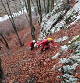
3          Characterization of speleothems from Floriilor Cave, Romania   Article no. 701

studies about the evolution of karst systems in the Carpathian Romanian are based
on stratigraphic and geomorphological evidence [31, 32].
        The Floriilor Cave is one of the most amazing wonders of nature from Romania.
It is an inactive cave, located in the upper basin of Jales, Valcan Mountains, Romania,
at an altitude of 600–620 m with the following coordinates: 45.212 latitude N and
23.132 longitude E, which is in conservation. The karstic landscape in the studied area
is conditioned by the presence of limestone and high amount of precipitations. The
geomorphologic process is given by the dissolution of water from precipitations, in
which a quantity of carbon dioxide is incorporated. Together they form a weak acid,
which, through the cracks along the cracks, takes up the calcium ions and favors its
widening. The geological and climatic conditions allow the development of both karst
relief groups: exokarst and endokarst. The most relevant aspect of the cave is the
abundance of different calcite speleothems covering the ceiling and walls, outstanding
for their beauty and uniqueness. This research was aimed at a preliminary study of
morphological and mineralogical aspects of speleothems collected from Floriilor Cave,
using non-invasive techniques, as a first survey for the future investigation regarding
the origin and genesis of this beautiful cave. It is necessary to highlight that the
Floriilor Cave is protected by Romanian legislation, being closed to tourism activities.
The samples were collected by a qualified speleologist and with the consent of the
authorities who manage this natural cave, using non-destructive methods, thus
preventing the destruction of the protected interior of the cave.

                                  2. SITE DESCRIPTION

       The Valcan Mountains are located near the Targu-Jiu town, located in the
centre-west part of the Southern Carpathians, between the valleys of the Motru
River (to the west) and Jiu River (to the east). In the north, it is bordered by the
Petrosani Depression, and in the south, it reaches the Subcarpathian Depression of
Oltenia. It measures 45 km long and on average 20 km wide (Fig. 1).
       Geologically, the outcropping area is located in the Danubian Autochthonous,
being uncovered from below the Getic Nappe by erosion, and emerges in the form
of a vast half-window in the south-west of the Meridional Carpathians, stretching
from the Oltet Valley to the Danube River. The dividing line between the Danubian
Autochthonous and the Getic Nappe goes northwards from the Polovragi village
and, after describing a circular arc in the area where the Lotru River springs, it goes
through the Petrosani Depression, north from the Retezat Mountains, and it bends
to the south, west from the Almaj Mountains, reaching the Danube River near the
Berzasca village. The surface boundaries of the Danubian unit are represented by
the Getic erosion outline and the edge of the Carpathian foredeep, whose deposits
cover Dacidic structures in a discordant manner.
       Geomorphologically, the major landforms overlapping the autochthonous are
the Parang, Valcan, Retezat, Cernei and Almaj mountains, the Petreanu and Tarcu
massifs and the Mehedinti Plateau. Certain areas of the half-window still preserve
Article no. 701                          Gica Pehoiu et al.                                        4

remnants of the Getic shell under the form of patches, which can be found in
Godeanu Mountains, Bahna, the Mehedinti Plateau and north from the Valari village.

 Fig. 1 – Geological map of the southwestern Southern Carpathian, including the Valcan Mountains
             and the location of the Flower Cave (modified after Michetiuc M.C. [33]).

       The stratigraphic profile of the Danubian Autochthonous consists mostly of
flaky crystalline lithological formations and magmatic bodies, which took form over
the course of several tectonic-magmatic pre-Alpine cycles. These hold pre-Alpine
and/or Alpine sedimentary formations. The flaky crystalline formations, which are
crossed by the magmatic bodies, make up the pre-Alpine basement units, and the
others compose the sedimentary cover. The pre-alpine basement includes two
generations (pre-Hercynian and Hercynian) of metamorphic rocks, crossed by
magmatic bodies consisting of granitoid rocks and basic and ultrabasic bodies. The
pre-Hercynian crystalline schists are the most developed and belong to two
metamorphic types: the mesometamorphic crystalline and epimetamorphic crystalline
schists. The pre-Baikal crystalline units include metamorphic rocks originating
from volcanogenic and terrigenous formations, which were metamorphosed under
almandine-amphibolite facies conditions and subsequently underwent retromorphic
phenomena. Specific to the pre-Hercynian basement are the numerous intrusions of
granitoid bodies, either syntectonic or posttectonic.
       The palynological and radiometric analyses have shown that the metamorphosis
and folding of the formations understudy took place during the Mid-Proterozoic,
as part of the pre-Baikal orogenetic processes. The pre-Hercynian mesometamorphites
belong to the groups Lainici-Paius, Dragsan, Poiana Mraconia and Neamtu. Of particular
interest in the region studied is the Group Lainici-Paius, which emerges in the
Cerna Mountains, on the southern side of Valcan Mountains, in Parang, and in
Retezat. The group is a metaclastic series, where the quartzite gneisses intercalated
5 Characterization of speleothems from Floriilor Cave, Romania Article no. 701
with micaceous schists, graphite shales and crystalline limestone are prevalent. In
many areas, it is affected by retromorphism. The Danubian sedimentary zone dates
back to the Upper Carboniferous, being superimposed by Permian (pre-Alpine)
deposits. At the end of the Palaeozoic Era, the area had risen above the sea level
and remained so in the first part of the alpine cycle as well. During the
sedimentation process (the first cycle), the accumulations consisted mainly of
calcareous (Liassic) deposits; in the second cycle, due to a powerful tectonic
instability (neotectonic movements), the deposits are of an arenaceous-turbiditic
type. Since the end of the Cretaceous, the area has evolved as a dryland undergoing
denudation, which has led to a marked erosion of the sedimentary zone. This
stratum is still seen in some areas, including the Cerna-Jiu, which stretches from
the Cerna Valley to Polovraci, on the southern side of the Valcan Mountains [28].
The morphological characteristic of the Valcan Mountains consists in the
presence of the three major height intervals corresponding to the three flattening areas
specific to the Meridional Carpathians: Borascu (750–900 m), Rau-Ses (450–600 m),
and Gornovita (250–400 m). The last-mentioned one has been shaping the limestone
deposits from the south, which allowed the formation of numerous exokarst forms. On
crossing the limestones, the rivers created gorges and ravines, and the infiltration of the
water coming from rainfalls and snowfalls, as well as the streams and rivers, generated
intense underground drainage, which helped the formation of numerous caves.
The Floriilor Cave was accidentally discovered by the speleologist Cornel Naidin
from Craiova, on the Palm Sunday of the year 1991 (hence the name Floriilor
Cave, which means Palm Sunday’s Cave), and is situated in the river basin of Jales
(better known by the local people under the name of Sohodol), on the left side of
the tributary Plesu (Macrisu, as the people in the area, call it), about 300 m up the
confluence of the two streams.
The Sohodol River (a tributary of the Oltenian Bistrita) springs from below the
Sigleul Mare Peak, carving, over a length that exceeds 12 km, the longest gorge-shaped
valley in the north of Oltenia, where numerous karst forms are to be found between
Luncile Contului and Runcu Village [29]. The karst landscape is well represented by
surface karst landforms (such as karrens and dolinas), as well as by numerous caves
that can be found near the riverbed and on the slopes bordering the valley terraces.
The first portion of the access, starting from the former ranger cabin Macrisul
(located at the junction of the two hydrographic arteries, reachable via DJ 672 C county
road), is easy, going along a forest road that runs parallel to the streamline, on the
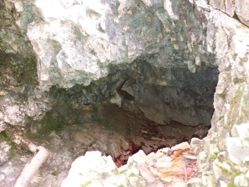
right side of the stream. After approximately 300 m, the route becomes extremely
difficult and the explorer must cross the stream (which is almost impossible when
the water level is high) (Fig. 2a); after that comes a portion of 50–60 m going up a
very steep slope (60–65 degrees); for this particular section, it is recommended to
use climbing ropes (Fig. 2b). The cave is situated at an altitude of 600–620 m and
has the following coordinates: 45.212 N and 23.132 E.
This is one of the over 70 caves in the Valcan Mountains. Being a small one,
it has not yet come to the attention of speleologists, and because of this, it was not
mapped. Floriilor Cave is in the custody of the Gorj Mountain Rescue Service,
Article no. 701                             Gica Pehoiu et al.                                     6

which considered it necessary to protect it by installing metal grilles (Fig. 2c) at the
entrance; access to the cave is permitted only with one of the members of this
service, but the cave is absolutely amazing (Fig. 2d).

                      (a)                                                   (b)

                      (c)                                                   (d)
       Fig. 2 – a) The access way to the Floriilor Cave; b) the slope before the Floriilor Cave,
                   inclination 60–65°; c) Floriilor Cave entrance, diameter ~ 50 cm;
                         d) stalactites and stalagmites inside of Floriilor Cave.

      The entrance to the cave resembles the den of a beast of prey. For the first 15 m,
the explorer must crawl, and then the cave opens up. It is an inactive (dead) cave,
in conservation under the protection of Gorj Mountain Rescue Service. The cave
measures 5 to 10 m in width, approximately 760 m in depth and 500 to 600 m in
height. After 760 m there is a very narrow and clogged gallery. It is possible that it
will develop further, but the speleologists did not have the opportunity to move
forward. The cave communicates with the outside and through other access roads,
possibly ditches or cracks in the limestone, due to the fact that inside there was an
intense air circulation (wind speed being over 1 m/s).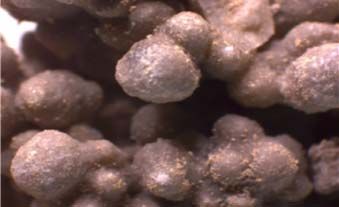
7               Characterization of speleothems from Floriilor Cave, Romania            Article no. 701

                                  3. MATERIALS AND METHODS

                   3.1. SAMPLING PROCEDURE AND SAMPLE PREPARATION

       Samples were carefully collected in autumn 2018, from the access points of
Floriilor Cave (Table 1) by qualified speleologists (see Acknowledgement),
without destroying the interior of the cave. The stalactite and stalagmite samples
studied were naturally detached from the ceiling or floor of the cave. All samples
presented in this article were non-invasively collected from a depth of 100–150 m,
only with the consent of the custodians (i.e., the representatives of the Gorj
Mountain Rescue Service).

                                                Table 1
       Samples collected from Floriilor Cave – photos of samples, sampling site inside of the cave

                       Sample                                        Sampling area

                                                                                             stalactites
                                                                                               Ceiling
    Stalactite *
       (C)

                                                                                         Floriilor Cave
                                                                                         rise from the
                                                                                          Stalagmites

                                                                                            floor of
    Stalagmite *
        (G)
                                                                                               Cave wall

       Rock
                                                                                           Cave sediments
                                                                                            and ancient
                                                                                               bones

     Sediment
Article no. 701                      Gica Pehoiu et al.                               8

      In the case of OM, SEM-EDS, ATR-FTIR and Raman investigations no
sample preparation was required. Before analysis by XRD and WDXRF, the samples
were ground using a vibratory disk mill, type LMWs (Testchem, Pszow, Poland)
equipped with a stainless-steel disk. This step aimed to reduce the particle size and to
improve the homogeneity of the compounds in the samples. Furthermore, 2 g from
each sample was mixed with 2 g Boreox® (Fluxana, Bedburg-Hau, Germany) and
were pressed using a manual laboratory press, type LPR 250 kN (Testchem, Pszow,
Poland) to form pellets in order to facilitate the analysis of the samples by WDXRF.
The obtained pellets (covered with PP Myler foil – thickness 6 μm) meet the
thickness criteria: X-ray intensity does not change with the thickness.

                            3.2. ANALYTICAL TECHNIQUES

                          3.2.1. Optical Microscopy (OM)

       Primo Star microscope (Carl Zeiss AG, Oberkochen, Germany) was chosen
for optic investigations due to its versatility and even though it can be used mainly
for biological samples, it can be adapted to the most sophisticated laboratory work
conditions. For inside laboratory researches, it offers the possibility to investigate
the samples in transmitted or reflected light at a magnification range up to 100x.
The microscope has a 5 megapixel HD digital video camera (Axiocam 105) attached
to it, through which the Zen software (Carl Zeiss AG, Oberkochen, Germany)
offers a real-time data acquisition. For this study, the images were obtained using
reflected light mode along with Plan-ACHROMAT dry objectives.

            3.2.2. Field Emission – Scanning Electron Microscopy coupled
                 with Energy Dispersive Spectrometry (FE-SEM-EDS)

       The geomorphological characterization of samples was performed using the
SU-70 microscope (Hitachi, Ibaraki, Japan). The scanning electron microscope is
the Field Emission (FE-SEM) type which operates under high vacuum (10–8 Pa) and
offers a high resolution of 1 nm at 15 kV acceleration voltages. SEM investigations
were performed under 5 kV accelerating voltage and 15–21 mm working distance
range; for EDS analysis the UltraDry detector (Thermo Fisher Scientific, Waltham –
Massachusetts, United States of America) was used coupled on SEM column, 20 kV
acceleration voltage and Phi-Rho-Z correction method available in NSS software
(Version 3.0).

   3.2.3. Attenuated Total Reflectance Fourier Transform Infrared Spectroscopy
                                   (ATR-FTIR)

      Molecular identification of chemical functional groups of inorganic compounds
in the solid samples was performed by Fourier Transform Infrared spectroscopy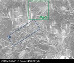
9           Characterization of speleothems from Floriilor Cave, Romania   Article no. 701

using Vertex 80v spectrometer (Bruker, Ettlingen, Germany), equipped with Attenuated
Total Reflectance (ATR) accessory and with HYPERION microscope. ATR-FTIR
spectroscopy has limited applications in quantitative researches of inorganic groups
of sediment or rock, as an example, since it has a penetration depth of only a few
microns, but for qualitative investigation, it could be a suitable technique. All spectra
were recorded in the range of 4000–400 cm–1, with 0.2 cm–1 spectral resolution and
0.1% T accuracy and 32 scans/spectra.

                               3.2.4. Raman Spectroscopy

       Raman spectra were recorded with a portable Xantus-2TM Raman analyzer
(Rigaku, Boston, United States of America) equipped with two laser sources (i.e.,
785 nm and 1064 nm) and two detectors (i.e., TE cooled CCD and TE cooled InGaAs).
For this study, the following parameters were used: 1064 nm excitation source, 400
mW laser power, 1000 ms integration time, and 3 scans/spectra. The spectral range was
200–2000 cm–1, with 15–18 cm–1 spectral resolution. Xantus-2TM reduces intrinsic
fluorescence issues and offers an extensive range of analysis capabilities.

           3.2.5. Wavelength Dispersive X-ray Fluorescence (WDXRF)

       WDXRF was used for determining the chemical composition of the
speleothems. For this purpose, a Supermini 200 system (Rigaku, Tokyo, Japan)
was employed. The spectrometer is equipped with a 200 W X-ray tube containing a
Pd target, two detectors (e.g., PC and SC) and 3 analyzer crystals (e.g., LiF, PET
and RX25 – with automatic exchange). The X-ray tube was operated at following
settings: 50 kV and 4 mA with vacuum for measurement of major and trace
elements. Supermini 200 allows WDXRF analyses with 0.1–1 ppm limit of
detection and 0.5% precision. For each sample the analysis was timed for 1500 s.

                            3.2.6. X-Ray Diffraction (XRD)

       The mineralogical composition of the speleothem was determined by X-ray
diffraction (XRD) in an Ultima IV diffractometer (Rigaku, Tokyo, Japan) using Cu
Kα radiation (λ = 1.54 Å), 40 kV accelerating voltage of the generator radiation,
30 mA emission current, step 1°, 60 s/° and scanning angular range 2θ from 10 to
100°. The obtained data were interpreted using the PDXL2.2 software and the
ICDD database PDF4 + release 2019.

                              4. RESULTS AND DISCUSSION

      In this respect, the morphological structure and mineral composition of
speleothems samples collected from the wild Floriilor Cave were investigated,
including stalactites and stalagmites, rocks and sediments. These analyses were
Article no. 701 Gica Pehoiu et al. 10
performed by three non-invasive techniques such as optical microscopy (OM), FE-
SEM-EDS, ATR-FTIR, Raman spectroscopy, WDXRF and XRD.
OM images (Fig. 3) indicated the presence of non-crystalline and crystalline
inorganic material, as well as some uniform structures.
(a) (b)
(c) (d)
Fig. 3 – Optical microscopy images: a) stalactite – 100 × magnification; b) stalagmite –
100 × magnification; c) rock – 10 × magnification; d) sediment – 40 × magnification.
FE-SEM observations of the speleothem samples (Fig. 4) showed some
crystalline mineral formations without impurities (i.e., microbial morphotypes, cells,
filaments etc.) [30, 34]. The stalactite has clear visible lamellar structure specific to
calcite, as well as few acicular Mg-Si structures and granular structure specific to
gypsum (Figs. 4 a–b) [31, 32]. As compared to the stalactite, in the stalagmite
sample a predominant porous structure with few integrated calcite crystals was
identified (Figs. 4c–d) [31, 35]. On the rock sample, the knobbly surface with small
cylindrical excrescences (Fig. 4e) was observed, with the structure similar to
triangular crystals interconnected by smaller cementing binders (Fig. 4f) [31, 36].
The sediments collected from the Floriilor Cave are characterized by very fine
granules (Fig. 4g) with lamellar structure (Fig. 4h) [31].
The results of the elemental composition achieved by EDS analysis are shown
in Table 2. The EDS analysis revealed a high amount of C, O and Ca and small
quantities of Mg, Al, Si, K, and Fe. In some samples, other elements were determined
(i.e., F, Na, P, and Cl). Data presented in Table 2 revealed good close similarities
between stalactite and stalagmite samples from the point of view of the elemental
content and important differences between rock and sediment, probably due to the fact
that sediments are the result of the disintegration of rocks, stalactite and stalagmite.11 Characterization of speleothems from Floriilor Cave, Romania Article no. 701
(a) (b)
(c) (d)
(e) (f)
(g) (h)
Fig. 4 – SEM photomicrographs of: a) stalactite (× 450) from the Floriilor Cave highlight the
presence of Mg-Si needles (green area) and gypsum (blue area) on calcite; b) surface of some calcite
crystals identified on stalactite (× 4 k); c) porous surface of stalagmite (× 150); d) some calcite
crystals integrated in gypsum identified on stalagmite (× 800); e) same structures identified by OM on
rock sample (× 30); f) triangular crystals interconnected by smaller cementing binders on rock sample
(× 2.5 k); g) fine granular structure of sediment sample (× 500); h) lamellar calcite structure identified
on sediment sample (× 5 k) (Color online).Article no. 701 Gica Pehoiu et al. 12
Table 2
EDS elemental content expressed in wt. [%], normalized to 100 wt.%.
Sample C O F Na Mg Al Si P Cl K Ca Fe
Stalactite 17.08 62.95 nd* nd* 0.53 0.32 0.34 nd* nd* 0.05 18.73 nd*
Stalagmite 17.23 56.91 2.09 nd* 0.76 1.7 2.59 nd* nd* 0.27 16.81 0.54
Rock 12.66 51.3 nd* 0.27 0.75 1.29 2.45 nd* 0.28 0.57 28.96 1.02
Sediment 16.42 56.76 nd* 0.03 0.35 1.41 5.18 1.32 nd* 0.33 17.47 0.73
Mean RSD
0.11 0.33 0.25 0.02 0.02 0.02 0.02 0.01 0.01 0.01 0.09 0.05
[%]
In order to also identify the qualitative confirmation of potential inorganic groups
in samples, Attenuated Total Reflection – Fourier Transform Infrared spectroscopy
was carried out. Fourier Transform Infrared spectra results of samples revealed the
occurrence of multiple functional groups in them (Table 3). Hence, functional
group analysis plays a vital role in understanding the overall physicochemical
properties of solid samples. Identification of solid phase using the fingerprint
region was based on the correlation between the peak pattern of the analysed
sample and the peak pattern of a standard material of known chemical composition
(i.e. NIST SRM 2710a: Montana Soil).
Table 3
Infrared spectra and absorption bands with tentative assignment
FTIR spectra /
Sample Tentative assignment
Wavenumber [c m – 1 ]
Stalactite
1793/1412/872/711/ (CO3)2– – calcite
1082/522/437/376/363/ Si-O asymmetrical bending vibration, quartz
Si-O stretching;
1008/470/
O-H deformation, kaolinite13 Characterization of speleothems from Floriilor Cave, Romania Article no. 701
Stalagmite
1795/1396/872/711/ (CO3)2– – calcite
Si-O stretching, kaolinite;
469/422/390/366/
Si-O asymmetrical bending vibration, quartz
Rock
1452/1163/795/ (CO3)2– ; C-O stretching
1082/1066/515/448/394/364/ Si-O asymmetrical bending vibration, quartz
777/693/ Si-O symmetrical stretching vibrations, feldspar
Sediment
3524/3397/ OHst from water
1683/1619/1106/ phosphate band 1106 cm–1 can be poorly
crystalline apatite
1428/873/672/ (CO3)2– group
599/468/371/ Si-O stretching, maybe kaoliniteArticle no. 701 Gica Pehoiu et al. 14
FTIR spectra have shown asymmetric bands and indicate if a solid sample is
an oxides mixture or if it has been modified by oxidation and so on. Farmer (1974)
and Derrick et al. (1999) in their publications [37, 38], studied absorption bands for
inorganic materials and reported that these are fewer in number, are broader, and
occur at lower wavenumbers than absorption bands for organic materials. This can
be attributed to external ion structure (i.e., solid or crystalline matrix) [39], as well
as to internal ion composition (i.e. functional groups) [40–42].
For all solid samples, a strong sharp band around 1000 cm–1 and a week but
large band around 1620 cm–1 were identified. First of all, carbonate (i.e., CaCO3
from calcite which has calcium ion, Ca2+) is one of the most complex inorganic
compounds classified as a complex anion (i.e., CO32–), because the anion is itself a
functional group. The covalent bonds in the carbonate tightly hold the anion
together. Thus, carbonate bending vibrations produce sharp bands in the region of
1793/1412/872/711 cm–l [43–45]. On the other hand, silicates, with a fully ordered
crystalline lattice structure, have a well-defined Si-O absorption band around
1082–1066 cm–l as well as. Also, weak or medium bending vibrations related to
SiO vibrations mainly from quartz [43–45], kaolinite and silica occurred around
600 cm–1 (Table 3). For sediment sample, two weak peaks were observed at 1683 and
1619/cm–1, which can be attributed to phosphate, and the strong peak at 1106 cm–1,
which can be poorly crystalline apatite.
In addition, the samples of the corresponding stalagmite, stalactite, rock cave
substrate, and floor cave sediments have been analysed by Raman spectroscopy
(Fig. 5). This non-destructive technique is able to characterize the chemical and
mineralogical composition of solid materials, thus the collected Raman spectra
being the ‘fingerprints’ of the investigated materials. In this regard, the Raman C-O
symmetric stretching band of the CO32– ion occurs at 283, 712 and 1086 cm–1,
according to several studies [46–50]. Both vibrational techniques (i.e., FTIR and
Raman spectroscopies) allow gathering information about major and minor
constituents of speleothems.
In addition, the results obtained by two destructive techniques such as X-Ray
Fluorescence for chemical elements analysis and X-Ray Diffraction for minerals
were correlated with preliminary data obtained by SEM-EDX, which widely combines
micromorphology and elemental analysis performed on collected speleothem
samples. In particular, Wavelength Dispersive X-Ray Fluorescence Spectroscopy
(WDXRF) detected the chemical constituents (Table 4) through the non-invasive
analysis of the fluorescence radiation emitted by the sample irradiated by the X-Rays
beam. The results concern mainly the comparison between the identification of the
chemical elements by using the X-Ray Fluorescence technique and the study of
their distribution in speleothem samples from X-Ray Diffraction analysis.
An essential point in the analyses by XRF and even XRD methods of
speleothems is to distinguish the compositional components and possible crusts due
to animals or birds’ excrements during a very long period. This is sometimes15 Characterization of speleothems from Floriilor Cave, Romania Article no. 701
extremely difficult, e.g. calcite as a major component in speleothems and calcareous
rock/sediments with possible phosphorite layers deposited above the speleothems.
In these cases, FTIR and Raman microanalyses of a cross-section of the speleothems
correlated with WDXRF and XRD analyses are of valuable assistance to determine
the in-depth distribution of the inorganic compounds. Phosphorite is a product of
degradation of apatite under the action of external agents, in the form of
hydroxyapatite, Ca5(PO4)3OH or Ca10 (PO4)6(OH)2, which is often dissolved from
vertebrate bones and teeth, mixed with carbonate-apatite, Ca3(PO4)2 ∙ Ca(HCO3)2,
and it is found in cavities in limestone rocks in the form of karstic phosphorites.
This can be an explanation of P2O5 presence in high amount in sediment (i.e.,
0.568 ± 0.026 %) as compared to the values obtained in stalactite, stalagmite and
even rock sample (Table 4).
sediment
200 700 1200 1700
Raman Shift [cm-1]
Intensity [a.u.]
rock
200 700 1200 1700
Raman Shift [cm -1]
stalagmite
200 700 1200 1700
Raman Shift [cm-1]
stalactite
200 700 1200 1700
Raman Shift [cm-1]
Fig. 5 – Overlapped Raman spectra of analysed samples.Article no. 701 Gica Pehoiu et al. 16
Table 4
Oxides composition of the speleothem samples determined by WDXRF,
expressed in mass [%] ± S.D. [%], normalized to 100%
Component Stalactite Stalagmite Rock Sediment
MgO 1.982±0.212 1.725±0.232 1.526±0.206 2.841±0.156
Al2O3 0.455±0.050 0.316±0.059 1.886±0.051 10.689±0.064
SiO2 1.679±0.049 1.968±0.047 4.202±0.055 25.918±0.086
P2O5 0.213±0.027 0.178±0.026 0.272±0.023 0.639±0.026
K2O 0.188±0.036 0.197±0.036 0.331±0.031 1.742±0.030
CaO 94.924±0.065 94.684±0.068 90.770±0.059 53.255±0.045
TiO2 nd2 nd2 nd2 0.556±0.059
MnO nd2 nd2 nd2 0.126±0.024
Fe2O3 0.481±0.048 0.931±0.053 0.955±0.041 4.172±0.029
SrO 0.078±0.018 nd2 0.058±0.015 0.021±0.009
ZrO2 nd2 nd2 nd2 0.041±0.010
In stalactite and stalagmite samples calcite (92.11–98.21%) is the main
mineral identified by XRD method. Actual results do not prove that other types of
minerals could not be present under the detection limit (LOD) of this method,
which is ~2%. On the other hand, the XRD results of rock and sediment samples
reveal small amounts close to LOD of quartz (2.23–4.81%), gypsum (1.81–2.95%)
or illite (1.02–1.85%).
Table 5
Lattice information for calcite in each sample
Sample a(Å) b(Å) c(Å)
Stalactite 4.982(3) 4.982(3) 17.046(10)
Stalagmite 4.9888(5) 4.9888(5) 17.050(4)
Rock 4.9812(19) 4.9812(19) 17.052(12)
Sediment 4.9875(7) 4.9875(7) 17.045(6)
The calculated unit cell parameters of calcite are presented in Table 5. A
small difference in the parameters of the unit cell of the calcite phases can be
explained by the presence of some impurities in the samples [51].
5. CONCLUSIONS
In this preliminary study regarding the characterization of different speleothems
collected non-destructively from the Floriilor Cave, one of the most beautiful
natural caves from Romania, not included in the touristic circuit, the main field-
based observations were highlighted in order to enhance understanding of the
micromorphology, elemental and mineralogy composition of this cave. Two
complementary non-destructive techniques (i.e., Optical Microscopy and Scanning17 Characterization of speleothems from Floriilor Cave, Romania Article no. 701
Electron Microscopy) were used for the morphological characterization of several
representative speleothems collected from the Floriilor Cave, Romania. Also, for
this study, vibrational spectroscopy (i.e., FTIR and Raman) was particularly used
for complementary identification of inorganic and organic compounds and partially
their chemical bonds in order to distinguish between organic and inorganic carbon,
an essential step for radiocarbon dating. In addition, destructive techniques such as
XRD for minerals and XRF for oxides composition were used. The collected data
have demonstrated the usefulness of the destructive techniques (i.e., X-ray Fluorescence
spectroscopy and X-Ray diffraction analysis) investigation, through which it has
been possible to reveal chemical elements undetectable by vibrational spectroscopy
and microscopy as well. Only calcite and gypsum were encountered in the cave.
The EDS elemental measurements identified C, O and Ca as main constituents of
the speleothems with some minor or trace elements being: Mg, Al, Si, K, and Fe.
The results are similar for stalagmites and stalactites, while the elemental content
of the host rock and sediments vary importantly. Raman and FTIR analysis
succeeded to identify the major and minor constituents of investigated speleothems
such as CO32– groups, phosphates, Si-O bounds calcite, and OH bounds. Further
on, in the next studies, the cave is planned to be mapped-out (when the custodians
will allow the access for further research); also, the already collected samples will
be the subject of a new round of more deep investigations (i.e., inductively coupled
plasma-mass-spectrometry for isotopic ratio, neutron tomography, neutron diffraction,
and 14C dating).
Funding. This work was supported by the project entitled “Health risk assessment associated
with abandoned copper and uranium mine tailings from Banat Region, Romania”, according to
Protocol no. 4748-4-2018/2020, the bilateral research project between Joint Institute for Nuclear
Research and “Valahia” University of Targoviste, on theme 03-4-1128-2017/2020.
Acknowledgements. The authors would like to acknowledge to Alin Marian Badea – teacher
of “Constantin Brailoiu” Arts Highschool of Targu Jiu, Darius Bistriceanu and Ion Negrea – members
of Gorj Mountain Rescue Service for the support provided in the sampling process of speleothems
fragments and for the information about the discovery of Floriilor Cave.
REFERENCES
1. D. Ford and P. Williams, Karst Hydrogeology and Geomorphology, John Wiley & Sons, Ltd.,
Chichester, 2007.
2. D. Ford and P. Williams, Karst Hydrogeology and Geomorphology, John Wiley & Sons Inc,
Hoboken, New Jersey, 2013.
3. C. Tolan-Smith, Human occupation of caves. In Encyclopedia of Caves and Karst Science, Gunn,
J. (Ed.), Taylor and Francis Group, New York, 2004, pp. 919–924.
4. D.S. Gillieson, Management of caves. In Karst management, van Beyen, P.E. (Ed.), Springer,
Dordrecht, 2011, pp. 141–158.
5. A.A. Cigna and P. Forti, Tourism Karst Area 6(1), 9–26 (2013).
6. K. Tomczyk-Zak, and U. Zielenkiewicz, Geomicrobiol. J. 33(1), 20–38 (2015).
7. M.J.Russell and V.L.MacLean, J. Environ. Manage. 87(3), 474–483 (2008).
8. C. Saiz-Jimenez, World J. Microbiol. Biotechnol. 28(7), 2453–2464 (2012).Article no. 701 Gica Pehoiu et al. 18
9. J.E. Brady and A. Scott, J. Cave Karst Stud. 59, 15–21(1997).
10. M. Ghinassi, A.C. Colonese, Z. Di Giuseppe, L. Govoni, D. Lo Vetro, G. Malavasi, F. Martini,
S. Ricciardi, and B. Sala, J. Quaternary Sci. 24(4), 383–398 (2009).
11. G.S. Springer, Clastic sediments in caves, In Encyclopedia of Caves, 3rd edition, W.B. White,
D.C. Culver and T. Pipan (Eds.), Academic Press, 2019, pp. 277–284.
12. Y.J. Wang, H. Cheng, R.L. Edwards, Z.S. An, J.Y. Wu, C.C. Shen, and J.A. Dorale, China. Sci.
294(5550), 2345–2348 (2001).
13. I.M. D’Angeli, M. Parise, M. Vattano, G. Madonia, S. Galdenzi, and J. De Waele, Geomorphology
333, 105–122 (2019).
14. S. Galdenzi and T. Maruoka, Geomorphology 328, 211–221 (2019).
15. E. Romano, L. Bergamin, G. Pierfranceschi, C. Provenzani, A. Marassich, Mar. Environ. Res.
133, 114–127 (2018).
16. E. Manzano, A. Garcia, S. Cantarero, D. Garcia, A. Morgado, and J.L. Vilchez, J. Archaeol. Sci.
Rep. 27, 101929 (2019).
17. L. Ojeda, I. Vadilo, G. Etiope, J. Benavente, C. Linan, Y. del Rosal, S.T. Tapia, M.A. Morinigo,
and F. Carrasco, Geochim. Cosmochim. Acta. 259, 302–315 (2019).
18. S.F.McGarry and C. Caseldine, Quat. Sci. Rev. 23(23–24), 2389–2404 (2004).
19. L.J. Maher Jr, Palaeogeogr. Palaeoclimatol. Palaeoecol. 237(1), 19–31 (2006).
20. C.O.Hunt and G. Rushworth, J. Archaeol. Sci. 32(3), 465–473 (2005).
21. T. Brad, C.A. Badaluta and A. Persoiu, Ice Caves in Romania, in Ice Caves, 1st edition, A. Persoiu
and S.E. Lauritzen (Eds.), Elsevier, Amsterdam, 2018, pp. 511–528.
22. P. Damm and V. Lascu, Speologia explorativă în Romania, available at https://www.frspeo.ro/
speologia-explorativa-in-romania/ (last accessed 15 March 2020).
23. C.M. Munteanu, Quat. Int. 279–280, 343–344 (2012).
24. A. Feurdean, A. Persoiu, A. Pazdur, and B.P. Onac, Rev. Palaeobot. Palynol. 165(1–2), 1–10
(2011).
25. B.P. Onac, J.G. Wynn and J.B. Sumrall, Chem. Geol. 288(3–4), 105–114 (2011).
26. S. Constantin, M. Robu, C.M. Munteanu, A.Petculescu, M. Vlaicu, I. Mirea, M. Kenesz, V. Dragusin,
D. Hoffmann, V. Anechitei, T. Timar-Gabor, R.D. Roban, and C.G. Panaiotu, Quat. Int. 339, 25–40
(2014).
27. T. Tamas and M. Vremir, Karstological investigations in the Middle Basin of Lada Valley
(Padurea Craiului Mountains, Romania), Proceedings of the 12th International Congress of
Speleology 1, 413–416 (1997).
28. V. Mutihac, M.I. Stratulat and R.M. Fechet, Geologia României, Edit. Didactică şi Pedagogică,
Bucharest, 2007.
29. M. Bleahu, V. Decu, S. Neagra, C. Plesa, I. Povara, I. Viehmann, Peşteri din România, Edit. Ştiintifică
şi Enciclopedică, Bucharest, 1976.
30. N.K. Dhami, A. Mukherjee and E.L.J. Watkin, Front. Microbiol. 9, 40 (2018).
31. B. Jones, Sedim. Geol. 226(1–4), 94–109 (2010).
32. P. Huerta, A. Martin-Perez, R. Martin-Garcia, A. Rodriguez-Berriguete, A. La Iglesia Fernandez,
A.M. Alonso-Zarza, Sediment. Geol. 383, 136–147 (2019).
33. M.C. Michetiuc, Calcarele Jurasicului superior–Cretacicului inferior din sudul Munţilor Vâlcan:
microfaciesuri, microfosile şi reconstituirea paleomediului depoziţional, PhD Thesis, Babes Bolyai
University of Cluj-Napoca, 2016, available at http://193.231.20.119/doctorat/teza/fisier/3478 (last
accessed 5 June 2020).
34. C.A. Hill and P. Forti, Cave Minerals of the World, 2nd Edition, National Speleological Society,
Huntsville, 1997.
35. K.S. Woo, D.W. Choi and K.C. Lee, Quatern. Int. 176–177, 82–95 (2007).
36. W. Wroblewski, M. Gradzinski, J. Motyka, and J. Stankovic, Quatern. Int. 437, 84–97 (2017).
37. M.R. Derrick, D. Stulik, J.M. Landry, Infrared spectroscopy in conservation science-scientific
tools for conservation, The Getty Conservation Institute, Los Angeles, 1999.19 Characterization of speleothems from Floriilor Cave, Romania Article no. 701
38. V.C. Farmer, The Anhydrous Oxide Minerals, in: V.C. Farmer (Ed.), The Infrared spectra of minerals,
Mineralogical Society, London, 1974, pp. 196.
39. G. Pehoiu, C. Radulescu, O. Murarescu, I.D. Dulama, I.A. Bucurica, S. Teodorescu, and
R.M. Stirbescu, Bull. Environ. Contam. Toxicol. 102, 504–510 (2019).
40. C. Radulescu, I. Ionita and A.M. Hossu, Dyes Pigm. 65(2), 175–177 (2005).
41. L. Barbes, C. Radulescu and C. Stihi, Rom. Rep. Phys. 66(3), 765–777 (2014).
42. C. Radulescu, C. Stihi, S. Iordache, D. Dunea, and I.D. Dulama, Rev. Chim. (Bucharest) 68(4),
805–810 (2017).
43. A. Bintintan, M. Gligor, I.D. Dulama, S. Teodorescu, R.M. Stirbescu, and C. Radulescu, Rev. Chimie
(Bucharest) 68(4), 847–852 (2017).
44. A. Bintintan, M. Gligor, I.D. Dulama, C. Radulescu, C. Stihi, R.M. Ion, S. Teodorescu, R.M. Stirbescu,
I.A. Bucurica, and G. Pehoiu, Rom. J. Phys. 64(5–6), 903 (2019).
45. A. Bintintan, M. Gligor, C. Radulescu, I.D. Dulama, R.L. Olteanu, S. Teodorescu, R.M. Stirbescu,
and I.A. Bucurica, Anal. Lett. 52(15), 2348–2364 (2019).
46. H.G.M. Edwards, I.B. Hutchinson, R. Ingley, J. Parnell, P. Vitek, and J. Jehlicka, Astrobiology,
13(6), 543 (2013).
47. G. Fiesta, C. Andreani, M. Baldoni, V. Cipollari, C. Martinez-Labarga, F. Martini, O. Richards,
M.F. Rolfo, L. Sarti, N. Volante, R. Senesi, F.R. Stasolla, S.F. Parker, A.R. Vassalo, A.P. Mamede,
L.A.E. Batista de Carvalho, and M.P.M. Marques, Sci. Adv. 5(6), eaaw1292 (2019).
48. R.L. Frost, S. Palmer, D.A. Henry, and R. Pogson, J. Raman Spectrosc. 42(6), 1447–1454 (2011).
49. A. Hernanz, M. Mas, B. Gavilan, and B. Hernandez, J. Raman Specrosc. 37(4), 492–497 (2006).
50. N. Zupančič, S. Šebela and M. Miler, Acta Carsologica 40(2), 307–317 (2011).
51. R.M. Ion, B.A. Bakirov, S.E. Kichanov, D.P. Kozlenko, A.V. Belushkin, C. Radulescu, I.D. Dulama,
I.A. Bucurica, A.I. Gheboianu, R.M. Stirbescu, S. Teodorescu, L. Iancu, M.E. David, R.M. Grigorescu,
Appl. Sci. 10, 3781 (2020).You can also read