Coding for Angioplasty & Stent Procedures - July 2020 - ADVOCATE ...
←
→
Page content transcription
If your browser does not render page correctly, please read the page content below

Coding for Angioplasty
& Stent Procedures
July 2020

Jennifer Bash, RHIA, CIRCC, RCCIR, CPC, RCC
           Director of Coding Education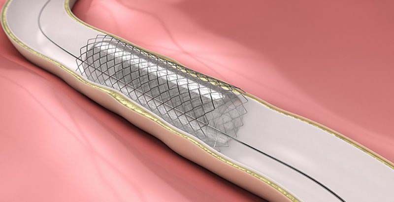
Agenda
• Introduction
• Definitions
• General Coding Guidelines
• Presenting Problems/Medical Necessity for
  Angioplasty & Stent
• General Angioplasty & Stent Procedures
• Cervicocerebral Procedures
• Lower Extremity Procedures

Disclaimer
The information presented is based on the experience
and interpretation of the presenters. Though all of the
information has been carefully researched and checked
for accuracy and completeness, ADVOCATE does not
accept any responsibility or liability with regard to
errors, omissions, misuse or misinterpretation.

CPT codes are trademark and copyright of the American
Medical Association.

Resources
• AMA
• CMS
• ACR/SIR
• ZHealth Publishing
Angioplasty & Stent Procedures
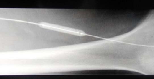
Angioplasty
Angioplasty, also known as balloon angioplasty and percutaneous
transluminal angioplasty, is a minimally invasive endovascular
procedure used to widen narrowed or obstructed arteries or veins,
typically to treat arterial atherosclerosis.
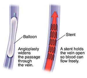
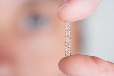
Vascular Stent
A stent is a tiny tube placed into the artery or vein used to treat
vessel narrowing or blockage. Most stents are made of a metal or
plastic mesh-like material.
General Angioplasty & Stent Coding
Guidelines
• Angioplasty is not separately billable when done with a
  stent
  • Pre-Dilatation
  • PTA converted to Stent
  • Prophylaxis
  • EXCEPTION-Complication extending to a different vessel
• Coded per vessel
• Codes include RS&I
• Territories
• Hierarchy
General Angioplasty & Stent Coding
Guidelines
• Bridging/Contiguous lesions
  • Short lesion between 2 vessels=1 intervention
  • Long lesion crossing entire length of 2 adjacent vessels with
    separate balloon or stent = 2 interventions
  • Two distinct lesions in adjacent vessels = 2 interventions
• Hemodynamically significant stenosis
• Angiography not separately billable
  • Exception:
     •   No prior angiographic imaging
     •   Prior imaging is suboptimal/inadequate visualization
     •   Change in patient condition/worsening of symptom
     •   New problem outside area of intervention
  • Should be modified (-59 or –XU)
Angioplasty & Stent Procedures PRESENTING PROBLEMS • Atherosclerosis • Stenosis • Occlusion • Blockage • Dissection • Vascular ischemia • Fibromuscular dysplasia MEDICAL NECESSITY • Hemodynamically significant stenosis • Medicare coverage
Angioplasty & Stent-General

• Sites Treated
   • Renal
   • Visceral
   • Aortic
   • Brachiocephalic/Upper Extremity

  INCLUDED                             SEPARATELY CODED

  RS&I                                 Selective cath placement*

  Stent inclusive of angioplasty       Medically necessary diagnostic imaging

  Closure Device (stent)               Vasc US guidance

                                       Moderate sedation

                                       Additional intervention (thrombectomy, infusion,
                                       etc.)
Angioplasty & Stent-General
• Angioplasty
• Arterial-37246-37247
• Venous-37248-37249
• Stent
• Arterial-37236-37237
• Venous-37238-37239
• Only 1 “initial” angioplasty/stent per session
• Codes are for open/percutaneous
• Renal Guidance/Exception
• Diagnostic renal angiography followed by angioplasty or stent, the catheter placement
is bundled
• If known lesion, catheter placement is separately codedAngioplasty-Case Study Exam: IR ANGIOGRAM RENAL UNILAT COMPLETE LT; IR TRLUML BALL PTA 1ST ART CLINICAL: Patient with a history of suspected renovascular hypertension. Was treated in the past of the left renal artery stent. Duplex follow-up has been equivocal at times and therefore we have been asked to evaluate stent angiographically. Expected benefits of the procedure and potential risks including the small risk of bleeding and infection were discussed and all questions were answered with the patient then gave oral and written informed consent. TECHNIQUE: Using modified Seldinger technique the right common femoral was punctured allowing placement of 6 sheath. Through this a Omni flush catheter was advanced just above the level of the renals for an aortogram. Subsequently the left renal artery was selectively catheterized and a pressure wire left across the stenosis. The FFR was approximately 0.92 normal being a value of 1.0. Subsequent ultrasound showed some narrowing in the region of the left renal artery stent. Subsequently angioplastied to 6 mm this was performed for follow-up FFR of 1.02. At this point the procedure was completed. The sheath was removed and hemostasis obtained using a Mynx closure device. This procedure was performed using all elements of maximal sterile barrier technique cap AND mask AND sterile gown AND sterile gloves AND a large sterile sheet AND hand hygiene AND 2%% chlorhexidine for cutaneous antisepsis STERILE TECHNIQUE WAS USED FOR ULTRASOUND GUIDANCE INCLUDING STERILE GEL AND STERILE PROBE COVER. Findings: In-stent restenosis on the left renal artery stent treated successfully with angioplasty. MEDICATIONS: Midazolam 1.0 mg, Fentanyl 50 mcg, heparin 5000 units, nitroglycerin 200 units intra-arterial CONTRAST: Omnipaque 85 ml RADIATION DOSE: Flouro Dose 780.62 dgy, Flouro Time 28.1 mins, Air Kerma 869 mGy Moderate Sedation was administered and I personally spent 55 minutes of face-to-face sedation time with the patient. A Registered Nurse was present throughout the procedure to serve as an independent trained observer to assist in the monitoring of the patient's level of consciousness and continuous monitoring of physiological status including pulse oximetry, heart rate, and blood pressure. Impression: Successful angioplasty of an in-stent restenosis of the left renal artery stent treated to 6 mm.
Angioplasty-Case Study
37246-Transluminal balloon angioplasty (except lower extremity artery(ies) for
occlusive disease, intracranial, coronary, pulmonary, or dialysis circuit), open or
percutaneous, including all imaging and radiological supervision and interpretation
necessary to perform the angioplasty within the same artery; initial artery

36251-Selective catheter placement (first-order), main renal artery and any accessory
renal artery(s) for renal angiography, including arterial puncture and catheter
placement(s), fluoroscopy, contrast injection(s), image postprocessing, permanent
recording of images, and radiological supervision and interpretation, including pressure
gradient measurements when performed, and flush aortogram when performed;
unilateral

99152-Moderate sedation services provided by the same physician or other qualified
health care professional performing the diagnostic or therapeutic service that the
sedation supports, requiring the presence of an independent trained observer to assist
in the monitoring of the patient's level of consciousness and physiological status; initial
15 minutes of intraservice time, patient age 5 years or older
Stent-Case Study
BILATERAL RENAL ANGIOGRAM AND BILATERAL RENAL ARTERY STENTS
INDICATION 58-year-old female smoker with history of diabetes who presents with difficult to control hypertension and ischemic
nephropathy. GFR measures 38. Renal artery Doppler performed on suggested a high-grade left renal artery stenosis. CT
angiography of the abdomen performed suggests right worse than left renal artery stenosis.
SURGICAL PROCEDURE After informed consent was obtained the patient was placed on the angiography table in the supine
position. The right groin was prepped with chlorhexidine and alcohol. Buffered lidocaine was utilized to anesthetize the skin
overlying the right common femoral artery. The right common femoral artery was punctured with a 19 gauge double wall needle
and a 5 French sheath was inserted into the right common femoral artery.
A 5 French pigtail catheter was advanced into the proximal abdominal aorta. Digital subtraction angiography of the abdomen was
performed. The catheter was exchanged to a 5 French C2 catheter. Selective catheterization of the right renal artery was
performed and digital subtraction angiography of the right renal artery was performed with magnification. A 0.035 Rosen guidewire
was advanced into a branch of the right renal artery. The 5 French sheath and 5 French C2 catheter was exchanged to a 6 French
Ansel 2 sheath. The sheath tip was positioned in the proximal right renal artery. 5000 units of heparin was delivered through the
sheath. 150 micrograms of nitroglycerin was delivered through the sheath into the right renal artery. A 6 mm x 17 mm Visipro
balloon expandable stent was advanced through the sheath and positioned across the stenosis at the origin of the low right renal
artery. The stent was deployed to 12 atmospheres of pressure. The results of the right renal artery stenting were assessed with
digital subtraction angiography through the 6 French sheath.
Multiple attempts were made to catheterize the left renal artery with the 5 French C2 catheter and an angled 0.035 hydrophilic
glidewire. These were unsuccessful due to a severe web-like stenosis at the origin of the left renal artery. The C2 catheter was
exchanged to a 5 French Sos catheter. The 5 French Sos catheter was eventually successful in catheterizing the left renal artery
and crossing the left renal artery stenosis.
Stent-Case Study
Digital subtraction angiography of the left renal artery was performed with magnification through the Sos catheter. The 0.035
Rosen guidewire was advanced into a branch of the left renal artery. The 6 French sheath was advanced across the left renal
artery stenosis. 150 micrograms of nitroglycerin was delivered into the left renal artery. A 5 mm x 17 mm Visipro balloon
expandable stent was advanced through the 6 French sheath into the proximal left renal artery. The stent was positioned across
the stenosis at the origin of the left renal artery and was successfully deployed to 12 atmospheres of pressure.

The results of the left renal artery stenting were assessed with digital subtraction angiography through the 6 French sheath. A 5
French pigtail catheter was advanced into the proximal abdominal aorta and digital subtraction angiography of the abdomen was
performed. At the completion of the procedure hemostasis was achieved in the right groin with the help of a Proglide closure
device. No hematoma was present. The patient tolerated the procedures very well.

FINDINGS: Digital subtraction angiography of the abdomen reveals mild atherosclerosis in the abdominal aorta and no aortic
stenosis or abdominal aortic aneurysm. The flush aortogram and selective right renal artery angiogram reveal a high-grade
stenosis of approximately 80 percent in the proximal right renal artery with poststenotic dilatation in the right renal artery. The flush
aortogram and selective left renal angiogram reveal a web-like high-grade stenosis at the origin of the left renal artery estimated at
90 percent. Renal artery branches are well-preserved bilaterally and the nephrograms appear promptly bilaterally. Following right
renal artery stenting an excellent angiographic result is seen with no residual stenosis and excellent positioning of the right renal
artery stent extending into the abdominal aorta for 1-2 mm. Following left renal artery stenting an excellent angiographic result is
seen with no residual stenosis and the left renal artery stent extending into the abdominal aorta for approximately 1 mm. There is
preservation of renal artery branches in both renal arteries.
CONCLUSION
1. High-grade proximal right renal artery stenosis. This responded well to 6 mm balloon expandable stenting.
2. High-grade proximal left renal artery stenosis. This responded well to 5 mm balloon expandable stenting.
3. The patient has been instructed to watch her blood pressure closely. She may need adjustment of her hypertensive medications
in light of the bilateral renal artery stents.
4. The patient will return for a follow-up renal artery Doppler and outpatient interventional radiology visit in 2 months.
Stent-Case Study
37236-Transcatheter placement of an intravascular stent(s) (except lower extremity
artery(s) for occlusive disease, cervical carotid, extracranial vertebral or intrathoracic
carotid, intracranial, or coronary), open or percutaneous, including radiological
supervision and interpretation and including all angioplasty within the same vessel,
when performed; initial artery

37237-Transcatheter placement of an intravascular stent(s) (except lower extremity
artery(s) for occlusive disease, cervical carotid, extracranial vertebral or intrathoracic
carotid, intracranial, or coronary), open or percutaneous, including radiological
supervision and interpretation and including all angioplasty within the same vessel,
when performed; each additional artery (List separately in addition to code for primary
procedure)

36245-50-Selective catheter placement, arterial system; each first order abdominal,
pelvic, or lower extremity artery branch, within a vascular family
Cervicocerebral Procedures
• Clinical Indications
    • Atherosclerosis/Stenosis
    • Vasospasm (PTA)
• Intracranial Territories
    • Right Hemisphere
    • Left Hemisphere
    • Posterior Fossa
 INCLUDED                                       SEPARATELY CODED
 RS&I                                           Cath placements outside of vascular family

 Ipsilateral catheter placement                 Diagnostic angio in other vascular family*

 Ipsilateral/same vascular family angiography   Arch angio*

 Closure Device                                 Vascular US guidance

 Stent inclusive of angioplasty                 Sedation

    *When medically necessary
Cervicocerebral Procedures-Angioplasty
• Intracranial:
• Atherosclerosis-61630
• Inpatient Only
• ≥50% stenosis
• FDA Approved Clinical Trial
• Restricted coverage
• Vasospasm:
• 61640/61641-same vascular territory
• 61642-ea add’l different vasc territory
• Cervical Carotid/Vertebral-Noncovered for CMS
• 37246/37247-Arterial
• -GZ modifierCervicocerebral Procedures-Stent
• Intracranial-61635
• Atherosclerosis
• Inpatient Only
• ≥50% stenosis
• FDA Approved Clinical Trial
• Restricted coverage
• Aneurysm/Embolization
• Same Day-Bundled
• Separate Date-61635 Non-Medicare ONLY (Non-Covered For Medicare)
• Carotid Cervical
• With Embolic Protection-37215
• W/O Embolic Protection-37216
• Intrathoracic Common Carotid/Innominate-37218
• Extracranial Vertebral-0075T/0076T
• Category IIICervicocerebral Procedures-Case Study PROCEDURE: Cerebral angiography Preprocedure diagnosis: stroke Post-procedure diagnosis: Same Indication: 63-year-old male with acute stroke presenting with a last known normal time of approximately 11:00 PM; complains of facial droop, confusion, and aphasia; CT angiogram shows complete occlusion left internal carotid artery with collateral reconstitution of the intracranial blood flow left anterior circulation; CT perfusion shows small core infarct left MCA distribution with larger area of at risk penumbra/ischemia; presents for angiography and attempted intervention PROCEDURE SUMMARY: - Arterial puncture with ultrasound guidance - Diagnostic angiography: Left common carotid artery; left internal carotid artery - Additional procedure(s): Balloon angioplasty left internal carotid artery using distal protection device PROCEDURE DETAILS: Pre-procedure Consent: Informed consent for the procedure including risks, benefits and alternatives was obtained and time-out was performed prior to the procedure. Preparation: The site was prepared and draped using maximal sterile barrier technique including cutaneous antisepsis. Anesthesia/sedation Level of anesthesia/sedation: Local 2%% lidocaine only. Access Local anesthesia was administered. The vessel was sonographically evaluated and judged to be patent. Real time ultrasound was used to visualize needle entry into the vessel and a permanent image was stored. A 8 French sheath was placed. Vessel accessed: Right common femoral artery Access technique: Micropuncture set with 21 gauge needle Aortography Vessel catheterized: Not performed Findings: Not applicable Diagnostic angiography The cervical arteries were selectively catheterized using a 0.035 Glidewire advantage, 6 Simmons 1 selective catheter, and 6 French neuron Max guide sheath. Vessel catheterized: Left common carotid artery Findings: There is tortuosity of the aortic arch noted with bovine variant and difficult origin angle of the left common carotid artery. The left common carotid and external carotid arteries are widely patent. There is total occlusion of the cervical portion left internal carotid artery. There is collateral reconstitution of the intracranial portion left internal carotid artery via external carotid artery branch collaterals. The left middle cerebral and anterior cerebral artery are patent proximally. They are suboptimally evaluated distally due to poor contrast opacification relating to collateral flow. Probable TICI 2B flow. Intervention Systemic heparinization performed. 6000 units administered intravenous with serial ACT measurements. Access was exchanged for a Fathom 0.016 microwire, Velocity microcatheter, and Jet7 aspiration catheter. Gentle wire manipulation at the level of the occlusion at the proximal internal carotid artery performed. The microwire and microcatheter were advanced across the occlusion into the distal intracranial portion left internal carotid artery. Angiography was performed through the microcatheter showing patency of intracranial branches and pullback angiography performed shows patency of the majority of the cervical left internal carotid artery with the exception of the proximal 2 cm obstructing lesion.
Cervicocerebral Procedures-Case Study
Decision was made to attempt stent placement. 3 mm Coyote balloon angioplasty performed for predilatation with concomitant administration of
0.5 mg atropine intravenous. Attempt to advance a 8-6mm Xact stent was unsuccessful after several attempts. There is tortuosity noted within
the neuron Max guide sheath at the level of the aortic arch. The stent could not be advanced across this location without risk of losing access
with the guide sheath. Decision was made to withdraw attempt at stent placement for now and proceed with balloon angioplasty. A Nav 6
Emboshield distal protection device was deployed successfully in the distal cervical internal carotid artery in the left. Balloon angioplasty was
performed using a 6 mm x 4 cm Sterling balloon again with concave mid and administration of 0.5 mg atropine intravenous. The distal protection
device was removed successfully. Follow-up completion angiography performed. There is a persistent 50-60%% stenosis (assessed by
NASCET criteria) proximal left internal carotid artery with much improved flow through the entire left internal carotid artery in the interval. The
carotid terminus is widely patent. The left anterior cerebral artery is widely patent. Proximal branches of the left middle cerebral artery are widely
patent. There are 2 discrete very distal filling defects identified distal M4 branches which are better delineated than preprocedure imaging. TICI
2B flow. No further intervention. Wires and catheters withdrawn. Closure Access site angiography performed: Yes
Findings: Patent vessel with appropriate access level Arterial closure technique: Angioseal Hemostasis achieved from closure technique: Yes
Duration of manual compression (minutes): 5 Contrast Contrast agent: Isovue-300 Contrast volume (mL): 50 Radiation Dose
Fluoroscopy time: (minutes): 23.1 Reference air kerma (mGy): 232 Additional Details Additional description of procedure: None Equipment
details:
None Specimens removed: None Estimated blood loss (mL): Less than 10
IMPRESSION: 1. Left carotid angiography confirms total occlusion with collateral reconstitution of intracranial branches.
2. Gentle wire and catheter manipulation was performed at the level of the occlusion and a wire was able to be advanced into the intracranial
portion left internal carotid artery. Pullback angiogram shows patency of the entire course of the left internal carotid artery with the exception of
short segment occlusion at the proximal aspect. This appears to reflect acute thrombotic occlusion of a pre-existing high grade or critical
stenosis of the proximal left internal carotid artery.
3. Carotid stent placement was attempted however tortuous anatomy including bovine arch variant precluded passage of a stent through the
guide sheath in the left common carotid artery. Decision was made to proceed with balloon angioplasty of t he proximal stenosis. 4. 6 mm
balloon angioplasty left proximal internal carotid artery using distal protection device with adequate angiographic results. There is a persistent
approximately 50-60%% stenosis proximally however there is good forward flow through the cervical left internal carotid artery. Intracranial
follow-through shows patent proximal branches of the anterior circulation however there are at least 2 distal M4 branch occlusions identified.
Overall TICI 2B flow.
Plan: ICU admission for post stroke care arranged. Recommend short-term follow-up evaluation for left carotid endarterectomy or stent
placement after management for acute stroke.
Cervicocerebral Procedures-Case Study

61630- Balloon angioplasty, intracranial (eg, atherosclerotic
stenosis), percutaneous
76937- Ultrasound guidance for vascular access requiring
ultrasound evaluation of potential access sites, documentation of
selected vessel patency, concurrent realtime ultrasound
visualization of vascular needle entry, with permanent recording
and reporting (List separately in addition to code for primary
procedure)
Lower Extremity Procedures
• Vascular Territories
   • Iliac
       Common, internal, external
   • Tibial-peroneal
       • Anterior tibial, posterial tibial, peroneal
   • Femoral-Popliteal
       • All vessels are considered a single vessel

• Hierarchy of Intervention
   • Stent w/atherectomy>Atherectomy>Stent>Angioplasty
Lower Extremity Procedures
• Codes are Unilateral
• Codes are open and percutaneous
• Lesion across territories
• Initial/Additional
INCLUDED SEPARATELY CODED
RS&I Medically Necessary* Diagnostic Imaging
Ipsilateral catheter placement Selective Cath placement outside of intervention
Closure Device Vascular US guidance
Intervention outside of area treated
Moderate SedationLower Extremity Procedures-Case Study Reason for Exam: pvd RIGHT COMMON ILIAC ARTERY STENT PLACEMENT, RIGHT COMMON FEMORAL ARTERY ANGIOPLASTY: INDICATION: Peripheral arterial disease, intermittent claudication, abnormal MRA showing lesions involving the: right common iliac artery and right common femoral artery. FLUOROSCOPY TIME: Total fluoroscopy time is 16.6 minutes. SURGICAL PROCEDURE: The risks, benefits and alternatives of the procedure were discussed with the patient, and informed consent was obtained. The patient was placed prone on the exam table with the right popliteal fossa widely prepped and isolated with sterile drapes. Non-ionic contrast was exclusively used. The right popliteal fossa was evaluated with ultrasound. The popliteal artery was identified. The overlying skin was anesthetized with lidocaine. A skin nick was made. Using continuous sonographic guidance, the popliteal artery was accessed with micropuncture technique. The access was upsized to allow placement of a 6 French sheath. Digital subtraction angiography was then performed of the popliteal artery and infrapopliteal runoff. 5000 units of heparin were then administered through the sheath. Retrograde injection was then used to perform DSA of the right SFA and right common femoral bifurcation. Over a wire, a 5 French Kumpe catheter was advanced into the proximal SFA. Several oblique angiograms were performed of the right common femoral bifurcation. A pathway for crossing the lesion was selected. A hydrophilic guidewire was adv anced. This did not work. Ultimately, I was able to cross the common femoral artery disease with a 0.016 Fathom guidewire. The Kumpe catheter followed the Fathom wire into the external iliac artery. This was confirmed by performing an angiogram with an oblique projection. Over a guidewire, the Kumpe catheter was exchanged for a straight flush catheter. This was advanced into the right common iliac artery. It would not advance beyond the common iliac artery into the aorta. Therefore a power injection was performed using digital subtraction technique of the aortic bifurcation. A hydrophilic guidewire was then advanced. The catheter was advanced across the aortic bifurcation. This was confirmed by injecting contrast into the aorta. Vessel measurements showed a diameter of the normal common iliac artery approximately 1 cm. A 10 mm x 37 mm balloon expandable stent was then advanced. The stent was deployed from the lower abdominal aorta across the right common iliac artery stenosis. The flush catheter was replaced and a repeat angiogram was performed. The wire was replaced. A 7 mm x 4 cm balloon was advanced across the lesion within the left common femoral artery. Angioplasty was performed. There was still not complete inflation to profile at maximal burst pressure. The balloon was deflated and advanced into the external iliac artery. DSA was performed of the right common femoral artery. The catheter was then removed. The runoff was then re- evaluated by a performing injection through the sheath. An ACT was measured and was almost 200 seconds. 50 milligrams of protamine sulfate were then administered intravenously. The sheath was removed. Hemostasis was achieved with manual compression. The wound was cleaned and sterilely dressed. The patient tolerated the procedure well and was discharged from the department in stable condition.
Lower Extremity Procedures-Case Study
FINDINGS: Initial retrograde injection through the popliteal access shows patency of the popliteal artery. The sheath is nearly
occlusive. There is a normal trifurcation. There is 2 vessel runoff through the anterior tibial and peroneal arteries. The posterior
tibial artery is patent up to the mid to distal calf. In the foot, the dorsalis pedis artery is visualized. There is partial visualization of
the arch in the foot. The SFA is normal in appearance.
Multiple images of the right common femoral bifurcation show rock like plaque at the common femoral bifurcation. There are 2
large plaques identified. This creates high-grade stenosis with at least 90 percent reduction in luminal diameter. The deep femoral
artery is patent with mild stenosis at its origin. The proximal common femoral artery is patent. The iliac circumflex and inferior
epigastric artery supply significant collateralization into the deep femoral system.
The external iliac artery is normal. The internal iliac artery is patent. There is atherosclerotic plaque involving the right common
iliac artery origin creating 80 percent stenosis just below the aortic bifurcation. The mid to distal common iliac artery is patent.
There is atherosclerotic plaque involving the lower abdominal aorta with mild luminal irregularity but no focal aortic stenosis. There
is a large lumbar collateral on the right side. The IMA is visualized and is patent. There is complete occlusion of the left common
iliac artery. After placement of a stent across the right common iliac artery stenosis, there was significantly improved luminal
diameter without residual stenosis or vessel injury. The right internal iliac artery remains patent with a stent ending well above the
origin. Interestingly, after placement of the stent flow is now visualized within the left common iliac artery. After angioplasty of the
right common femoral artery, there was no significant improvement in luminal diameter. There was no vessel injury. Repeat
evaluation of the runoff now shows short segmental occlusion of the dorsalis pedis artery. This may be an embolic event.
CONCLUSION:
1. High-grade stenosis of the right common iliac artery origin was successfully treated with a balloon expandable stent with
resolution of stenosis. 2. High-grade stenosis of the right common femoral artery was treated with balloon angioplasty. However
there was no significant improvement in luminal diameter as the lesion consists of 2 focal calcified rock like plaques. These are at
the common femoral bifurcation. The deep femoral artery is patent with mild stenosis at its origin and there are multiple collaterals
arising from the iliac circumflex and the inferior epigastric artery. 3. There is no significant femoropopliteal disease. There is 2
vessel tibial runoff through the anterior tibial and peroneal arteries. The posterior tibial artery occludes in the mid to distal calf. In
the foot, there is a patent dorsalis pedis artery. However, after intervention there is short segmental occlusion of the dorsalis pedis
which may be embolic. 4. Interestingly, after stenting of the right common iliac artery origin, there is now flow observed in the
previously occluded left common iliac artery. 5. These findings were discussed with the patient and her husband following the
procedure. She is already on anti-platelet therapy. She will return in 1 month for repeat noninvasive arterial exam and clinic visit. If
there has been clinical improvement, no additional intervention will be necessary.
Lower Extremity Procedures-Case Study
37221-Revascularization, endovascular, open or percutaneous, iliac artery,
unilateral, initial vessel; with transluminal stent placement(s), includes
angioplasty within the same vessel, when performed

37224-Revascularization, endovascular, open or percutaneous, femoral,
popliteal artery(s), unilateral; with transluminal angioplasty
Q&A
Thank you!
Jennifer Bash, RHIA, CIRCC, RCCIR, CPC, RCC
Director of Coding Education
ADVOCATE Revenue Cycle Management
5475 Rings Road, Suite 300 | Dublin, Ohio 43017
O: 614.210.1885 | F: 614.210.1874
jennifer.bash@advocatercm.com| www.radadvocate.com
You can also read