Cognitive therapy - Screening: Campimetry www.rehacom.com - HASOMED GmbH
←
→
Page content transcription
If your browser does not render page correctly, please read the page content below
Cognitive therapy

           Screening:
           Cam pim etry

www.rehacom.com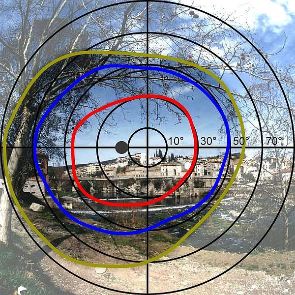
Cognitive therapy
                                                                                 by RehaCom GmbH

                This manual contains information about using the RehaCom
                therapy system.

                Our therapy system RehaCom delivers tested methodologies
                and procedures to train brain performance.
                RehaCom helps patients after stroke or brain trauma with the
                improvement on such important abilities like memory, attention,
                concentration, planning, etc.

                Since 1986 we develop the therapy system progressive. It is our
                aim to give you a tool which supports your work by technical
                competence and simple handling, to support you at clinic and
                practice.

                User assistance information:

                Please find help on RehaCom website of your country. In case of
                any questions contact us via e-mail or phone (see contact
                information below).

CAUTION   Risk of misdiagnosis. Screening for use of RehaCom only. Use standardized tests for diagnostic.

          Germany / Europe / Worldw ide:                      USA:
          HASOMED GmbH                                        Pearson Clinical Assessment
          Paul-Ecke-Str. 1                                    19500 Bulverde Road, Suite 201
          D-39114 Magdeburg                                   San Antonio, TX 78259-3701

          Tel: +49 (391) 610 7645                             Phone: 1-888-783-6363
          w w w .rehacom.com                                  w w w .pearsonclinical.com/RehaCom
          info@rehacom.com                                    rehacominfo@pearson.com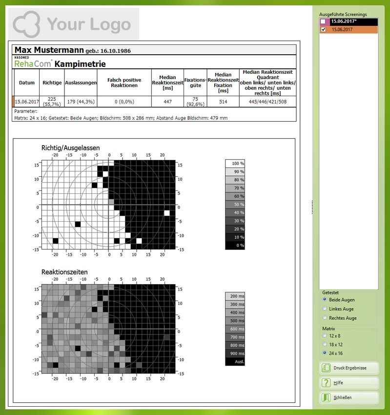
Contents                 I

Dear user,
please read the entire instruction manual before trying to operate RehaCom.
It's unsafe to start using RehaCom without reading this manual.
This manual includes lots of advice, supporting information and hints in order to reach
the best therapy results for the patients.

Table of contents
   Part 1 Disorders of the visual field                                                                                                          1
          1 PC-supported
               ...................................................................................................................................
                                detection of visual field deficits                                                                                 5

   Part 2 Field of application                                                                                                                   8

   Part 3 Test description                                                                                                                       9
          1 Settings
               ................................................................................................................................... 9
          2 Testing
              ...................................................................................................................................
                       process                                                                                                                    12
          3 Evaluation
              ................................................................................................................................... 16

   Part 4 Bibliography                                                                                                                         20

             Index                                                                                                                               0

© 2019 HASOMED GmbH
1      Campimetry

1       Disorders of the v isual field
        Basic information on the data analysis of screening results is available in the
        RehaCom manual, Chapter "Screening and Diagnostics".

        Impairments of vision, visual-spatial perception, and attention are common results of
        brain injuries. According to Zihl (2006), 20–40% of patients with an acquired brain
        injury have visual problems, 61.7% of those patients have restrictions or losses in the
        visual field.

        The ability to see is a very important sensory function. Disorders affecting the visual
        field impact orientation and, depending on the degree of disturbance, have an effect
        on everyday actions (e.g. searching and finding objects when eating or getting
        dressed, safely navigating a room without bumping into something, recognizing
        faces, reading the newspaper, watching TV, using the computer). Visual field is
        vitally important, especially when driving a motor vehicle. Disorders of the field of
        view and visual field affect participation in many aspects of life according to ICF and
        therefore require therapy and rehabilitation to improve the participation.

        Body-Head-Eye system

        A persons entire body is actively involved in the visual orientation and visual
        cognition, including the orientation and position of the body, the head, and the eyes
        in space; the performance capability of the eyes (e.g. visual acuity or agility), and
        eventually the central information processing of all involved sensomotoric processes
        in the brain. Our normal behavior is that we constantly move our body or very rarely
        remain motionless. We constantly explore objects and our surrounding world with
        countless eye movements geared to our current goal of action, or briefly turn to
        peripheral new stimuli and assess them. The field of vision solely by eye movements
        (without moving one’s head) is thereby smaller (about 50° one-sided) than the
        peripheral visual field in central fixation (about 90° one-sided). The combination of
        the field of vision and the visual field extends the visual range up to 115° (one-sided).
        The field of vision can be shifted sideways even further by head and body
        movements.

                                                                               © 2019 HASOMED GmbH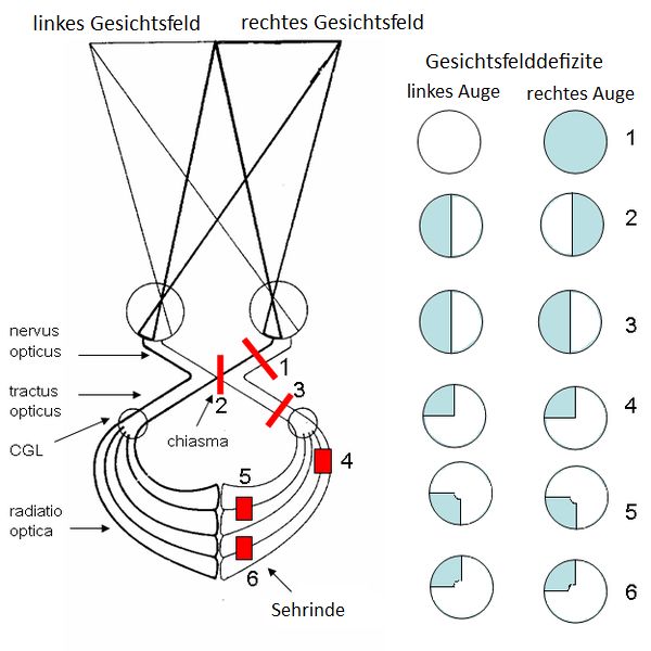
Disorders of the visual field   2

                       Fig. 1: Full visual field of both eyes
        Shape recognition is quite possible in the central field of vision (about 5° on one side
        unilateral) with fixation; in the peripheral visual field without fixation, even color
        perception (see fig. 1) and especially motion perception are possible. In order to
        reliably recognize and differentiate shapes and patterns, focusing on the stimulus
        visually (fixation) is necessary.

        Figure 2 shows the qualitative visual field limits in the normal left visual field.

             Fig. 2: qualitative visual field limits in the normal left visual field
        Fig. 2 shows a polar diagram of visual field of the left eye [Wikipedia].
        red and blue circles: sensitive area for colors;
        yellow circle: entire visual field; black spot: blind spot

        Consideration of further cognitive performances

        The ability to see is an integral part of our interactions with the world around us.

© 2019 HASOMED GmbH
3      Campimetry

    Because the ability to see is so important, further cognitive performances, especially
    attention, are always involved.
    When testing a patient’s visual field after an acquired brain injury, you should also
    take into account other performance functions, such as attention and concentration,
    memory and executive functions. The interpretation of results depends on many
    factors, particularly in the early phase after a brain injury (1-4 months), where a
    characteristic instability for many functions will be found.
    You should make sure to have pauses during the implementation of therapy. Also
    ensure the comprehension of the instructions by repetition (in case of memory
    losses) (Zihl & von Cramon, 1985).

    Triad of the limitations of the visual field

    In disorders of the visual field and the visual perception, there are three limitations
    (Zihl & von Cramon, 1985) for which we may collect measurements:
      limitations of the visual field that affect stimulus detection at a central fixation (visual
      field limitation)
      limitations of spontaneous amplitudes and systematics of eye movements in the
      affected visual field (narrowing of the field of vision)
      limitations of spontaneous exploratory searching and noticing of stimuli (spatial
      attention restriction)

    Limitations of the visual field and saccadic movements / gaze strategies can occur,
    especially in a patient with homonymous hemianopsia without disorders of the
    attention field.

                                                                              © 2019 HASOMED GmbH
Disorders of the visual field 4
Fig. 3: Visual field deficits depending on the location of the lesion
o 3) homonymous hemianopsia (right or left): In both eyes, the same side is affected by loss
(patients see only the left or right side of an image section - occurs frequently)
o 2) heteronymous (binasal oder bitemporal) hemianopsia: On both eyes the opposite side is
affected by loss (patients suffer from “tunnel vision” - occurs rarely)
o 4 - 6) Quadrantanopsia is characterized by the loss of a quarter of the visual field. Like the
hemianopsia the quadrantanopsia is mostly homonymous, both eyes are affected by the loss in
the same quadrant.
Fig. 4 : Restricted visual field in hemianopsia to the left
© 2019 HASOMED GmbH5 Campimetry
Disorders of the visual-spatial attention (neglect)
In case of severe neglect, the three limitations must be considered. Neglect
phenomena can be assumed if spontaneous orientation, stimulus discovery, and
exploration by saccades are disturbed. The patient might not detect people, items,
or test stimuli in the affected part of the visual field (Heilmann, 1979; Karnath, 2003).
Fig. 5: Restricted field of perception in neglect to the left
In patients with neglect, indicators for neglect can often be found in other sensory
modalities, such as extinction at double simultaneous stimulation, visual-spatial
disorders, and improvements in performance on conscious attention allocation.
Patients with hemianopsia without neglect can't improve visual field deficits by
conscious attention allocation (Kerkhoff, 2004).
Therefore, a competent differential diagnosis is important, especially as lesions are
not uncommon where hemianopic vision deficits and neglect phenomena can be
found in the acute phase and can only be differentiated in the chronic phase ( > 3
months after lesion).
Through spontaneous remissions in the acute phase, neglect disorders can almost
halve to about persistent 33% of patients with right hemispheric lesions and
approximately 13% of patients with left hemispheric lesions (Stone et.al., 1991, as
cited by Kerkhoff, 2004). In the clinical practice, it is not uncommon for patients to
show a quadrantanopsia that was previously hidden by the neglect.
In the event that you suspect a patient having a disorder combined with neglect and
hemianopsia/quadrantanopsia, both defects should be treated.
1.1 PC-supported detection of visual field deficits
The ophthalmologists use perimetry to measure the visual field by varying either
position or intensity of a stimulus (e.g. Goldmann perimeter or Tübinger perimeter).
Under standardized stimulus conditions, the ophthalmologist can examine both the
expansion of the general visual field (stimulus detection) and, if required, the
limitations for the light sensitivity, color, and shape recognition accurately.
© 2019 HASOMED GmbHDisorders of the visual field 6
In the neurological clinic, rehab clinic, or outpatient neuropsychological practice,
cost-intensive perimetry is (usually) not available. It is therefore necessary to resort to
simple testing, using paper-pencil methods and PC-based visual field screenings.
However, this diagnostic instrument allows an imaging of visual field deficits, neglect
symptoms and reading disorders that is sufficiently precise for therapeutic
indications.
In the PC-supported measurement of the visual field, which is used as a screening,
limitations towards a perimeter are existing in the methods that were available up to
now:
as a result of the limitation of the monitor, the covered visual angle area is much
smaller
the programs do not allow an evaluation of the contrast sensitivity, color or
shape perception
the fixation constancy can not be observed in confrontation
By the use of modern flatscreen monitors (24'' or 27'') or TV (32'' or 34''), PC-
supported coverages are already able to capture a binocular visual field up to 30°.
When using a projector with a rear projection screen, the coverage can be expanded
up to 60° (that corresponds to the required visual field for the driving ability).
Central and peripheral task
In the perimetry, the fixation constancy is controlled by a person who observes the
patient from the front and reacts accordingly when leaving the fixation. This possibility
is missing when measuring the visual field on the PC. Besides the frequency of
measured points, the fixation constancy as a critical indicator of a precise
measurement is of particular importance.
To control the fixation constancy, a central task was therefore introduced into PC
visual field tests, which can only be solved by visual focusing. In parallel, the actual
measurement of the perception of stimuli in the visual field was defined as a
peripheral task. This created a dual task design with requirements on increased
attentional performances. In addition to the sustained attention, the selective and
divided attention performances mainly belong to it.
For example, the original version of a common PC visual field testing consists of a
visual-verbal central task (recognize letters and name them verbally) and a visual
discovery task of peripheral kind (notice flicker stimuli and press the button). The
transition between the central and the peripheral task requires adaption ability and
sequencing (verbal naming, nonverbal keystokes). The stimuli are presented for a
maximum of 3000 ms. The presentation time is also the maximum reaction time.
The peripheral stimulus is displayed over a "longer" period (max. 3000 ms) and can
cause an automatic orienting response, an eye movement up to the Flicker stimulus.
The shortest trial version runs for about five minutes, the longer version more than ten
minutes without pause.
In the new variant, the central task has been replaced by a nonverbal visual
discrimination task with requirements on selective attention (answer 4 target stimuli
out of 8 stimuli). The response reaction in the central task is nonverbal in the form of
© 2019 HASOMED GmbH7 Campimetry
a keystroke and has been aligned to the output of the peripheral task. The
presentation time of the central stimuli is 400 ms. The presentation time of the
peripheral stimuli is 3000 ms. The test duration is five minutes without a pause.
Another newer screening design for driving ability diagnostics combines the
measurement of the visual field with distractor stimuli, which are used in neglect
examinations. The screen is filled with grey dots. One dot at a time can flash up
brightly (peripheral task). The presentation time for peripheral stimuli was shortened
to 800 ms. The central task again consists of a nonverbal visual discrimination task
with requirements on selective attention (answer 4 target stimuli out of 8 stimuli). It
has to be reacted to both tasks non-verbally with a keystroke. The number of central
stimuli and thus the requirements on the visual focusing was significantly increased
(ratio stimuli central / peripheral: 470/85).
Requirements on selective and divided attention
Attentional processes and requirements for greater attention performance outside
the spatial attention are involved in all previous designs for PC-based visual field
tests and may affect the actual intended visual stimulus perception and response
action in disorders of attention performance.
To reduce the impact of attention deficits, the discrimination tasks should be as
simple as possible (selective attention).
The central and peripheral task should be of the type and modality as similar as
possible in dual-task design to keep the demands on the shared attention low.
To relieve the focused attention and sustained attention, pauses during the
implementation should be made possible.
Visual field tests enable conclusions about the patient's performance and a
probability indication of the available field of view, but they are not a direct
measurement of the underlying cerebral failure or neuronal damage.
Directly observable and computerizedly measurable are only behavioral responses
to the stimuli of the task. An omission or incorrect reaction may also have other
causes than immediate vision loss. The interpretation of the overall result therefore
requires professional expertise.
Other situational conditions such as daytime and lighting conditions, psychological
state or medication can have an effect on the result. Therefore, care should be taken
to standardize the examination situation (e.g. darkened room).
© 2019 HASOMED GmbHField of application 8
2 Field of application
Basic information about the screening can be found in the RehaCom manual, Kapitel
"Screening und Diagnostik".
The "Campimetry" screening is a grid test to monitor and evaluate computer
supported training. It includes the modules saccade training, exploration training, and
restitutive field of vision training. At the first screening the limitations of the field of
vision are measured and the severity is determined. The results of all examinations,
e.g. the neglect examination, should always be considered when evaluating the field
of vision.
Target Patients
The screening is suitable in case of suspected neurological vision defects, e.g.
hemianopsia or quadrantanopia. The goal is to examine the field of vision for defects
through static stimuli with fixation control, and to determine the where training is
needed as well as the areas in which there is potential for restitution.
For clients who suffer from additional cognitive deficits such as attention- and
concetration disorders or a slowed down perception and motor skills, another
screening with different settings or adjusted parameters can be done. The same is
true for vision defects such as cataract or disordered sensitivity for coulour and
contrast. Children whose speech and understanding of language and words is
limited can usually do the screening without any problems.
© 2019 HASOMED GmbH9 Campimetry
3 Test description
In the campimetry screening, the field of vision can be examined binocularly or
monocularly. Unlike the three-dimensional perimetry, campimetry is two-dimensional.
The client's task is to react to stimuli within his field of vision under fixation control.
Fig. 6: Campimetry screening w ith central fixation point and stimulus in the periphery.
Before beginning a screening parameters need to be adjusted, such as the size of
the monitor, the distance between the monitor and the eyes, the size of the matrix,
and the client's eyes. The client should be positioned in front of the monitor in a set
distance, comfortable but fixed. We recommend using a chin rest/head rest for
training of the visual field.
During the screening the client's gaze should stay fixed on a point on the screen. To
ensure this the client has to react to the point changing in shape and colour in
irregular intervals.
In the test, stimuli appear in random intervals and on random places on the screen.
Position and number of stimuli are calculated based on the parameters set in the
beginning. Every stimulus and change of colour should be acknowledged as fast as
possible by pushing the "answer" key.
3.1 Settings
To conduct a campimetry screening it is necessary to enter several parameters.
They are asked for before beginning the screening.
In the parameter menu the program will ask you for settings such as screen height-
© 2019 HASOMED GmbHTest description 10
and width, distance patient to screen, size of matrix, and eye to check. Without them,
no usable results are attainable.
Please note: Only tests which use the same parameters can be compared. Tests
with differently sized matrices, e.g. 24x16 and 18x12, cannot be shown layered over
each other. If screen height, -width, and/or distance are different layering is not
possible.
Fig.7: Advice to entering parameters and comparability of results.
© 2019 HASOMED GmbH11 Campimetry
Fig.8: The parameter menu w ith boxes to enter required parameters.
Parameters of campimetry:
Eye to Please choose which of the client's eyes should be tested:
check Both eyes
Left eye
Right eye
Size of Choose a grid in which the field of vision should be measured.
matrix The following options are available depending on your selection in the
parameter "eye to check":
Both eyes:
o 24 x 16 (404 stimuli*)
o 18 x 12 (236 stimuli*)
o 12 x 8 (100 stimuli*)
Left or right eye:
o 16 x 16 (276 stimuli*)
o 12 x 12 (160 stimuli*)
o 8 x 8 (68 stimuli*)
ScreenwidthEnter the width of the entire used part of the monitor.
Screenheig Enter the height of the entire used part of the monitor.
ht
Distance Enter the distance of the client's eyes to the screen after placing your
patient to client in front of the centre of the screen.
screen
© 2019 HASOMED GmbHTest description 12
* stimuli to check the field of vision, fixation controls excluded.
In addition, the optimal distance between the client and the screen is shown below
the parameter boxes.
The parameters are only saved after clicking "continue". If you click "reset", all
changes will be discarded and the client's last parameters/ the default parameters
will be restored.
3.2 Testing process
Procedure
1. Entering parameters
Before conducting the test the required Settings need to be adjusted.
2. Instruction and Testing
The test begins with a short practice run. The different symbols are introduced and
the procedure is explained. After that the practice run takes place and can be
repeated if needed.
The test uses 3 different symbols. It is the client's task to react to the stimuli as fast
as possible by pressing the OK-button.
Abb. 9: Fixationspunkt Abb.10: Fixationskontrollpunkt Abb.11: Stimulusreiz
The fixation point is in the centre of the screen and will occasionally change its
shape/colour. This is necessary in order to fix the gaze and to ensure the field of
vision stays statically examinable. If there is a reaction to a change of colour/shape
within the designated time window of 1000 ms, a feedback sound plays.
The stimulus appears anywhere on the screen. It appears in a random order. When a
stimulus is seen, the OK-button should be pressed as fast as possible. If there is a
reaction within the designated time window, a feedback sound plays.
© 2019 HASOMED GmbH13 Campimetry
3. Conducting the test
After the practice run the test takes place.
Every stimulus (or fixation control) appears after a random amount of time. Total
number and position of the stimuli are calculated based on the parameters. After 500
to 1000 ms have passed, the stimulus is shown for a short amount of time and then
hidden again. Afterwards, the client has 1000 ms to react to the stimulus.
The positions around the point of fixation are checked multiple times, depending on
the size of matrix selected.The following images show how often every positon in a
matrix is checked.
Fig. 12 Matrix 8x8
© 2019 HASOMED GmbHTest description 14
Fig. 13 Matrix 12x12
Fig. 14 Matrix 16x16
© 2019 HASOMED GmbH15 Campimetry
Fig. 15 Matrix 12x8
Fig. 16 Matrix 18x12
© 2019 HASOMED GmbHTest description 16
Fig. 17 Matrix 24x16
The fixation control happens randomly at a chance of 15% after each stimulus. Every
fixation control should be answered by pressing the OK-button. If the fixation control
is ignored 3 times in a row, a signaling sound will play to alert the client to it.
If the OK-button is pressed continuously, the test is interrupted. A signal sound will
play and the test will be paused as long as the button is pressed. When the button is
released, the test will continue.
Duration
Depending on the size of matrix chosen and on which eye to check, the test should
take between 2 and 15 minutes.
3.3 Evaluation
Basic information about the result evaluation of the screening results can be found in
the RehaCom manual, chapter "Screening Results".
Overview of results
For the campimetry screening the Z-norm is taken into consideration. The Z-norm
© 2019 HASOMED GmbH17 Campimetry
states how many stimuli were overlooked in relation to the total number of stimuli.
Fig.18: Overview of the results of a campimetry screening.
Detailed information about the screening process can be found in "Details" (see Fig.
18).
Details
The details page shows the evaluation parameters for all tests conducted.
On the right is a list of all tests in order of the time they were conducted. Entries that
are marked with an asterisk (*) show tests that weren't completeted.
Every result selected (in the list on the right) is assigned a column in the table. The
results are distinguishable by their different colours. The background colour of the
entries on the list of all the tests (on the right) is the same as the colour of the column
of the result in the table (middle).
The evaluation of the details shows a maximum of 3 results at the same time. The
first and last test conducted are chosen automatically.
© 2019 HASOMED GmbHTest description 18
Fig.19: Detailed results of the campimetry results, w ith the list of tests on the right and evaluation tables and charts in the
centre.
Colums of the results table:
Date Time at which the screening was conducted
Correct Number of correct reactions to a stimulus
Omissions Number of non-reactions to a stimulus
False positives Number of reactions before 150 ms
© 2019 HASOMED GmbH19 Campimetry
Median Response Median of all response times in ms
Time (ms)
Fixation quality Number of reactions to central fixation stimulus
Median Response Median of all response times to central fixation stimulus in ms
Time Fixation
Median Response Median of all reaction times in every quadrant:
Time Quadrant o upper left
o lower left
o upper right and
o lower right
Furthermore, there are two charts showing the reaction time and the quality of the
reactions.
The evaluation of the respective eyes and the different matrix sizes can be checked
in the boxes on the right. The charts will change according your selections.
The "Correct/Omission" chart shows which point was responded to at what rate. The
rate is realized in a grey scale. Matrix fields that are filled in with a dot signify false
positives. Matrix fields that are grey show points that were evaluated more than once
during the test. Those points produced both correct reactions and omissions.
The "Response time" chart shows how quickly a point was reacted to. The response
times are indicated by the grey scale. To interpret the shades of grey you can look at
the key on the side. The faster the response time, the lighter the matrix field will
appear. The times (ms) shown on the key always represent the shortest time of the
shade shown. In the chart of the response times intermediate values are calculated
for the shades according to the response time. For example, a response time of 250
ms would have a shade of grey in between the shades of 200 and 300 ms as they
are shown on the key.
Layering of screenings
A maximum of three results can be layered over each other. To do this choose the
screenings from the "Completed screenings" list. In the "Correct/Omissions" and the
"Response time" charts, the average calculated will be shown.
Please note: Only those results that were attained using the same parameters
(identical screen sizes, same distance of the client to the screen, same matrix) can
be layered.
© 2019 HASOMED GmbHBibliography 20
4 Bibliography
Schmielau, Wong (2007) Recovery of visual fields in brain-lesioned patients by
reaction perimetry treatmen
Kasten, Wüst, Behrens-Baumann, Sabel (1998). Computer based training for the
treatment of partial blindness. Nature Medicine. Nr. 4, S. 1083-1087
Poggel, Müller, Kasten, Sabel (2008). Multifactorial predictors and outcome
variables of vision restoration training in patients with post-geniculate visual field
loss. Restorative Neurology and Neuroscience. Nr. 26, S. 321-339
Schlüter, Schulz, Kenkel, Romano (2009). Functional Improvements after a Visual
Rehabilitation Intervention for Patients with Homonymous Visual Field Defects.
Poster presented at the Annual Meeting of the American Academy of Neurology,
Seattle, April 26-May 2, 2009
de Haan GA, Melis-Dankers B, Brouwer WH, Tucha O, Heutink J.( 2016) The Effects
of Compensatory Scanning Training on Mobility in Patients with Homonymous Visual
Field Defects: A Randomized Controlled Trial. PLoS One. 2015 Aug
14;10(8):e0134459. doi: 10.1371/journal.pone.0134459. eCollection 2015.
Überblicksartikel:
Kerkhoff, Oppenländer, Finke, Bublak (2007). Therapie cerebraler visueller
Wahrnehmungsstörungen. Der Nervenarzt, Nr. 78, S. 457–470
© 2019 HASOMED GmbHYou can also read