Comparison of the Type and Severity of Nasal Septal Deviation between Chronic Rhinosinusitis Patients Undergoing Functional Endoscopic Sinus ...
←
→
Page content transcription
If your browser does not render page correctly, please read the page content below
Hindawi
International Journal of Dentistry
Volume 2022, Article ID 2925279, 6 pages
https://doi.org/10.1155/2022/2925279

Research Article
Comparison of the Type and Severity of Nasal Septal
Deviation between Chronic Rhinosinusitis Patients Undergoing
Functional Endoscopic Sinus Surgery and Controls

          Nafiseh Nikkerdar ,1 Atena Karimi ,1 Fatemeh Bazmayoon,2 and Amin Golshah                                              3

          1
            Department of Oral and Maxillofacial Radiology, School of Dentistry, Kermanshah University of Medical Sciences, Kermanshah,
            P.O. Code: 6715847141, Iran
          2
            Students Research Committee, School of Dentistry, Kermanshah University of Medical Sciences, Kermanshah,
            P.O. Code: 6715847141, Iran
          3
            Department of Orthodontics, School of Dentistry, Kermanshah University of Medical Sciences, Kermanshah,
            P.O. Code: 6715847141, Iran

          Correspondence should be addressed to Amin Golshah; amin.golshah@gmail.com

          Received 26 May 2021; Revised 11 January 2022; Accepted 6 April 2022; Published 25 April 2022

          Academic Editor: Rossana Izzetti

          Copyright © 2022 Nafiseh Nikkerdar et al. This is an open access article distributed under the Creative Commons Attribution
          License, which permits unrestricted use, distribution, and reproduction in any medium, provided the original work is
          properly cited.

          Objectives. Some correlations have been proposed between chronic rhinosinusitis (CRS) and type and severity of nasal
          septal deviation. This study sought to compare the type and severity of nasal septal deviation between CRS patients
          undergoing functional endoscopic sinus surgery (FESS) and asymptomatic controls using cone-beam computed to-
          mography (CBCT). Materials and Methods. This prospective case-control study evaluated 49 CRS patients who did not
          respond to pharmaceutical therapy and were candidates for FESS and 49 asymptomatic controls. All participants un-
          derwent CBCT and were inspected for septal deviation type and severity. Data were analyzed by the independent t-test and
          chi-square test. Results. The study population comprised of 58.25% males and 41.8% females, with a mean age of
          33.74 ± 11.78 years. Significant correlations were noted between the presence of CRS and severity of septal deviation
          (P  0.007). Type of septal deviation had no significant correlation with the presence of CRS (P  0.443). Conclusion.
          Patients with CRS have significantly more severe nasal septal deviation. However, type of septal deviation is not correlated
          with CRS.

1. Introduction                                                        obstruction/congestion or nasal discharge (anterior/
                                                                       posterior nasal drip) ± facial pain/pressure ± reduction
Rhinosinusitis is a common condition worldwide, im-                    or loss of smell for ≥12 weeks [1]. CRS is a significant
posing a significant burden on the healthcare systems. It              health problem that affects 5–12% of the general pop-
is among the most common reasons for antibiotic pre-                   ulation [1]. It also affects as high as 10.9% of the pop-
scription, and its proper management is highly impor-                  ulation of Europe [3]. The prevalence of sinusitis is 53%
tant considering the emerging resistance to antibiotics                in Iran [4]. Patients with CRS often have a much lower
[1]. Chronic rhinosinusitis (CRS) refers to complex in-                health-related quality of life compared with healthy
flammation of the nose and paranasal sinuses. Despite                  controls [5], and their health status has been claimed to
medical care, its signs and symptoms often last for about              be comparable to that of cancer, arthritis, and asthmatic
12 weeks or longer [2]. CRS (with or without nasal                     patients [6].
polyps) in adults is defined as the presence of two or more                Treatment of CRS is costly for both patients and the
symptoms, one of which should be either nasal blockage/                healthcare system. In the United States, the total cost of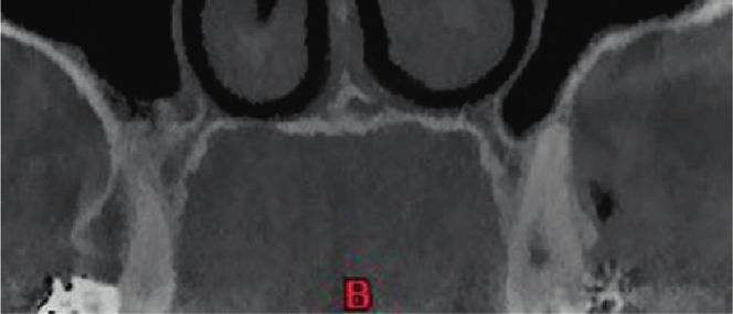
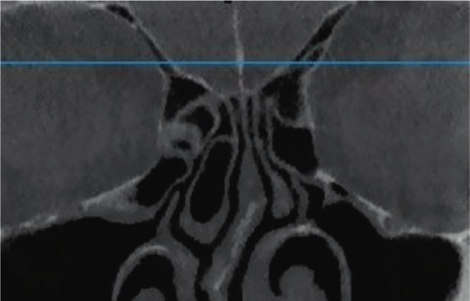
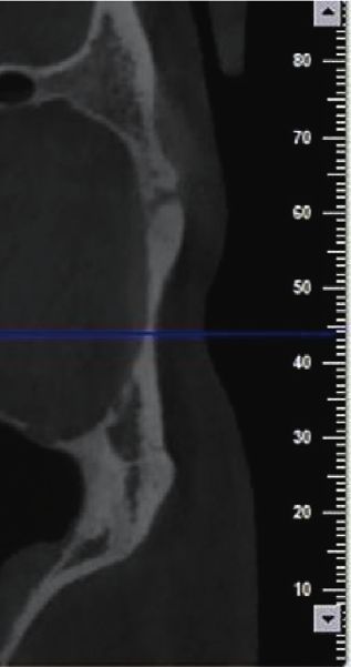
2                                                                                           International Journal of Dentistry

healthcare related to CRS was estimated to be 6.9–9.9 billion    CBCT scans for purposes other than the ear, nose, and throat
dollars/year in 2014 [7]. In case of failure of pharmaceutical   problems.
therapy, functional endoscopic sinus surgery (FESS) is often         The inclusion criteria were as follows: confirmed diag-
performed for treatment of CRS [8]. An endoscope is used in      nosis of CRS according to the European Position Paper on
FESS instead of open surgery. This procedure aims to clean       Rhinosinusitis and Nasal Polyps (EPOS), 2020, characterized
the paranasal sinuses under direct vision while preserving       by two or more symptoms, one of which should be either
the physiological health of the sinuses and the nasal cavity     nasal blockage/obstruction/congestion or nasal discharge
[9]. Nasal septal deviation can impair the normal nasal          (anterior/posterior nasal drip): ± facial pain/pressur-
physiology. Also, it can narrow the middle meatus by             e ± reduction or loss of smell, the patients had to have this
moving the nasal concha laterally [10]. A recent study           condition for more than 12 weeks with no response to
showed septal deviation resulting in a concavity on the other    medical and pharmaceutical therapy [22], and patients who
side of the septum and narrowing of the contralateral side       had indication for medical imaging (CT/CBCT) and FESS.
[11]. In addition to nasal obstruction, septal deviation can     Patients also had mucosal changes within the ostiomeatal
obstruct the path of nasal drainage and impair the muco-         complex and/or sinuses on their CT scans. The exclusion
ciliary clearance, leading to nasal congestion and secondary     criteria for CRS patients were sinus malignancies, preg-
infection of the sinuses [10]. Nasal septal deviation has been   nancy, immunodeficiency, cystic fibrosis, age 15°
private ear, nose, and throat clinic to undergo FESS. The
patients underwent CBCT prior to their surgical procedure.           Nasal septal deviation was also divided into three main
Control individuals were selected among those presenting to      types as shown in Figure 1 (in the coronal and axial planes)
a private oral and maxillofacial radiology clinic to obtain      [25].
International Journal of Dentistry                                                                                                          3

                                (a)                                (b)                                  (c)

                                       (d)                                              (e)

Figure 1: Types of septal deviation: (a) C-shaped in the coronal plane, (b) reverse C-shaped in the coronal plane, (c) reverse S-shaped in the
coronal plane, (d) reverse S-shaped in the coronal plane, and (e) S-shaped in the coronal plane.

2.4. Sample Size Estimation. The minimum sample size was                 3. Results
calculated to be 21 in each group according to a previous
study [26], assuming the effect size of 0.713, alpha � 0.05, a            The minimum sample size was calculated to be 21 in each
level of significance of 0.05, and study power of 90% using               group, but 49 patients were enrolled in each group to ensure
PASS (version 11; NCSS, Kaysville, UT, USA). To increase                 the reliability of the results.
the reliability of the results, 49 patients were enrolled in each            Of all participants, 41 (41.8%) were females and 57
group.                                                                   (58.2%) were males. The mean age of the participants was
                                                                         33.74 ± 11.78 years (range 22–46 years).
                                                                             The intraexaminer (ICC � 0.963, kappa score � 0.86) and
2.5. Interexaminer Reliability. To assess the intraexaminer              interexaminer reliability coefficients were found to be ex-
reliability, the CBCT scans were reevaluated again after 2               cellent (ICC � 0.952, kappa score � 0.89). The total duration
weeks, and to evaluate the interexaminer reliability between             of the study was approximately 12 months.
the radiologists, the intraclass correlation coefficient (ICC)                 Table 1 provides the severity of septal deviation in the
was calculated for the severity of nasal septal deviation, and           two groups. A significant correlation existed between the
the kappa score was computed for the type of nasal septal                severity of nasal septal deviation and presence of CRS, such
deviation.                                                               that septal deviation was significantly more severe in the
                                                                         CRS group compared with the control group (chi-square
                                                                         test, P � 0.007).
2.6. Statistical Analysis. The data were entered into SPSS for               The mean degree of nasal septal deviation was
Windows version 20 (SPSS Inc., IL, USA) by a third person                13.97 ± 4.92 degrees in the CRS group and 10.77 ± 4.12
who was blinded to the group allocation of patients.                     degrees in the control group. The CRS and control groups
Normal distribution of data was evaluated by the Kol-                    had a significant difference in the mean degree of nasal septal
mogorov–Smirnov test. Since the data were normally                       deviation, such that the mean degree of nasal septal devi-
distributed, the severity of nasal septal deviation was                  ation was significantly greater in the CRS group (inde-
compared between the two groups using the independent                    pendent samples t-test, P � 0.001).
sample t-test. The chi-square test was applied to analyze the                Table 2 provides the frequency of different types of nasal
correlation between the qualitative variables. Level of                  septal deviation in the CRS and control groups. Type of
significance was set at 0.05.                                             septal deviation in the horizontal plane had no significant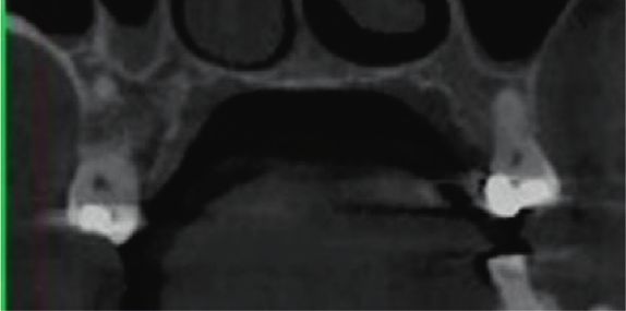
4                                                                                                      International Journal of Dentistry

                                          Table 1: Severity of septal deviation in the two groups.
                                                                                       Group
                                                                      CRS                                Control                P value†
                                                          Count        Column N (%)            Count       Column N (%)
                                            Mild            8              16.3                 22               44.9
Severity of nasal septal deviation        Moderate         21              42.9                 16               32.7             0.007
                                           Severe          20              40.8                 11               22.4
†
    Chi-square.

       Table 2: Frequency of different types of nasal septal deviation in the coronal and axial planes in the CRS and control groups.
                                                                                      Group
                                                                          CRS                              Control              P value†
                                                              Count       Column N (%)     Count             Column N (%)
                                           S-shaped             8             16.3          10                     20.4
                                       Reverse S-shaped         5             10.2          10                     20.4
Types of nasal septal deviation                                                                                                   0.443
                                          C-shaped             22             44.9          17                     34.7
                                       Reverse C-shaped        14             28.6          12                     24.5
†
    Chi-square.

correlation with the presence of CRS (chi-square test,                  quantification of the severity of septal deviation since
P � 0.443). The frequency of S-shaped deviation in the                  Verhoeven and Schmelzer [30] measured this parameter
control group was higher than that in the CRS group, while              clinically while application of CT would be more appropriate
the frequency of C-shaped deviation was higher in the CRS               for this purpose [21]. In this study, CBCT was used due to its
group, but not significantly (P > 0.05).                                 advantages over CT.
                                                                            C-shaped and reverse C-shaped deviations were the
4. Discussion                                                           most common types of septal deviations in this study and
                                                                        previous investigations [31, 32]. The nasal airway and
The nasal airway is influenced by the degree, location, and              probably the discharge of mucosa into the nasal cavity are
type of nasal septal deviation [27]. This study sought to               influenced by the type of nasal septal deviation [27, 33]. In a
compare the type and severity of nasal septal deviation                 study by Alharethy et al. [33], most patients with normal
between CRS patients undergoing FESS and controls using                 discharge had S-shaped or reverse S-shaped deviations,
CBCT. The results showed that the severity of nasal septal              while the most common types of septal deviation in pa-
deviation was significantly greater in CRS patients who were             tients who were candidates for surgery were C-shaped and
candidates for FESS compared with controls. However, no                 reverse C-shaped deviations. Prasad et al. [34] observed
other significant correlations were noted between CRS and                that patients with C-shaped and reverse C-shaped devia-
type of nasal septal deviation.                                         tions were more susceptible to sinusitis because a C-shaped
    Taghiloo and Halimi [28] reported a significant corre-               septum can cause involvement of the space below the
lation between the severity of nasal septal deviation and               lateral superior nasal cartilage and upper surface of the
increased mucosal thickness of the maxillary sinuses. Kumar             inferior concha. The current results indicated the higher
et al. [27] reported that nasal septal deviation would make             frequency of C-shaped and reverse C-shaped deviations in
the nasal mucosa susceptible to chronic inflammation and                 the CRS group and higher frequency of S-shaped and re-
squamous metaplasia. These changes can make the patients                verse S-shaped deviations in the control group; however,
susceptible to CRS [27]. Gregurić et al. [17] stated that only         these differences were not statistically significant. This
severe nasal septal deviation had a significant correlation              finding may be due to our small sample size, which was a
with the severity of sinusitis. Poorey and Gupta [29]                   limitation of this study.
demonstrated that increased angle of septal deviation further               Considering the current findings and those of Janovic
increased the changes in the maxillary sinus mucosa. Also,              et al. [35] and Verhoeven and Schmelzer [30], no significant
they showed that increased angle of septal deviation in-                correlation exists between the type of septal deviation and
creased the prevalence and severity of CRS. Thus, it may be             nasal congestion. Thus, it appears that classification (type) of
stated that more severe septal deviations can increase the risk         septal deviation is not useful for prediction of the symptoms
of CRS.                                                                 of CRS. Further studies are recommended on a larger sample
    In contrast to the abovementioned findings, Verhoeven                size from different racial and ethnic groups to acquire a
and Schmelzer [30] discussed that the severity of nasal septal          better perspective of the issue.
deviation cannot predict the severity of nasal congestion,                  One limitation of this study was that only CRS patients
which is a symptom of CRS. This controversy can be due to               who were candidates for surgery were enrolled. Future
the differences in the methods of assessment and                         studies are required to compare the type and severity of nasal
International Journal of Dentistry                                                                                                           5

septal deviations between CRS patients not eligible for                    [10] Y. J. Jang, N.-H. Myong, K. H. Park, T. W. Koo, and
surgery and asymptomatic controls.                                              H.-G. Kim, “Mucociliary transport and histologic charac-
                                                                                teristics of the mucosa of deviated nasal septum,” Archives of
5. Conclusion                                                                   Otolaryngology—Head and Neck Surgery, vol. 128, no. 4,
                                                                                pp. 421–424, 2002.
Patients with CRS have significantly more severe nasal septal               [11] W. Espinosa, R. Genito, and R. Z. Ramos, “Anatomic vari-
deviation. However, the type of septal deviation is not                         ations of the nasal cavity and paranasal sinus and their
correlated with CRS.                                                            correlation with chronic rhinosinusitis using Harvard staging
                                                                                system,” Journal of Otolaryngology-ENT Research, vol. 10,
                                                                                no. 4, pp. 190–193, 2018.
Data Availability                                                          [12] L. Rudmik, J. Mace, B. J. Ferguson, and T. L. Smith, “Con-
The data used to support the findings of this study are                          current septoplasty during endoscopic sinus surgery for
                                                                                chronic rhinosinusitis: does it confound outcomes assess-
available from the corresponding author upon request.
                                                                                ment?” The Laryngoscope, vol. 121, no. 12, pp. 2679–2683,
                                                                                2011.
Disclosure                                                                 [13] S. Collet, B. Bertrand, S. Cornu, P. Eloy, and P. Rombaux, “Is
                                                                                septal deviation a risk factor for chronic sinusitis? review of
This study was derived from a thesis submitted to Ker-
                                                                                literature,” Acta Oto-Rhino-Laryngologica Belgica, vol. 55,
manshah University of Medical Sciences, School of                               no. 4, pp. 299–304, 2001.
Dentistry.                                                                 [14] L. Li, D. Han, L. Zhang et al., “Aerodynamic investigation of
                                                                                the correlation between nasal septal deviation and chronic
Conflicts of Interest                                                           rhinosinusitis,” The Laryngoscope, vol. 122, no. 9,
                                                                                pp. 1915–1919, 2012.
The authors declare that there are no conflicts of interest.                [15] S. A. Madani, S. A. Hashemi, and M. Modanluo, “The inci-
                                                                                dence of nasal septal deviation and its relation with chronic
Acknowledgments                                                                 rhinosinusitis in patients undergoing functional endoscopic
                                                                                sinus surgery,” JPMA. The Journal of the Pakistan Medical
This study was financially supported by Kermanshah Uni-                          Association, vol. 65, no. 6, pp. 612–614, 2015.
versity of Medical Sciences, Kermanshah, Iran.                             [16] R. R. Orlandi, “A systematic analysis of septal deviation as-
                                                                                sociated with rhinosinusitis,” The Laryngoscope, vol. 120,
References                                                                      no. 8, pp. 1687–1695, 2010.
                                                                           [17] T. Gregurić, T. Baudoin, D. Tomljenović, M. Grgić,
 [1] W. J. Fokkens, V. J. Lund, C. Hopkins et al., “European                    M. Štefanović, and L. Kalogjera, “Relationship between nasal
     position paper on rhinosinusitis and nasal polyps 2020,”                   septal deformity, symptoms and disease severity in chronic
     Rhinology, vol. 58, no. Suppl S29, pp. 1–464, 2020.                        rhinosinusitis,” European Archives of Oto-Rhino-Laryngology:
 [2] I. Brook, “The role of antibiotics in pediatric chronic rhi-               Official Journal of the European Federation of Oto-Rhino-
     nosinusitis,” Laryngoscope Investigative Otolaryngology, vol. 2,           Laryngological Societies (EUFOS): Affiliated with the German
     no. 3, pp. 104–108, 2017.                                                  Society for Oto-Rhino-Laryngology—Head and Neck Surgery,
 [3] D. Hastan, W. J. Fokkens, C. Bachert et al., “Chronic rhi-                 vol. 273, no. 3, pp. 671–677, 2016.
     nosinusitis in Europe—an underestimated disease. a GA2LEN             [18] N. S. Jones, A. Strobl, and I. Holland, “A study of the CT
     study,” Allergy, vol. 66, no. 9, pp. 1216–1223, 2011.                      findings in 100 patients with rhinosinusitis and 100 controls,”
 [4] S. Andy, D. Sarookhani, and M. R. Tavirany, “Prevalence of
                                                                                Clinical Otolaryngology and Allied Sciences, vol. 22, no. 1,
     Sinusitis in Iran: a systematic review and meta-analysis study,”
                                                                                pp. 47–51, 1997.
     Der Pharmacia Lettre, vol. 8, no. 5, p. 31, 2016.
                                                                           [19] S. J. Zinreich, “Rhinosinusitis: radiologic diagnosis,” Oto-
 [5] K. I. Macdonald, J. D. McNally, and E. Massoud, “The health
                                                                                laryngology—head and neck surgery: Official Journal of
     and resource utilization of Canadians with chronic rhinosi-
                                                                                American Academy of Otolaryngology-Head and Neck Surgery,
     nusitis,” The Laryngoscope, vol. 119, no. 1, pp. 184–189, 2009.
 [6] E. Newton, A. Janjua, E. Lai, G. Liu, T. Crump, and                        vol. 117, no. 3 Pt 2, pp. S27–S34, 1997.
     J. M. Sutherland, “The impact of surgical wait time on patient        [20] N. Nikkerdar, N. Eivazi, M. Lotfi, and A. Golshah, “Agree-
     reported outcomes in sinus surgery for chronic rhinosinu-                  ment between cone-beam computed tomography and func-
     sitis,” International Forum of Allergy & Rhinology, vol. 7,                tional endoscopic sinus surgery for detection of pathologies
     no. 12, pp. 1156–1161, 2017.                                               and anatomical variations of the paranasal sinuses in chronic
 [7] S. Marcus, L. T. Roland, J. M. DelGaudio, and S. K. Wise, “The             rhinosinusitis patients: a prospective study,” Imaging Science
     relationship between allergy and chronic rhinosinusitis,”                  in Dentistry, vol. 50, no. 4, pp. 299–307, 2020.
     Laryngoscope Investigative Otolaryngology, vol. 4, no. 1,             [21] S. Lata, S. K. Mohanty, S. Vinay, A. C. Das, S. Das, and
     pp. 13–17, 2018.                                                           P. Choudhury, “Is cone beam computed tomography (CBCT)
 [8] E. Sivasli, A. Sirikçi, Y. A. Bayazýt et al., “Anatomic variations        a potential imaging tool in ENT practice?: a cross-sectional
     of the paranasal sinus area in pediatric patients with chronic             survey among ENT surgeons in the state of odisha, India,”
     sinusitis,” Surgical and Radiologic Anatomy: SRA, vol. 24,                 Indian Journal of Otolaryngology and Head & Neck Surgery,
     no. 6, pp. 400–405, 2003.                                                  vol. 70, no. 1, pp. 130–136, 2018.
 [9] T. M. Jones, J. M. D. Almahdi, R. K. Bhalla, H. Lewis-Jones,          [22] D. C. Lanza and D. W. Kennedy, “Adult rhinosinusitis de-
     and A. C. Swift, “The radiological anatomy of the anterior                 fined,” Otolaryngology—Head and Neck Surgery: Official
     skull base,” Clinical Otolaryngology and Allied Sciences,                  Journal of American Academy of Otolaryngology-Head and
     vol. 27, no. 2, pp. 101–105, 2002.                                         Neck Surgery, vol. 117, no. 3 Pt 2, pp. S1–S7, 1997.
6 International Journal of Dentistry
[23] R. Zojaji, M. Naghibzadeh, M. Mazloum Farsi Baf, S. Nekooei,
B. Bataghva, and S. Noorbakhsh, “Diagnostic accuracy of
cone-beam computed tomography in the evaluation of
chronic rhinosinusitis,” ORL, vol. 77, no. 1, pp. 55–60, 2015.
[24] N. H. Al-Rawi, A. T. Uthman, E. Abdulhameed, A. S. Al
Nuaimi, and Z. Seraj, “Concha bullosa, nasal septal deviation,
and their impacts on maxillary sinus volume among Emirati
people: a cone-beam computed tomography study,” Imaging
Science in Dentistry, vol. 49, no. 1, pp. 45–51, 2019.
[25] J. W. Lee and S. R. Baker, “Correction of caudal septal de-
viation and deformity using nasal septal bone grafts,” JAMA
Facial Plastic Surgery, vol. 15, no. 2, pp. 96–100, 2013.
[26] J. J. Rao, E. C. V. Kumar, K. R. Babu, V. S. Chowdary, J. Singh,
and S. V. Rangamani, “Classification of nasal septal devia-
tions-relation to sinonasal pathology,” Indian Journal of
Otolaryngology and Head & Neck Surgery, vol. 57, no. 3,
pp. 199–201, 2005.
[27] L. Kumar, B. P. Belaldavar, and H. Bannur, “Influence of
deviated nasal septum on nasal epithelium: an analysis,” Head
and Neck Pathology, vol. 11, no. 4, pp. 501–505, 2017.
[28] H. Taghiloo and Z. Halimi, “The frequencies of different types
of nasal septum deviation and their effect on increasing the
thickness of maxillary sinus mucosa,” Journal of Dental Re-
search, Dental Clinics, Dental Prospects, vol. 13, no. 3,
pp. 208–214, 2019.
[29] V. K. Poorey and N. Gupta, “Endoscopic and computed
tomographic evaluation of influence of nasal septal deviation
on lateral wall of nose and its relation to sinus diseases,”
Indian Journal of Otolaryngology and Head & Neck Surgery,
vol. 66, no. 3, pp. 330–335, 2014.
[30] S. Verhoeven and B. Schmelzer, “Type and severity of septal
deviation are not related with the degree of subjective nasal
obstruction,” Rhinology Journal, vol. 54, no. 4, pp. 355–360,
2016.
[31] S. K. Koo, J. D. Kim, J. S. Moon, S. H. Jung, and S. H. Lee, “The
incidence of concha bullosa, unusual anatomic variation and
its relationship to nasal septal deviation: a retrospective ra-
diologic study,” Auris Nasus Larynx, vol. 44, no. 5,
pp. 561–570, 2017.
[32] V. Periyasamy, S. Bhat, and M. N. Sree Ram, “Classification of
naso septal deviation angle and its clinical implications: a CT
scan imaging study of palakkad population, India,” Indian
Journal of Otolaryngology and Head & Neck Surgery: Official
Publication of the Association of Otolaryngologists of India,
vol. 71, no. Suppl 3, pp. 2004–2010, 2019.
[33] S. Alharethy, M. Al-Amro, and S. Al-Angari, “Nasal septal
deformity in relation to the mode of delivery,” Journal of
Craniofacial Surgery, vol. 28, no. 5, pp. e503–e505, 2017.
[34] S. Prasad, S. Varshney, S. S. Bist, S. Mishra, and N. Kabdwal,
“Correlation study between nasal septal deviation and rhi-
nosinusitis,” Indian Journal of Otolaryngology and Head &
Neck Surgery, vol. 65, no. 4, pp. 363–366, 2013.
[35] N. Janovic, A. Janovic, B. Milicic, and M. Djuric, “Is computed
tomography imaging of deviated nasal septum justified for
obstruction confirmation?” Ear, Nose, & Throat Journal,
vol. 100, no. 2, pp. NP131–NP136, 2021.You can also read