Defibrillate you later, alligator; Q10 scaling and refractoriness keeps alligators from fibrillation
←
→
Page content transcription
If your browser does not render page correctly, please read the page content below
Defibrillate you later, alligator; Q10 scaling and refractoriness keeps
                         alligators from fibrillation
                         C. Herndon1 , H. C. Astley2 , T. Owerkowicz3 , F. H. Fenton1 *
                             1

                                                                                                                                                Downloaded from https://academic.oup.com/iob/advance-article/doi/10.1093/iob/obaa047/6120966 by guest on 12 March 2021
                       School of Physics, Georgia Institute of Technology, GA
    2
        Biomimicry Research & Innovation Center, Dept. of Biology, University of Akron, OH
                3
                  Dept. of Biology, California State University, San Bernardino, CA

*Corresponding author: Flavio H. Fenton (flavio.fenton@physics.gatech.edu)

Running Title: Antiarrhythmic protection in alligators

Keywords: Cardiac, electrophysiology, arrhythmia, alligator, trade-offs

                                                                   Abstract
            Effective cardiac contraction during each heartbeat relies on the coordination of an electrical wave of exci-
        tation propagating across the heart. Dynamically induced heterogeneous wave propagation may fracture and
        initiate reentry-based cardiac arrhythmias, during which fast rotating electrical waves lead to repeated self-
        excitation that compromises cardiac function and potentially results in sudden cardiac death. Species which
        function effectively over a large range of heart temperatures must balance the many interacting, temperature-
        sensitive biochemical processes to maintain normal wave propagation at all temperatures. To investigate how
        these species avoid dangerous states across temperatures, we optically mapped the electrical activity across
        the surfaces of alligator (Alligator mississippiensis) hearts at 23◦ C and 38◦ C over a range of physiological
        heart rates and compare them with that of rabbits (Oryctolagus cuniculus). We find that unlike rabbits, alli-
        gators show minimal changes in wave parameters (action potential duration and conduction velocity) which
        complement each other to retain similar electrophysiological wavelengths across temperatures and pacing
        frequencies. The cardiac electrophysiology of rabbits accommodates the high heart rates necessary to sustain
        an active and endothermic metabolism at the cost of increased risk of cardiac arrhythmia and critical vulner-
        ability to temperature changes, whereas that of alligators allows for effective function over a range of heart
        temperatures without risk of cardiac electrical arrhythmias such as fibrillation, but is restricted to low heart
        rates.

  © The Author(s) 2021. Published by Oxford University Press on behalf of the Society for Integrative and Comparative Biology.
  This is an Open Access article distributed under the terms of the Creative Commons Attribution License
  (http://creativecommons.org/licenses/by/4.0/), which permits unrestricted reuse, distribution, and reproduction in any medium, provided the
  original work is properly cited.
Antiarrhythmic protection in alligators

Introduction
   Variations in temperature can have large effects on the dynamics of many chemical processes, including

those underlying biological systems (Hegarty 1973). A decrease in temperature can dramatically lower rate-

dependent processes such as muscle power and activation time, heart rate, immune function, and digestive

rate (Bennett 1985; Driedzic and Gesser 1994; Le Morvan et al. 1998; Wang et al. 2002). These reaction

rate changes can also lead to changes at much higher scales, including whole-animal performance, behavior,

                                                                                                                      Downloaded from https://academic.oup.com/iob/advance-article/doi/10.1093/iob/obaa047/6120966 by guest on 12 March 2021
ecology, and phenology (Bennett and John-Alder 1984; Greenwald 1974; John-Alder et al. 1988; Marsh and

Bennett 1986; Waldschmidt et al. 1986). However these variations due to temperature changes are not equal

across processes or species, as changes of 10◦ C can lead to losing performance rapidly for some, while others

can have minimal performance loss, even across a broad range of temperatures (Bennett 1985; James 2013).

   For the heart, cardiac function across temperatures presents a particularly interesting problem due to

complex interactions between multiple physiological processes. Effective cardiac function relies on rhythmic

contraction, which is orchestrated by a coordinated propagation of electrical excitation across the heart.

Wave dynamics emerge from complex ionic exchange both across the membrane and within cells (Fenton and

Cherry 2008; Hodgkin and Huxley 1952), and the reaction rates of underlying physiological processes affect

wave speed (Veeraraghavan et al. 2014), refractory period, and action potential duration (Mines 1913). The

propagation of an action potential across the heart is followed by a spatially extended region of refractory

tissue, within which attempted re-excitations fail to evoke an action potential. The spatial wavelength of an

action potential and its subsequent refractory region safeguard against reentrant-based arrhythmia, wherein

a fragmented excitation wavefront forms a spiral wave of electrical activity (Cherry and Fenton 2008; Davi-

denko et al. 1992) that has the potential to repeatedly turn and re-excite post-refractory tissue (Allessie et al.

1976, 1977; Mines 1913). Dynamically induced heterogeneities in refractoriness due to complex wave dynam-

ics (Qu et al. 2000; Watanabe et al. 2001) can lead to wavebreak and the initiation of multiple re-entrant

spiral waves (Fenton et al. 2002) which produce irregular spatiotemporal dynamics that compromise cardiac

function and drive the heart toward ventricular fibrillation and sudden cardiac death (Gray et al. 1998).

   One of the ways species may accommodate increased metabolic demand is through an increased heart

rate (i.e., a reduced cardiac cycle length). This reduction in time that the heart may receive and eject blood

introduces concomitant challenges which necessitate a variety of anatomical and physiological modifications,

many of which are themselves paired with trade-offs and consequences (Burggren et al. 2014). One such

requirement for a reduced cardiac cycle length is an accompanying reduction of the action potential duration,

which exhibits cycle length dependent shortening known as restitution (Elharrar and Surawicz 1983). If the

action potential wavelength (the safeguard against reentrant-based arrhythmia) shrinks to a spatial extent

comparable to the heart, there is a risk of sudden cardiac death (Allessie et al. 1976, 1977; Klebér and Rudy

2004; Mines 1913). The cardiac electrophysiology in species which demand high metabolic performance is

therefore challenged to coordinate its variation in wave properties across cardiac cycle lengths to maintain

sufficiently large action potential wavelengths while minimizing the risk of arrhythmia.

   Temperature affects heart rate across many species (DeFur and Mangum 1979; Driedzic and Gesser 1994;
Antiarrhythmic protection in alligators

Lillywhite et al. 1999; Wilber 1960). If different physiological processes show different Q10 values, these crucial

parameters may become mismatched and adversely affect coordinated wave dynamics as temperature changes.

These risks are further exacerbated by Q10 values which themselves may vary across heart rates (Shah et al.

2006). In mammals, these temperature-induced mismatches can lead to wave instabilities and fibrillation

at low temperatures (Fenton et al. 2013; Filippi et al. 2014; Smeets et al. 1986), but many poikilothermic

(“cold-blooded”) animals must operate across a wide range of core body temperatures and, within each

temperature, at a wide range of heart rates (from rest to maximal exertion). Consequently, we hypothesize

                                                                                                                      Downloaded from https://academic.oup.com/iob/advance-article/doi/10.1093/iob/obaa047/6120966 by guest on 12 March 2021
that species which must function effectively across a range of heart temperatures will display lower thermal

sensitivity of cardiac wave properties and a broader range of heart rates without unstable or deleterious wave

patterns compared to species which maintain constant heart temperature. Locomotor muscles of poikilother-

mic species with a preference for higher body temperatures in the wild (Bennett 1985) and metabolically

warmed muscles of regional heterotherms (Donley et al. 2007) show greater sensitivity to temperature than

muscles of species with preference for cooler and more variable body temperatures; data are too sparse to

reach a definite conclusion on whether the same effect holds true in homeothermic endotherms (James 2013).

To test this hypothesis, we examine how temperature induces wave property mismatch in domestic rabbits

(Oryctolagus cuniculus) and juvenile captive American alligators (Alligator mississippiensis).

Methods

Experimental animals
   We quantified the transmembrane voltage dynamics across the hearts of New Zealand white rabbits

(Oryctolagus cuniculus, N=3, 2-3 kg, age >6 months) and American alligators (Alligator mississippiensis,

N=5, age 4 months, ∼300 g) via optical mapping (Uzelac et al. 2017). Both species have four-chambered

hearts of similar size (∼3 cm); however, while rabbits maintain a constant heart temperature of 38◦ C, the

body temperature of active, wild alligators ranges from 10-37◦ C (Brattstrom 1965; Lewis and Gatten 1985).

Unfortunately, only three alligators could be fully analyzed since heart motion could not be suppressed with

blebbistatin, an issue which has also been reported elsewhere recently (Jensen et al. 2018). All experimental

procedures were approved by the office of Research Integrity Assurance of Georgia Tech under IACUC no.

A15034. Alligator eggs were collected at the Rockefeller Wildlife Refuge in Grand Chenier, Louisiana, and

incubated (at 30◦ C) and hatched at CSUSB before being transferred to GT. Alligators were maintained

communally in environments with access to water and dry land with ambient temperature maintained at

25◦ C.

Anesthesia and euthanasia
   Alligators were captured and manually restrained by an experienced handler (HCA) and given an in-

tramuscular injection of ketamine/xylazine (7.5-10/1-2 mg kg−1 , respectively) for sedation. Once sedated,

heparin (300 U kg−1 ) was injected via the occipital sinus, followed by a short delay for circulation and an

overdose of sodium pentobarbital (150 mg kg−1 ), after which the heart was removed for experimentation.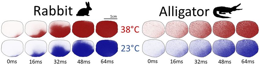
Antiarrhythmic protection in alligators

Rabbits were anesthetized with ketamine/xylazine/acepromazine (17/9/0.9 mg kg−1 , respectively) and in-

jected with heparin (300 U kg−1 ). After five minutes, euthanasia was induced with pentobarbital (120 mg

kg−1 ), and hearts were quickly excised via left thoracotomy.

Optical mapping
   Immediately following extraction, retrograde perfusion via the aorta in rabbits and right (systemic)

aorta in alligators with cardioplegic solution (in mM: NaCl 110, KCl 16, NaHCO3 10, MgCl2 ·6H2 O 16,

                                                                                                                    Downloaded from https://academic.oup.com/iob/advance-article/doi/10.1093/iob/obaa047/6120966 by guest on 12 March 2021
CaCl2 ·2H2 O 1.2) allowed transport to the lab (less than 10 minutes), where hearts were immersed in a

temperature-controlled chamber and perfused with Tyrode’s solution (in mM: NaCl 124, KCl 4, NaHCO3

24, NaH2 PO4 ·H2 O 0.9, MgCl2 ·6H2 O 0.7, CaCl2 ·2H2 O 2, Dextrose 5.5) gassed with 95% O2 and 5% CO2

at a pressure of approximately 60 mmHg controlled by peristaltic pump. Hearts were stained for 15-20 min

with 40 µM of voltage-sensitive dye Di-4-ANBDQPQ (Matiukas et al. 2007), previously dissolved in ethanol

(24.4 mg mL−1 ). Heart motion was suppressed by perfusion with 2-5 µM blebbistatin 20-30 min prior data

acquisition. Blebbistatin was ineffective in suppressing contraction in the alligator hearts, which required

an increased pressure to further prevent motion of the heart and provide stable images. Transmembrane

voltages across the surfaces of the hearts were obtained via fluorescence imaging using an EMCCD camera

at a frame rate of 250 or 500 Hz and spatial resolution of 128×128 pixels, corresponding to approximately

250 µm pixel−1 .

Electrical restitution
   Hearts were stimulated with a AgCl bipolar lead current source at the apex separated by approximately

1 mm, delivering at least 4× the current necessary to induce excitation. Steady-state cardiac dynamics were

recorded over a range of cardiac cycle lengths by following the steady-state restitution pacing protocol (El-

harrar and Surawicz 1983), whereby the heart is stimulated at a constant period, referred to as the basic

cycle length (BCL), and allowed to equilibrate for a minimum of 30 beats before recording. Hearts were

first stimulated at the largest BCL that prevented self-pacing followed by incremental reductions of 50 ms

(alligator) and 10 ms (rabbit) until either the ventricular functional refractory period (VFRP) was reached or

ventricular fibrillation (VF) occurred. Experiments were first conducted at 23 ± 1◦ C followed by replication

at 38 ± 1◦ C.

Analysis
   Fluorescent signals were smoothed in time (moving average, window size of 2 frames) and in space (5×5

pixel Gaussian kernel, σ=3 pixels), and signals were detrended by removing linear fits to signal baselines

in each pixel. Action potential duration (APD) was defined as the amount of time that transmembrane

voltage (Vm) was maintained above 70% repolarization over the course of an action potential. For clarity in

comparison between hearts, the values we report for each BCL are the median of APDs following equilibration

over a range of 20×20 pixels near the apex of each heart with bars signifying 1 s.d. For a general view of how

action potentials varied across BCLs and temperature, a single representative from each is selected and all

are plotted together in Fig. 1. Likewise, the qualitative effect of temperature on action potential propagation
Antiarrhythmic protection in alligators
across the heart is shown in Fig. 2 through snapshots of the heart’s surface over the course of a single action
potential.
The speed of wave propagation across the heart, or conduction velocity (CV), was calculated by first
assigning for each (x, y) coordinate the time at which the corresponding pixel achieved its maximum upstroke
dVm
velocity dt max (Laughner et al. 2012) over the course of each action potential propagation. We define CV as
the inverse of the magnitudes of this surface’s gradient vectors. A corresponding action potential wavelength
(λAPD ) may then be calculated as the product of APD and CV (i.e., λAPD = APD · CV). Since all hearts were
Downloaded from https://academic.oup.com/iob/advance-article/doi/10.1093/iob/obaa047/6120966 by guest on 12 March 2021
stimulated from the apex, we report only apex-to-base CV and do not report anisotropy ratio, the quotient
of longitudinal to transverse CV. For short BCLs, action potentials may exhibit a beat-to-beat alternation
in APD (Laurita and Rosenbaum 2008) and CV (Frame and Simson 1988) known as alternans. Data at
each BCL were thus separated by even and odd beats and analyzed separately to account for alternans. For
clarity, we report only the set of action potentials exhibiting the lower value of APD and CV in the figures,
as our focus is on the risk of arrhythmia brought about by the shortening of action potential wavelength.
Values for APD, CV, and λAPD are shown in Fig. 3 for a single rabbit and alligator (others are provided in
Supplementary Material).
The impact of temperature on tissue scale parameters such as APD and CV can be quantified by a
temperature coefficient Q10 (Shah et al. 2006). The Q10 value associated with some measured dynamic
property K is defined such that its values KT1 and KT2 at temperatures T1 and T2 , respectively, are related
by
◦
KT2 = KT1 · Q10 (T2 −T1 )/10 C
.
The Q10 values for cardiac wave dynamics were calculated for each animal (shown in Fig. 4 for a single rabbit
and alligator; others are provided in Supplementary Material) as a function of BCL across the range for which
hearts at both 23◦ C and 38◦ C could be stimulated, with standard deviation δQ10 calculated in terms of the
standard deviations δKT1 and δKT2 as
s 2 2
10◦ C
δKT1 δKT2
δQ10 = Q10 + .
T2 − T1 KT1 KT2
Results
Susceptibility to ventricular fibrillation
All rabbit hearts exhibited ventricular fibrillation (VF) at 38◦ C when the basic cycle length (BCL) of
stimulation was shorter than approximately 150 ms. When temperature was reduced in rabbit hearts to
23◦ C, VF occurred for BCLs as large as 340 ms. No alligator hearts were observed to fibrillate under any
circumstances. BCLs in alligator hearts were reduced until reaching the ventricular functional refractory
period (VFRP), at which point stimulations could not elicit excitations. The VFRPs in alligators were
approximately 860 ms and 1000 ms at 38◦ C and 23◦ C, respectively. Attempts to initiate VF in alligator
hearts using the standard 9V battery protocol (Bishop et al. 2014) consistently failed, including during in-Antiarrhythmic protection in alligators
situ manipulation (data not shown).
Downloaded from https://academic.oup.com/iob/advance-article/doi/10.1093/iob/obaa047/6120966 by guest on 12 March 2021
Fig. 1: Effect of temperature and basic cycle length (BCL) on action potential duration (APD) and morphology in
rabbit (A, C) and alligator (B, D). (A, B): Representative APs for all measured BCLs overlaid to accentuate BCL
and temperature dependence. As BCL is shortened, APD likewise decreases. (C, D): Two subsequent APs from one
BCL are overlaid to show beat-to-beat variation in AP morphology at 38◦ C (left) and 23◦ C (right). Note that x-axis
scales are different for the two species.
Action potential morphology and propagation
Temperature reduction in the rabbit heart substantially increased action potential duration (APD) and
decreased conduction velocity (CV), in agreement with prior studies (Fedorov et al. 2008). When action
potentials from all measured BCLs and both animals are compared (Fig. 1A, B), the two species exhibit
clear differences in action potential morphology beyond APD. At both temperatures depolarization in the
alligator action potential was observed to last approximately 100 ms, about five times the duration seen
in the rabbit, for which depolarization elapsed within 20 ms. Beat-to-beat alternation in action potential
morphology, or alternans, may be observed by overlaying successive action potentials as in Fig. 1C, D.
While alternans occurred at both temperatures in the rabbit for short BCLs, alternans magnitude increased
prominently at 23◦ C and over a larger range of BCLs consistent with other mammalian studies (Filippi et al.
2014; Laurita and Rosenbaum 2008). Alligators did not exhibit alternans regardless of temperature or BCL.
Tissue-scale dynamics in the alligator were mostly unaffected by temperature but were even more sensitive
in the rabbit. Snapshots during the propagation of an action potential from apex to base, shown in Fig. 2,
accentuate the visibly appreciable differences in the temperature sensitivity of CV. Furthermore, the increased
wavefront curvature observed in the hypothermic rabbit heart implies an increased anisotropy ratio, which
has been correlated with increased risk of arrhythmia (Fedorov et al. 2008).
At all BCLs measured at both temperatures, the rabbit heart experienced an increase in APD ranging
from 5 − 50% when temperature was reduced from 38◦ C to 23◦ C (Fig. 3A, left), whereas the alligator only
experienced between a 5−10% increase in APD when temperature was reduced (Fig. 3A, right). Temperature
had some effect on reducing CV in the alligator (Fig. 3B); however, while apex-to-base CV decreased by
30 − 67% in rabbits, alligators experienced a 20% reduction at most.Antiarrhythmic protection in alligators
Downloaded from https://academic.oup.com/iob/advance-article/doi/10.1093/iob/obaa047/6120966 by guest on 12 March 2021
Fig. 2: Snapshots of the propagation of an action potential across similar sized regions of tissue in rabbit (left) and
alligator (right) at 38◦ C (top) and 23◦ C (bottom). The difference in wave propagation between temperatures is nearly
indistinguishable in the alligator heart, whereas the rabbit heart displays a clear conduction velocity reduction in
hypothermia. The wavefront of excitation in the alligator is not as discernible as in the rabbit due to a nearly order of
magnitude longer depolarization, which results in a “smearing” of the action potential’s wavefront over the alligator’s
heart.
Fig. 3: Cardiac electrical wave characteristics in rabbit and alligator at 38◦ C (red) and 23◦ C (blue) across basic
cycle lengths (BCLs). (A): Action potential duration (APD). APDs in rabbit exhibit an increased temperature-
dependent divergence at larger BCLs, but alligator APDs are consistently altered by temperature across all BCLs. (B):
Conduction velocity (CV). Reduction of CV at 23◦ C is substantial in rabbit but negligible in alligator. (C): Action
potential wavelength λAPD . Ventricular fibrillation occurred in rabbit hearts when stimulated at BCLs for which λAPD
fell below the ventricular length scale (yellow region below horizontal dashed line). The long ventricular functional
refractory period in the alligator prohibited λAPD from approaching this critical zone. Insets are magnifications of
correspondingly colored regions.Antiarrhythmic protection in alligators
Downloaded from https://academic.oup.com/iob/advance-article/doi/10.1093/iob/obaa047/6120966 by guest on 12 March 2021
Fig. 4: Temperature coefficients Q10 for cardiac dynamic properties in rabbit and alligator across basic cycle lengths
(BCLs) for which hearts could be stimulated at both 23◦ C and 38◦ C. (A): The Q10 of action potential duration (APD)
in alligator approaches unity across all BCLs. In rabbit, APD is more greatly affected by temperature at larger BCLs.
(B): Conduction velocity (CV) in rabbit is increasingly sensitive to temperature as BCL is reduced, and CV in alligator
shows small temperature sensitivity across all BCLs. (C): Action potential wavelength λAPD in alligator is insensitive
to temperature across all BCLs. This wavelength in rabbit exhibits strong temperature sensitivity, exacerbated further
as BCL is reduced. (D, E): Q10 for all coefficients overlaid for each species. In rabbit (D), QCV APD
10 and Q10 become
increasingly unbalanced at shorter BCLs, whereas in alligator (E), temperature sensitivities of APD and CV coordinate
and preserve a temperature insensitive λAPD across BCLs. Error bars denote 1 s.d. Colored traces in A-C denote linear
regressions with slopes listed in plots.
Action potential wavelength
Although temperature reduction from 38◦ C to 23◦ C likewise reduced action potential wavelengths λAPD
at corresponding BCLs in each species (Fig. 3C), the minimum achieved action potential wavelength was
unchanged (3±1 cm) in the rabbit but increased (6±1 cm to 10±1 cm) in the alligator. At both temperatures,
the onset of VF in rabbits occurred at BCLs for which λAPD became comparable to heart size (3 cm, dotted
horizontal line). At both temperatures, the large VFRP in the alligator maintained a λAPD far greater than
the length of the heart.
Temperature coefficients
Calculations of Q10 (see Fig. 4) show that the temperature sensitivities of cardiac wave parameters in the
alligator are low, with averages of QAPD
10 = 0.97 ± 0.10 and QCV
10 = 1.08 ± 0.23. Interestingly, these tempera-
ture sensitivities were largely independent of BCL as well. Linear regressions across BCLs (Fig. 4A-C colored
lines) show that a 1000 ms change in BCL translates to changes of 0.02 (QAPD
10 , R2 = 31%) and 0.15 (QCV
10 ,Antiarrhythmic protection in alligators
R2 = 53%). Furthermore, these changes were complementary, such that the temperature sensitivity of action
potential wavelength averaged over all BCLs was low (Q10λAPD = 1.04 ± 0.26), and its linear regression shows
a change in 0.17 for every 1000 ms changed in BCL (R2 = 65%). In contrast, rabbits showed much more
sensitive CV, with an average QCV APD
10 = 1.95 ± 0.16, while APD appeared less sensitive (Q10 = 0.86 ± 0.19).
Both, however, were strongly affected by BCL, showing changes of 0.74 (QAPD
10 , R2 = 93%) and 3.49 (QCV
10 ,
R2 = 91%) over a 1000 ms change in BCL according to best fit regression. As a result, the temperature
sensitivity of action potential wavelength was large (Q10λAPD = 1.70 ± 0.59) and strongly dependent on BCL,
Downloaded from https://academic.oup.com/iob/advance-article/doi/10.1093/iob/obaa047/6120966 by guest on 12 March 2021
changing in value by 4.10 across a 1000 ms change in BCL (R2 = 86%).
Discussion
The crocodilian heart serves as an interesting example of ectothermic anatomy and physiology. Apart
from a fully divided ventricle (Beddard and Mitchell 1895), which may be a vestige of the endothermic past in
crocodilian ancestors (Hillenius and Ruben 2004; Seymour et al. 2004), they retain the reptilian style outflow
tract which allows for a pulmonary bypass shunt (Eme et al. 2009; Jones and Shelton 1993). Compared to
other reptiles of similar size, they also appear to be more restricted in their maximal heart rates (Boukens
et al. 2019; Joyce et al. 2018). Recent investigations into the alligator’s cardiac electrophysiology have likewise
revealed the existence of a conduction pathway for electrical excitations, reminiscent of the atrioventricular
conduction system found in birds and mammals (Jensen et al. 2018) which depends on the formation of a
complete ventricular septum during development (Kvasilova et al. 2020). And although electrocardiographic
deflections in alligators vary across temperatures in a manner similar to heterothermic endotherms (Egorov
et al. 2012; Fedorov et al. 2008; Huggins et al. 1969; Jensen et al. 2018; Wilber 1960), there are distinct
differences which are not attributable to temperature or conduction system alone (Boukens et al. 2019). How
these differences in cardiac electrical properties vary simultaneously across both temperature and cardiac
cycle has before now been unknown.
APD and CV in the rabbit at 38◦ C are well matched to maintain a safe value for λAPD for all but the
shortest of BCLs. And although the rabbit heart permits stimulation at these shorter BCLs (or equivalently,
higher heart rates), there is a trade-off with an increased risk of cardiac arrhythmia as λAPD quickly reduces
to a critical length. Alligator hearts resisted stimulation at these short BCLs, and values of λAPD were main-
tained well above dangerous levels.
Reducing temperature from 38◦ C to 23◦ C generally decreases CV and increases APD in the hearts of both
alligators and rabbits, but the magnitude of these effects in the alligator are considerably lower and more
similar to each other, which further helps safeguard the alligator from cardiac electrical arrhythmias. The
high temperature sensitivity of CV in rabbit hearts greatly impacts susceptibility to arrhythmia, as the action
potential wavelength λAPD becomes dangerously short at a much larger BCL in 23◦ C than in 38◦ C, providing
a deadly substrate for functional reentry and fibrillation (Allessie et al. 1976, 1977). In contrast, functional
and electrical dynamics in alligator hearts remain coordinated due to similar Q10 values of relevant variables
across the entire range of measured BCLs. This coordination in the alligator is visible in Fig. 4 (right), whereAntiarrhythmic protection in alligators
the temperature sensitivities of APD (A) and CV (B) maintain a balanced insensitivity of λAPD (C) across
BCLs. Alternatively, the rabbit (Fig. 4 left) demonstrates the consequences of a mismatched balance in
temperature sensitivities. Although the opposing effects of temperature on APD and CV reasonably preserve
λAPD at the largest of BCLs, there is no coordination as BCL is reduced. The trade-off in the rabbit to
permit stimulation at shorter BCLs becomes increasingly dangerous as the reduction of temperature further
exacerbates risk of λAPD approaching a critical length.
The alligators’ large VFRP prevented stimulation at the short BCLs achievable in the rabbit, and the
Downloaded from https://academic.oup.com/iob/advance-article/doi/10.1093/iob/obaa047/6120966 by guest on 12 March 2021
increase in VFRP at 23◦ C more than compensates for any increased risk of arrhythmia introduced by changes
in APD or CV. Consequently, the alligator’s maximum heart rate is nearly an order of magnitude lower than
that of the rabbit, severely limiting peak cardiac output and the ability of the circulatory system to sup-
port elevated aerobic metabolism. This relationship underlies the fundamental trade-off between the species:
alligators can function at a wide range of heart temperatures without risk of cardiac arrhythmia but are
restricted to lower heart rates, while rabbits can achieve high heart rates necessary to sustain an active and
endothermic metabolism but are critically vulnerable to temperature changes.
This thermal insensitivity and large VFRP in the alligator heart are not traits shared by all ectotherms.
For example, cardiac wave properties in zebrafish (Rayani et al. 2018), frogs (Goto et al. 1976, 1978; Mashima
and Matsumura 1964), and turtles (Stecyk et al. 2007) show significant thermal sensitivity. In the case of
zebrafish, maintaining wave parameters across temperatures and cycle lengths is unnecessary since their small
hearts preclude any risk of reentry-based arrhythmia. And although larger species with low heart rates (large
BCLs) do not risk reentry during healthy function, the absence of a sufficiently large VFRP can permit
fibrillation in the event of an unfortunately timed ectopic beat. The susceptibility to fibrillation observed in
frogs (Savino and Valentinuzzi 1988) and turtles (Hoffman et al. 1951) serves to illustrate the threatening
alternative in this trade-off.
The sensitivity of the domestic rabbit heart to low temperature may be a consequence of its stable core
body temperature in all conditions. Although rabbits do not hibernate, a variety of other mammals do
enter periods of decreased metabolic rate and body temperature, and consequently these species exhibit an
assortment of antiarrhythmic measures in hypothermic conditions, including a consistent λAPD and increased
VFRP (Duker et al. 1983; Egorov et al. 2012; Fedorov et al. 2008; Johansson 1996). Similarly, non-hibernating
but poikilothermic mammals, such as sloths, are better at resisting induced VF (Oliveira et al. 1980; Valentin-
uzzi et al. 1984). The similar properties we have observed in the alligator heart likewise enable survival over
a wide range of temperatures, suggesting that the fundamental mechanism either has been retained from
archosaur ancestors or has been lost in modern birds. Detailed characterization of heart electrophysiology in
other reptile species is needed to answer the evolutionary history of arrhythmic properties. The fundamental
trade-off with heart rate is irrelevant to ectothermic species, due to their reliance on anaerobic metabolism
for “bursts” of high performance, making sustained high heart rates superfluous even when active (Bennett
1982; Joyce et al. 2018; Seymour 2013).
More broadly, these results highlight the perils of temperature-induced mismatch between interacting,Antiarrhythmic protection in alligators
dynamic processes and the need for robust solutions. With minimal cardiac innervation, the propagation
of electrical waves across heart muscle is driven entirely by intrinsic biochemical processes which, combined
with the ability of these waves to interact, leaves cardiac dynamics uniquely vulnerable to disruption by
temperature-induced mismatch of reaction rates (Fenton et al. 2013; Filippi et al. 2014). It remains uncertain
to what extent the nervous system may be able to compensate for these mismatches in other muscular sys-
tems which show strong temperature effects, such as locomotion and feeding, particularly since the nervous
system itself may be vulnerable to temperature induced changes (Montgomery and Macdonald 1990), causing
Downloaded from https://academic.oup.com/iob/advance-article/doi/10.1093/iob/obaa047/6120966 by guest on 12 March 2021
performance loss in tasks requiring coordination (Greenwald 1974).
Acknowledgements
We thank Dr. Ilija Uzelac for construction and maintenance of the optical mapping apparatus. We
are grateful to Dr. Rush Elsey and staff at the Rockefeller Wildlife Refuge (Louisiana Dept. Wildlife and
Fisheries) for donation of alligator eggs for research. We thank Zofia Owerkowicz for the cover photo. We
are grateful for the translations of our abstract performed by Valentin Krinski, Jean Bragard, Ulrich Parlitz,
Alessio Gizzi, Flavio H. Fenton, Batoul Makki, Suleiman Al-Sabah, Charlotte Meyer, Lise Dalton, Panagiotis
Vagenas, Nicole Bournias, Ke-li Tsai, Mike Chao, Yanyan Claire Ji, Tomasz Owerkowicz, Michal Jasieński,
and István Szűcs.
Competing Interests
Conflict of Interest Statement: none declared.
References
Allessie, M. A., Bonke, F. I. and Schopman, F. J. (1976), ‘Circus movement in rabbit atrial muscle as a mech-
anism of tachycardia. II. The role of nonuniform recovery of excitability in the occurrence of unidirectional
block, as studied with multiple microelectrodes’, Circulation Research 39(2), 168–177.
Allessie, M. A., Bonke, F. I. and Schopman, F. J. (1977), ‘Circus movement in rabbit atrial muscle as a
mechanism of tachycardia. III. The “leading circle” concept: a new model of circus movement in cardiac
tissue without the involvement of an anatomical obstacle.’, Circulation Research 41(1), 9–18.
Beddard, F. E. and Mitchell, P. C. (1895), ‘On the structure of the heart of the alligator’, Proceedings of the
Zoological Society of London pp. 343–349.
Bennett, A. F. (1982), The energetics of reptilian activity, in C. Gans and P. H. Pough, eds, ‘Biology of the
reptilia’, Vol. 13, Academic Press, New York, pp. 155–199.
Bennett, A. F. (1985), ‘Temperature and muscle’, Journal of Experimental Biology 115, 333–344.
Bennett, A. F. and John-Alder, H. B. (1984), ‘The effect of body temperature on the locomotory energetics
of lizards’, Journal of Comparative Physiology B 155, 21–27.
Bishop, M. J., Burton, R. A. B., Kalla, M., Nanthakumar, K., Plank, G., Bub, G. and Vigmond, E. J. (2014),
‘Mechanism of reentry induction by a 9-V battery in rabbit ventricles’, American Journal of Physiology:
Heart and Circulatory Physiology 306(7), H1041–H1053.Antiarrhythmic protection in alligators
Boukens, B. J., Kristensen, D. L., Filogonio, R., Carreira, L. B., Sartori, M. R., Abe, A. S., Currie, S., Joyce,
W., Conner, J., Opthof, T., Crossley, D. A., Wang, T. and Jensen, B. (2019), ‘The electrocardiogram of
vertebrates: Evolutionary changes from ectothermy to endothermy’, Progress in Biophysics and Molecular
Biology 144, 16 – 29.
Brattstrom, B. H. (1965), ‘Body temperatures of reptiles’, American Midland Naturalist 73, 376–422.
Burggren, W. W., Christoffels, V. M., Crossley II, D. A., Enok, S., Farrell, A. P., Hedrick, M. S., Hicks,
J. W., Jensen, B., Moorman, A. F. M., Mueller, C. A., Skovgaard, N., Taylor, E. W. and Wang, T. (2014),
Downloaded from https://academic.oup.com/iob/advance-article/doi/10.1093/iob/obaa047/6120966 by guest on 12 March 2021
‘Comparative cardiovascular physiology: future trends, opportunities and challenges’, Acta Physiologica
210(2), 257–276.
Cherry, E. M. and Fenton, F. H. (2008), ‘Visualization of spiral and scroll waves in simulated and experimental
cardiac tissue’, New Journal of Physics 10(12), 125016.
Davidenko, J. M., Pertsov, A. V., Salomonsz, R., Baxter, W. and Jalife, J. (1992), ‘Stationary and drifting
spiral waves of excitation in isolated cardiac muscle’, Nature 355(6358), 349–351.
DeFur, P. L. and Mangum, C. P. (1979), ‘The effects of environmental variables on the heart rates of inverte-
brates’, Computational Biochemistry and Physiology A: Molecular & Integrative Physiology 62(2), 283–294.
Donley, J. M., Shadwick, R. E., Sepulveda, C. S. and Syme, D. A. (2007), ‘Thermal dependence of contractile
properties of the aerobic locomotor muscle in the leopard shark and shortfin mako shark’, Journal of
Experimental Biology 210, 1194–1203.
Driedzic, W. R. and Gesser, H. (1994), ‘Energy metabolism and contractility in ectothermic vertebrate hearts:
hypoxia, acidosis, and low temperature’, Physiological Reviews 74(1), 221–258.
Duker, G. D., Olsson, S. O., Hecht, N. H., Senturia, J. B. and Johansson, B. W. (1983), ‘Ventricular fibrillation
in hibernators and nonhibernators’, Cryobiology 20(4), 407–420.
Egorov, Y. V., Glukhov, A. V., Efimov, I. R. and Rosenshtraukh, L. V. (2012), ‘Hypothermia-induced spatially
discordant action potential duration alternans and arrhythmogenesis in nonhibernating versus hibernating
mammals’, American Journal of Physiology: Heart and Circulatory Physiology 303(8), H1035–H1046.
Elharrar, V. and Surawicz, B. (1983), ‘Cycle length effect on restitution of action potential duration in dog
cardiac fibers’, American Journal of Physiology: Heart and Circulatory Physiology 244(6), H782–H792.
Eme, J., Gwalthney, J., Blank, J. M., Owerkowicz, T., Barron, G. and Hicks, J. W. (2009), ‘Surgical removal
of the right-to-left cardiac shunt in the american alligator (Alligator mississippiensis) causes ventricular
enlargement but does not alter apnoea or metabolism during diving’, Journal of Experimental Biology
212, 3553–3563.
Fedorov, V. V., Glukhov, A. V., Sudharshan, S., Egorov, Y., Rosenshtraukh, L. V. and Efimov, I. R. (2008),
‘Electrophysiological mechanisms of antiarrhythmic protection during hypothermia in winter hibernating
versus nonhibernating mammals’, Heart Rhythm 5(11), 1587 – 1596.
Fenton, F. H. and Cherry, E. M. (2008), ‘Models of cardiac cell’, Scholarpedia 3(8), 1868.
Fenton, F. H., Cherry, E. M., Hastings, H. M. and Evans, S. J. (2002), ‘Multiple mechanisms of spiral wave
breakup in a model of cardiac electrical activity’, Chaos 12(3), 852–892.Antiarrhythmic protection in alligators
Fenton, F. H., Gizzi, A., Cherubini, C., Pomella, N. and Filippi, S. (2013), ‘Role of temperature on nonlinear
cardiac dynamics’, Physical Review E 87, 042717.
Filippi, S., Gizzi, A., Cherubini, C., Luther, S. and Fenton, F. H. (2014), ‘Mechanistic insights into hypother-
mic ventricular fibrillation: the role of temperature and tissue size’, EP Europace 16(3), 424–434.
Frame, L. H. and Simson, M. B. (1988), ‘Oscillations of conduction, action potential duration, and refractori-
ness. A mechanism for spontaneous termination of reentrant tachycardias’, Circulation 78(5), 1277–1287.
Goto, M., Saito, M., Ikemoto, Y. and Tsuda, Y. (1976), ‘Effects of temperature on membrane currents of the
Downloaded from https://academic.oup.com/iob/advance-article/doi/10.1093/iob/obaa047/6120966 by guest on 12 March 2021
frog myocardium’, Proceedings of the Japan Academy 52(7), 389–392.
Goto, M., Tsuda, Y., Yatani, A. and Saito, M. (1978), ‘Effects of low temperature on the membrane currents
and tension components of bullfrog atrial muscle’, The Japanese Journal of Physiology 28(2), 211–224.
Gray, R. A., Pertsov, A. M. and Jalife, J. (1998), ‘Spatial and temporal organization during cardiac fibrilla-
tion’, Nature 392(6671), 75–78.
Greenwald, O. E. (1974), ‘Thermal dependence of striking and prey capture by gopher snakes’, Copeia
1974(1), 141–148.
Hegarty, T. W. (1973), ‘Temperature coefficient (Q10), seed germination and other biological processes’,
Nature 243, 305–306.
Hillenius, W. J. and Ruben, J. A. (2004), ‘Getting warmer, getting colder: Reconstructing crocodylomorph
physiology’, Physiological and Biochemical Zoology 77(6), 1068–1072. PMID: 15674776.
Hodgkin, A. L. and Huxley, A. F. (1952), ‘A quantitative description of membrane current and its application
to conduction and excitation in nerve’, Journal of Physiology 117(4), 500–544.
Hoffman, B. F., Gorin, E. F., Wax, F. S., Siebens, A. A. and Brooks, C. M. (1951), ‘Vulnerability to fibrillation
and the ventricular-excitability curve’, American Journal of Physiology-Legacy Content 167(1), 88–94.
PMID: 14885473.
Huggins, S. E., Hoff, H. E. and Peña, R. V. (1969), ‘Heart and respiratory rates in crocodilian reptiles under
conditions of minimal stimulation’, Physiological Zoology 42(3), 320–333.
James, R. S. (2013), ‘A review of the thermal sensitivity of the mechanics of vertebrate skeletal muscle’,
Journal of Comparative Physiology B 183(6), 723–733.
Jensen, B., Boukens, B. J., Crossley, Dane A, I., Conner, J., Mohan, R. A., van Duijvenboden, K., Postma,
A. V., Gloschat, C. R., Elsey, R. M., Sedmera, D., Efimov, I. R. and Christoffels, V. M. (2018), ‘Specialized
impulse conduction pathway in the alligator heart’, eLife 7, e32120.
Johansson, B. W. (1996), ‘The hibernator heart - nature’s model of resistance to ventricular fibrillation’,
Cardiovascular Research 31, 826–832.
John-Alder, H. B., Morin, P. J. and Lawler, S. (1988), ‘Thermal physiology, phenology, and distribution of
tree frogs’, American Naturalist 132(4), 506–520.
Jones, D. R. and Shelton, G. (1993), ‘The physiology of the alligator heart: Left aortic flow patterns and
right-to-left shunts’, Journal of Experimental Biology 176(1), 247–270.
Joyce, W., Elsey, R. M., Wang, T. and Crossley, D. A. (2018), ‘Maximum heart rate does not limit cardiacAntiarrhythmic protection in alligators
output at rest or during exercise in the american alligator (Alligator mississippiensis)’, American Journal
of Physiology – Regulatory, Integrative and Comparative Physiology 315(2).
Klebér, A. G. and Rudy, Y. (2004), ‘Basic mechanisms of cardiac impulse propagation and associated ar-
rhythmias’, Physiological Reviews 84(2), 431–488. PMID: 15044680.
Kvasilova, A., Olejnickova, V., Jensen, B., Christoffels, V. M., Kolesova, H., Sedmera, D. and Gregorovicova,
M. (2020), ‘The formation of the atrioventricular conduction axis is linked in development to ventricular
septation’, Journal of Experimental Biology 223.
Downloaded from https://academic.oup.com/iob/advance-article/doi/10.1093/iob/obaa047/6120966 by guest on 12 March 2021
Laughner, J. I., Ng, F. S., Sulkin, M. S., Arthur, R. M. and Efimov, I. R. (2012), ‘Processing and analysis of
cardiac optical mapping data obtained with potentiometric dyes’, American Journal of Physiology: Heart
and Circulatory Physiology 303(7), H753–H765.
Laurita, K. R. and Rosenbaum, D. S. (2008), ‘Cellular mechanisms of arrhythmogenic cardiac alternans’,
Progress in Biophysics & Molecular Biology 97(2), 332 – 347.
Le Morvan, C., Troutaud, D. and Deschaux, P. (1998), ‘Differential effects of temperature on specific and
nonspecific immune defences in fish’, Journal of Experimental Biology 201(2), 165–168.
Lewis, L. Y. and Gatten, R. E. (1985), ‘Aerobic metabolism of american alligators, Alligator mississippiensis,
under standard conditions and during voluntary activity’, Computational Biochemistry and Physiology A:
Molecular & Integrative Physiology 80(3), 441–447.
Lillywhite, H. B., Zippel, K. C. and Farrell, A. P. (1999), ‘Resting and maximal heart rates in ectothermic ver-
tebrates’, Computational Biochemistry and Physiology A: Molecular & Integrative Physiology 124(4), 369
– 382.
Marsh, R. L. and Bennett, A. F. (1986), ‘Thermal dependence of sprint performance of the lizard Sceloporus
occidentalis’, Journal of Experimental Biology 126(1), 79–87.
Mashima, H. and Matsumura, M. (1964), ‘The effect of temperature on the mechanical properties and action
potential of isolated frog ventricle’, The Japanese Journal of Physiology 14(4), 422–438.
Matiukas, A., Mitrea, B. G., Qin, M., Pertsov, A. M., Shvedko, A. G., Warren, M. D., Zaitsev, A. V.,
Wuskell, J. P., de Wei, M., Watras, J. and Loew, L. M. (2007), ‘Near-infrared voltage-sensitive fluorescent
dyes optimized for optical mapping in blood-perfused myocardium’, Heart Rhythm 4(11), 1441 – 1451.
Mines, G. R. (1913), ‘On dynamic equilibrium in the heart’, Journal of Physiology 46(4-5), 349–383.
Montgomery, J. C. and Macdonald, J. A. (1990), ‘Effects of temperature on nervous system: implications
for behavioral performance’, American Journal of Physiology – Regulatory, Integrative & Comparative
Physiology 259(2), R191–R196.
Oliveira, L. H. A., da Costa, C. P. and Huggins, S. E. (1980), ‘Cardiac mass, blood temperature and ventricular
fibrillation: a study of the comparative physiology of the three toed sloth and domestic cat’, Comparative
Biochemistry and Physiology A 67, 483–490.
Qu, Z., Garfinkel, A., Chen, P.-S. and Weiss, J. N. (2000), ‘Mechanisms of discordant alternans and induction
of reentry in simulated cardiac tissue’, Circulation 102(14), 1664–1670.
Rayani, K., Lin, E., Craig, C., Lamothe, M., Shafaattalab, S., Gunawan, M., Li, A. Y., Hove-Madsen, L.Antiarrhythmic protection in alligators
and Tibbits, G. F. (2018), ‘Zebrafish as a model of mammalian cardiac function: Optically mapping the
interplay of temperature and rate on voltage and calcium dynamics’, Progress in Biophysics and Molecular
Biology 138, 69 – 90. The Use of Zebrafish for Cardiac Research.
Savino, G. V. and Valentinuzzi, M. E. (1988), ‘Ventricular fibrillation-defibrillation in the toad Bufo parac-
nemis’, International Journal of Cardiology 19(1), 19 – 25.
Seymour, R. S. (2013), ‘Maximal aerobic and anaerobic power generation in large crocodiles versus mammals:
Implications for dinosaur gigantothermy’, PLOS ONE 8(7).
Downloaded from https://academic.oup.com/iob/advance-article/doi/10.1093/iob/obaa047/6120966 by guest on 12 March 2021
Seymour, R. S., Bennett-Stamper, C. L., Johnston, S. D., Carrier, D. R. and Grigg, G. C. (2004), ‘Evidence
for endothermic ancestors of crocodiles at the stem of archosaur evolution’, Physiological and Biochemical
Zoology 77(6), 1051–1067.
Shah, U., Bien, H. and Entcheva, E. (2006), ‘Cardiac arrhythmogenesis and temperature’, Proceedings of the
28th IEEE EMBS Annual International Conference pp. 841–844.
Smeets, J., A Allessie, M., Lammers, W., Bonke, F. and Hollen, J. (1986), ‘The wavelength of the cardiac
impulse and reentrant arrhythmias in isolated rabbit atrium. The role of heart rate, autonomic transmitters,
temperature, and potassium’, Circulation Research 58, 96–108.
Stecyk, J. A. W., Paajanen, V., Farrell, A. P. and Vornanen, M. (2007), ‘Effect of temperature and pro-
longed anoxia exposure on electrophysiological properties of the turtle (trachemys scripta) heart’, Ameri-
can Journal of Physiology-Regulatory, Integrative and Comparative Physiology 293(1), R421–R437. PMID:
17442785.
Uzelac, I., Ji, Y. C., Hornung, D., Schröder-Scheteling, J., Luther, S., Gray, R. A., Cherry, E. M. and Fenton,
F. H. (2017), ‘Simultaneous quantification of spatially discordant alternans in voltage and intracellular
calcium in Langendorff-perfused rabbit hearts and inconsistencies with models of cardiac action potentials
and Ca transients’, Frontiers in physiology 8, 819.
Valentinuzzi, M., Ruiz, E. and da Costa, C. (1984), ‘Ventricular fibrillation threshold in the three-toed sloth
(Bradypus tridactylus)’, Acta Physiologica et Pharmacologica Latino-Americana 34, 312–322.
Veeraraghavan, R., Gourdie, R. G. and Poelzing, S. (2014), ‘Mechanisms of cardiac conduction: a history of
revisions’, American Journal of Physiology: Heart and Circulatory Physiology 306(5), H619–H627.
Waldschmidt, S. R., Jones, S. M. and Porter, W. P. (1986), ‘The effect of body temperature and feeding
regime on activity, passage time, and digestive coefficient in the lizard Uta stansburiana’, Physiological
Zoology 59(3), 376–383.
Wang, T., Zaar, M., Arvedsen, S., Vedel-Smith, C. and Overgaard, J. (2002), ‘Effects of temperature on
the metabolic response to feeding in Python molurus’, Computational Biochemistry and Physiology A:
Molecular & Integrative Physiology 133(3), 519 – 527.
Watanabe, M. A., Fenton, F. H., Evans, S. J., Hastings, H. M. and Karma, A. (2001), ‘Mechanisms for
discordant alternans’, Journal of Cardiovascular Electrophysiology 12(2), 196–206.
Wilber, C. G. (1960), ‘Effect of temperature on the heart in the alligator’, American Journal of Physiology
198(4), 861–863.You can also read