Demodectic mange in threatened southern sea otters (Enhydra lutris nereis)
←
→
Page content transcription
If your browser does not render page correctly, please read the page content below
Vet Dermatol 2021                                                                                                   DOI: 10.1111/vde.12947

Demodectic mange in threatened southern sea otters
(Enhydra lutris nereis)
Nadia Javeed*, Janet Foley*   , Arturo Oliver-Guimera†, Verena K. Affolter† , Michael K. Keel†,
                                 draig J. Duignan§, Michael Murray††, MT Tinker** and
Angelina Reed‡, Risa Pesapane*, Pa
Melissa A. Miller‡,¶
*Departments of Medicine and Epidemiology, School of Veterinary Medicine, University of California Davis, Davis, CA 95616, USA
†Departments of Pathology, Microbiology, and Immunology, School of Veterinary Medicine, University of California Davis, Davis, CA 95616, USA
‡Marine Wildlife Veterinary Care and Research Center, California Department of Fish and Wildlife, 151 McAllister Way, Santa Cruz, CA 95060,
USA
§The Marine Mammal Center, 2000 Bunker Road Fort Cronkhite Sausalito, Sausalito, CA 94965, USA
¶Karen C. Drayer Wildlife Health Center, 1089 Veterinary Medicine Drive, Davis, CA 95616, USA
**Nhydra Ecological Research, 11 Parklea Drive, Head of St Margarets Bay, Davis, NS B3Z 2G6, Canada
††Monterey Bay Aquarium, 886 Cannery Row, Monterey, CA 93940, USA
Correspondence: Janet Foley, 1320 Tupper Hall, Davis, CA 95616, USA. E-mail: jefoley@ucdavis.edu

     Background – Southern sea otters (Enhydra lutris nereis) rely on intact pelage for thermoregulation, and thus cli-
     nically significant demodicosis and associated alopecia can cause morbidity and death.
     Hypothesis/Objectives – This study aimed to describe lesions associated with follicular Demodex sp. infesta-
     tion, estimate the prevalence and intensity of infestation, describe mite distribution across key anatomical
     regions, and assess mite presence or absence in relation to lesions and host risk factors.
     Animals – Twenty necropsied, wild southern sea otters that stranded along the central California coast from
     2005 to 2018.
     Methods and materials – Grossly normal and abnormal integument from the head, perineum, genitals, mami-
     llary papillae and limbs was assessed microscopically for mites and mite-associated pathological findings.
     Results – Intrafollicular mites were observed in the integument of 55% of otters and 20% had clinical demodico-
     sis. Demodicosis was considered to be contributory to death or euthanasia in two cases. Although Demodex sp.
     mites often were observed microscopically in grossly normal skin, the presence of multiple densely-packed intra-
     follicular mites generally was associated with pigmentary incontinence, ectatic follicles, lymphoplasmacytic peri-
     folliculitis, and neutrophilic and lymphoplasmacytic, dermal inflammation. Other findings included epidermal
     hyperplasia, orthokeratotic hyperkeratosis of epidermis and follicular epithelium, concurrent pyoderma and cell
     necrosis. Perioral integument, especially of the chin, had the highest prevalence of mites and the highest mite
     density, suggesting facial contact as a means of mite transmission.
     Conclusions and clinical importance – Our research confirmed demodectic mange as a contributor to morbi-
     dity and mortality in sea otters, with important implications for clinical care, rehabilitation and conservation.

                                                                          include folliculitis, pigmentary incontinence, epidermal
Introduction
                                                                          hyperplasia, hyperkeratosis, furunculosis and perifollicular
Mites of the genus Demodex (Acari: Demodicidae) inhabit                   granulomas,10–15 often exacerbated by secondary or con-
the skin of virtually all mammals.1 Although morphologi-                  current bacterial or fungal infections.11,16,17 The pathogen-
cally similar, these mites are highly host-specific, inhabit-             esis of clinically significant demodicosis is poorly
ing hair follicles or sebaceous glands and feeding on                     understood, although it is postulated to be associated with
sebum or epithelial cells.2,3 Local or generalized mite prolif-           deficient immune responses.18
eration in the skin of the host has been reported in dogs,                   Among aquatic species, Demodex spp. infestation
cats, cattle and wildlife.4–9 Associated microscopic lesions              and/or lesions have been reported in California sea
                                                                          lions (Zalophus californianus),16,19 harbour seals (Phoca
                                                                          vitulina)14,20 and European otters (Lutra lutra).21
                                                                          Lesions include scaling, alopecia and deep ulcers.14,16
Accepted 27 September 2020                                                Most descriptions are individual case reports, so little
Sources of Funding: The California Department of Wildlife, Mar-           is known about the prevalence and population-level
ine Wildlife Veterinary Care and Research Center (MWVCRC),
                                                                          impacts of Demodex spp. among marine mammals
Office of Spill Prevention and Response, and The Marine Mam-
                                                                          including southern sea otters (Enhydra lutra nereis).
mal Center provided support for sea otter necropsy and
histopathology.                                                           Southern sea otters are listed as threatened under the
Conflicts of Interest: All authors declare no conflict of interest        US Endangered Species Act, and their population has
with this work.                                                           been slowly recovering from near-extinction with

© 2021 the European Society of Veterinary Dermatology and the American College of Veterinary Dermatology.                                      1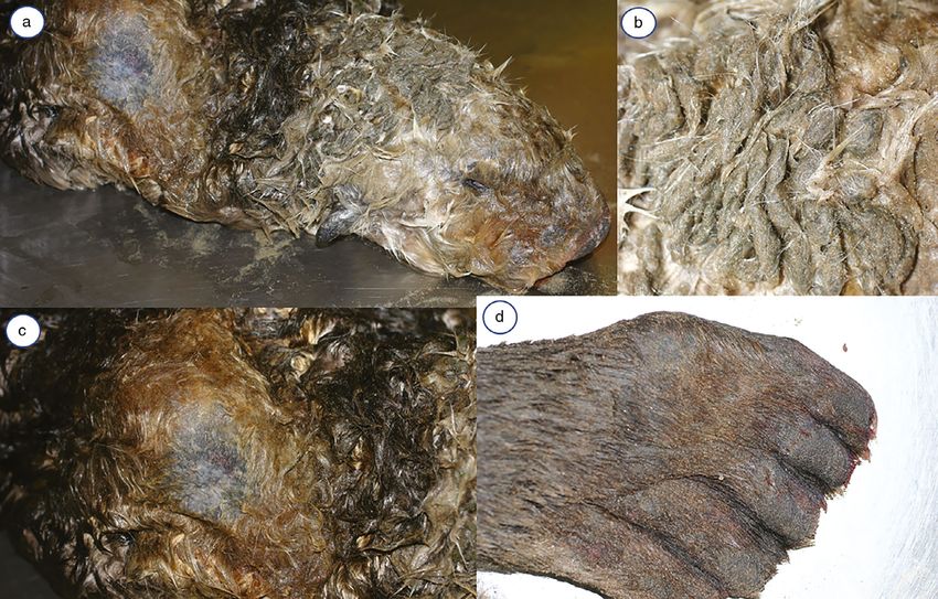
Javeed et al.

disease remaining an important threat.22 Because sea                       DNA sequencing
otters have high energetic requirements23 and depend                       Transverse 25 mg slices were collected from frozen skin with a clean
on an intact, healthy and dense pelage for thermoregu-                     razor blade for DNA extraction using the DNeasy Blood and Tissue kit
                                                                           (Qiagen; Redwood City, CA, USA). DNA also was extracted from 5–
lation,24 Demodex-associated skin pathological conse-
                                                                           10 lm thickness scrolls from five paraffin blocks with high mite den-
quences may contribute to morbidity and death.
                                                                           sity (≥10 mites per tissue section on histopathological evaluation)
   We examined skin samples from 20 stranded southern                      using a QIAamp DNA FFPE kit (Qiagen). An internally transcribed
sea otters in central California from 2005 through 2018.                   spacer (ITS)1–2 fragment of rDNA was amplified using previously
Our goals were to describe Demodex sp. distribution and                    published primers: forward 5’-AGAGGAAGTAAAAGTCGTAACAAG-3’
density across key anatomical regions, estimate the                        and reverse 5’-ATATGCTTAAATTCAGGGGG-3’25 in a 25 uL PCR
prevalence of mite infestation in southern sea otters, and                 reaction containing GoTaq Green Master Mix (Promega; Madison,
                                                                           WI, USA). Cycling conditions were: 95°C for 2 min followed by 35
assess mite presence in relation to defined lesions and
                                                                           cycles of 94°C for 30 s, 54°C for 30 s and 72°C for 2 min, with a final
host risk factors. We also characterized the mites mor-                    extension step of 7 min at 72°C. PCR products were visualized on a
phologically and using DNA sequencing.                                     1% agarose gel and purified using ExoSAP-IT (ThermoFisher; West
                                                                           Sacramento, CA, USA) before sequencing on an ABI 3730 sequencer
                                                                           (Davis Sequencing; Davis, CA, USA). Sequenced amplicons were
Methods and materials                                                      evaluated by BLAST (NCBI; http://blast.ncbi.nlm.nih.gov/Blast.cgi).

Sample collection and necropsy
Samples were collected during necropsies of 20 wild southern
                                                                           Statistical methods
sea otters that stranded along the central California coast from           Summary data were compiled in EXCEL (Microsoft; Redmond, WA,
2005 through 2018. After combing the pelage to assess the pre-             USA) and analysed using R.26 Fisher’s exact tests were used to
dominant direction of hair growth, approximately 4 cm 9 2 cm               examine associations between mite presence/absence and sea
rectangular strips of haired skin were collected from defined              otter sex, county of stranding and age class (pups, 0–6 months;
anatomical locations. To facilitate tissue processing and mite             subadults, 1–4 years; adults, >4–10 years; and aged adults,
detection, the long axis of each sample was orientated in the              >10 years). The mean numbers of mites in mite-positive male and
direction of hair growth; this optimized longitudinal sectioning of        female otters of any age class were compared using a two-tailed
the compound hair follicles.                                               Student’s t-test. Fisher’s exact tests also were used to assess
   Skin samples were collected from perioral, perinasal, periaural and     whether mite presence/absence was associated with the lesions
periocular regions, plantar aspects of the rear flippers, plantar sur-     described above. Tests were considered significant if the P-value
faces of the paws, perivaginal and mammary peripapillary skin in           was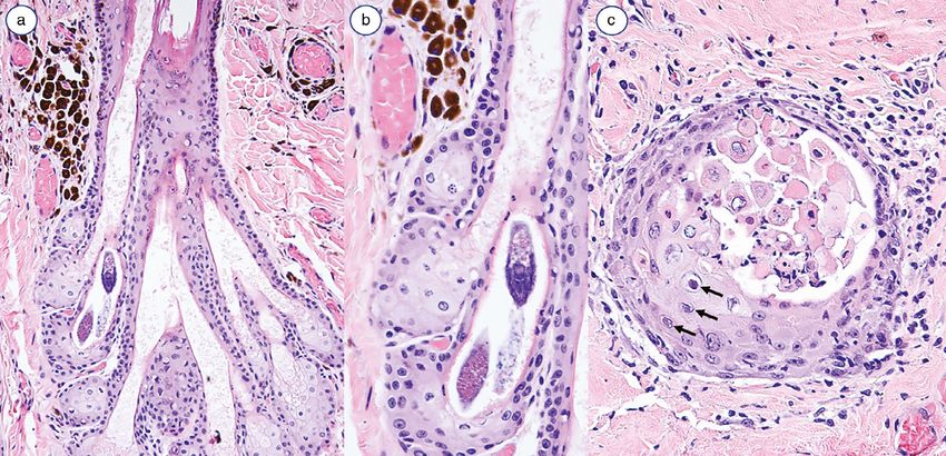
Demodectic mange in southern sea otters

Table 1. Demographic data for 20 necropsied southern sea otters (Enhydra lutris nereis) that were examined microscopically for Demodex sp.
mite infestation
Age class                       Estimated age                     Sex                            County                             Mite number
Pup                             1–2 weeks                         Male                           Monterey                           None
Subadult                        3–4 years                         Male                           Marin                              High†
Subadult                        1–3 years                         Female                         Monterey                           High
Subadult                        Unknown                           Female                         Monterey                           High
Subadult                        Unknown                           Female                         San Luis Obispo                    None
Adult                           4 years                           Male                           San Luis Obispo                    Low*
Adult                           5–6 years                         Female                         Monterey                           None
Adult                           7–8 years                         Female                         Monterey                           None
Adult                           7–8 years                         Female                         Monterey                           None
Adult                           7–8 years                         Female                         Monterey                           Low
Adult                           8 years                           Male                           San Luis Obispo                    None
Adult                           4–6 years                         Male                           San Luis Obispo                    High
Adult                           5–7 years                         Female                         Monterey                           None
Adult                           Unknown                           Female                         Monterey                           Low
Aged adult                      12–14 years                       Male                           Monterey                           High
Aged adult                      14 years                          Male                           Santa Barbara                      Low
Aged adult                      10+ years                         Female                         Monterey                           High
Aged adult                      13–14 years                       Female                         Monterey                           None
Aged adult                      12–14 years                       Female                         Santa Barbara                      None
Aged adult                      12–14 years                       Female                         Monterey                           Low
*Small number of mites, 0.44–1.2 mites/cm basement membrane.
†
Large number of mites, >1.2 mites/cm basement membrane.

Table 2. Anatomical distribution and density of Demodex sp. mite infestation in integument from southern sea otters (Enhydra lutris nereis)
                                      Number      Number      Number of                    Mean mite density/cm     Mean mite
                          Number of   of          of          otters       Proportion of   basement membrane        density for each   Maximum
Anatomical site of        sections    blocks      otters      positive     mite-positive   of mite-positive         mite-positive      mites/
sampled integument        examined    examined    examined    for mites    otters          sections                 section             section
Perioral*                 26          15          14          7            50%†            0.84                     7.2                60
Perinasal                  7           7           7          0             0%†            0                        0                   0
Periocular                11           8           8          0              0%            0                        0                   0
Periaural                 15          12          10          1            10%†            4.1                      22                 30
Top of head                0           0           0          0             0%†            0                        0                   0
Front paw                 14          12           9          0             0%†            0                        0                   0
Rear limb/ Flipper        12          11          10          1            10%†            8.5                      47                 47
Main body                  2           1           1          1             50%            1                        3                   3
Mammary                    1           1           1          0              0%            0                        0                   0
Perivulvar/ Perineal/      7           7           4          1             25%            5.8                      6                   6
 Periscrotal/ Preputial
Tail                       0           0           0          0              0%            0                        0                   0
Unknown anatomic          25          16          14          3            21%†            1.8                      2.7                 6
 location ‡
*Six of seven sections of integument from the chin were mite-positive, with a mean of 10.9 mites/section.
†
  Includes samples of grossly apparent skin lesions (alopecia, dermatitis, dermal hyperplasia and/or excoriation).
‡
  Some unknown sections may be from the ventral abdominal mid-line (a common sample site for sampling sea otter integument during pelting).

   All intrafollicular mites were elongate and fusiform,                    and highest mite density occurred in chin with
with an average length of 164  3.67 µm (Figure 1b).                        ≤60 mites/section (Table 2). For slides that lacked Demo-
The scaled, semi-transparent bodies were composed of                        dex mites, subsequent tissue sections of the same paraf-
two fused segments with four pairs of short, stout appen-                   fin blocks also were mite-negative (data not shown). The
dages arising from the anterior body segment. Mites                         number of mites varied between 0.44 and 8.5 mites/cm
were orientated with their head towards the hair bulb.                      of basement membrane with no significant difference
They were located in the follicular infundibulum near the                   (P = 0.61) between males [average 3.17 mites/cm; stan-
opening of the sebaceous gland ducts (79%), within the                      dard deviation (SD) 1.56] and females (average
ducts or the sebaceous gland (20%), or in the follicular                    3.80 mites/cm; SD 3.50).
isthmus (1%). In otters with a high mite density, up to                        Gross lesions were most common on the face and
seven Demodex mites were visible within each follicle,                      head, especially around the mouth, and were character-
often clustered in adjacent follicles.                                      ized by brownish-orange exudate, matted hair, orange
   The most commonly infested locations were the face                       discolouration of perilesional hair shafts (interpreted as
and head (73% of all mites seen), followed by the hind                      salivary staining), alopecia and lichenification. A small
flippers (22%), prepuce (3%) and shoulders (2%). Perioral                   number of papules and excoriations also were evident
skin had the highest proportion of mite-positive sections                   (Figures 2a–d, 3a–d).
© 2021 the European Society of Veterinary Dermatology and the American College of Veterinary Dermatology.                                     3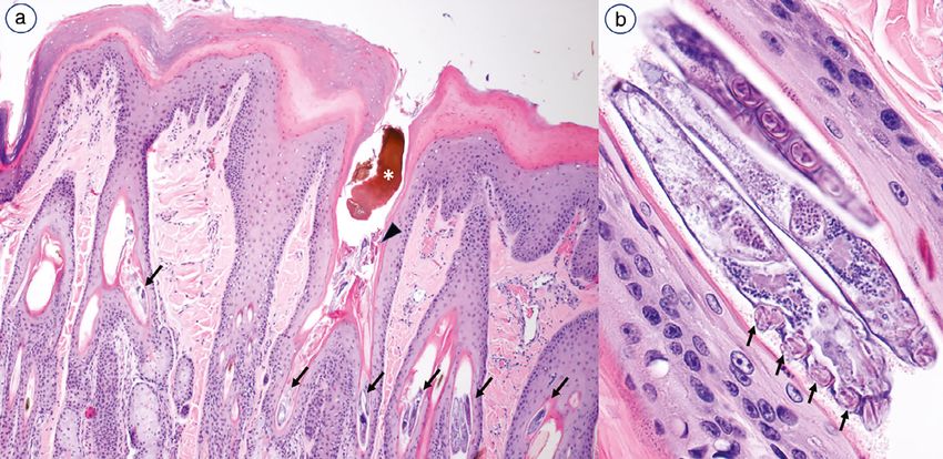
Javeed et al.

Figure 1. Demodicosis and bacterial pyoderma in an aged adult female southern sea otter (Enhydra lutris nereis: case 6734-13) (euthanized due
to severe chronic dermatitis).
(a) Dorsal surface of the nose, showing marked dermal hyperplasia characterized by prominent ridges and deep tissue folds, plus moderate alope-
cia and patchy hypo/hyperpigmentation. The skin and surrounding hair are lightly coated with orange-brown viscous material (sebum, salivary stain-
ing and/or exudate from concurrent bacterial dermatitis). (b) Ventral flipper, showing chronic severe proliferative dermatitis, lichenification,
alopecia, patchy hypo/hyperpigmentation and mild excoriation. Scant viscous orange-brown material is visible on the skin surface. (c) Dorsal tail,
showing lesions similar to those on the flipper, with proliferative dermatitis, alopecia, patchy hypo/hyperpigmentation and surface excoriation. (d)
Dorsal flipper, showing marked tissue thickening and blunting visible at the edge of each digit, along with patchy hypo/hyperpigmentation, excoria-
tion and alopecia. The skin and hair are coated with moderate orange-brown viscous material.

Figure 2. Follicular and surface epithelial histopathological findings for an aged adult female southern sea otter (Enhydra lutris nereis: case 6734-
13) (euthanized due to severe chronic dermatitis).
(a) Haired skin from the left hind flipper, showing superficial and follicular hyperkeratosis. The follicular lumenae are variably dilated, and the ostium
of one follicle is plugged with dark brown material (sebum: asterisk) and keratin debris (arrowhead). Follicles and associated sebaceous gland ducts
contain numerous Demodex sp. mites (arrows). (b) Higher magnification view of a single follicle (the second follicle from the right in Figure 2a),
showing longitudinal profiles of three cigar-shaped mites orientated head-down in the lumen. Four short, stout legs are visible on the anteroventral
body surface of the lefthand mite (arrows). Haematoxylin and eosin.

  Histological changes associated with intrafollicular mite                      P = 0.0003), follicular ectasia (P = 0.0009), superficial
presence included perifollicular perivascular dermatitis                         and follicular hyperkeratosis, and dermal fibrosis and plug-
(P = 0.03), pigmentary incontinence (19 of 20 sections;                          ging of follicular ostia with admixed sebum and keratin

4                             © 2021 the European Society of Veterinary Dermatology and the American College of Veterinary Dermatology.
Demodectic mange in southern sea otters

Figure 3. Follicular mural and perifollicular histopathological findings for an aged adult female southern sea otter (Enhydra lutris nereis: case 6734-
13) (euthanized due to severe chronic dermatitis).
(a) Integument from the chin: longitudinal section through a hair follicle, showing partial profiles of mites within a sebaceous gland. The sebaceous
gland ducts and follicular lumen are moderately dilated. The adjacent dermis contains abundant perifollicular and perivascular macrophages with
abundant dark brown cytoplasmic granules (melanin; pigmentary incontinence: top left and top right). (b) Higher magnification detail of a portion of
the previous image to show the mites and dermal pigmentary incontinence. (c) Hind flipper, transverse section of a hair follicle at the level of the
infundibulum: some epithelial cells contain swollen, pale nuclei with central amphophilic intranuclear inclusions (presumptive herpesvirus: arrows)
accompanied by epithelial apoptosis and mild perifollicular and follicular mural suppurative and lymphoplasmacytic dermatitis. All photos: Haema-
toxylin and eosin.

debris (10 of 20 sections) (Table 3; Figure 1a,b). These                       (6734-13) necessitated humane euthanasia. In addition to
lesions were not present in nine mite-negative otters, and                     demodicosis, she had epidermal ulceration associated
lesions suggestive of infestation were significantly less                      with herpesvirus-like intranuclear inclusions in ker-
common and less severe in mite-negative tissue sections                        atinocytes (Figures 2a–d, 3a–c). Bacterial cocci were pre-
(Table 3). The perifollicular infiltrate was lymphoplasma-                     sent on the skin surface and within some affected
cytic (P = 0.02), whereas interstitial and perivascular der-                   follicles. Aerobic bacterial culture of affected skin and axil-
mal infiltrates consisted of lymphocytes, plasma cells and                     lary and inguinal lymph nodes yielded Staphylococcus
neutrophils (P = 0.03). Mural folliculitis (P = 0.39) and                      intermedius, S. pseudintermedius, Streptoccocus pho-
furunculosis (P = 0.42) were not significantly associated                      cae and Archanobacterim phocae. This animal also had
with mite presence. Marked epidermal hyperplasia and                           chronic cerebral lymphoplasmacytic meningoencephalitis
compact orthokeratotic hyperkeratosis indicated chronic                        with intralesional tissue cysts compatible with Toxo-
irritation. Epithelium of heavily affected follicles and asso-                 plasma gondii. The intestines were heavily infected with
ciated sebaceous glands had apoptotic keratinocytes,                           Corynosoma enhydri acanthocephalans.
some distorted follicles and some cell necrosis. Furuncu-                         The second case with severe demodicosis, an aged
losis was attributed to the effects of secondary bacterial                     adult male (6397-12), died from dilated cardiomyopathy,
folliculitis.                                                                  with secondary acute hepatic vein thrombosis and mas-
    Two otters (10%) had chronic, severe generalized der-                      sive hepatic parenchymal infarction. Chronic severe
matitis and alopecia that contributed to death or euthana-                     alopecia and dermatitis of the shoulder, head, extremities
sia. Severe chronic skin lesions in an aged adult female                       and thorax associated with demodicosis and secondary

Table 3. Univariate associations between histologically confirmed Demodex sp. mite infestation in skin of necropsied southern sea otters (Enhy-
dra lutris nereis) and potential mite-associated skin lesions
                                        Number of mite-positive     Number of mite-negative      Number of mite-positive     Number of mite-negative
Lesion                       P-value    sections with lesions       sections with lesions        sections without lesions    sections without lesions
Pigmentary incontinence      0.0003*    10                          12                           10                          88
Ectatic follicles            0.0009*    10                          14                           10                          86
Perifolliculitis             0.02*       9                          19                           11                          81
Dermal inflammation          0.03*       8                          17                           12                          83
Mural folliculitis           0.39        3                           8                           17                          92
Furunculosis                 0.42        1                           2                           19                          98
Perivascular inflammation    0.46       10                          61                           10                          39
P-values obtained via Fisher’s exact test: *indicates statistically significant difference between mite-positive and mite-negative integument.

© 2021 the European Society of Veterinary Dermatology and the American College of Veterinary Dermatology.                                            5
Javeed et al.
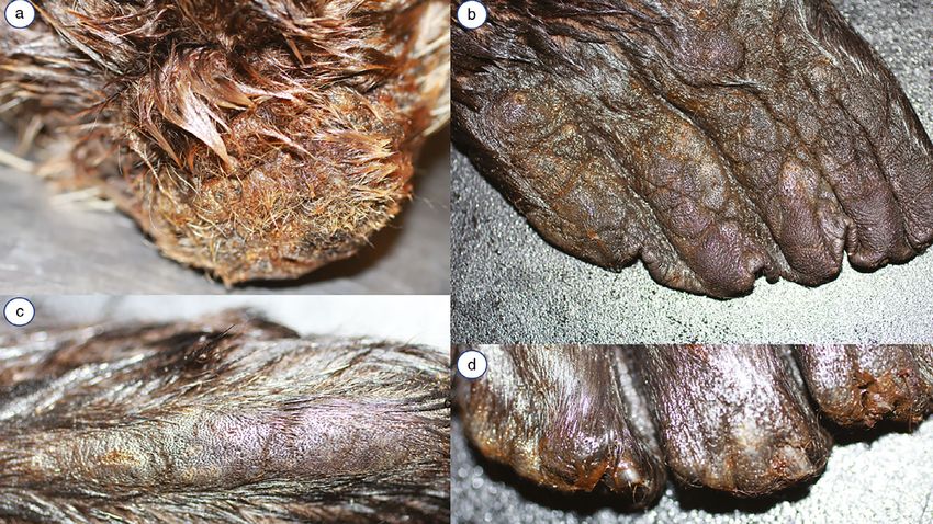
Figure 4. Demodicosis and bacterial pyoderma in an aged adult male southern sea otter (Enhydra lutris nereis: case 6397-12) (dermatitis as a con-
tributing cause of death).
(a) Head and right shoulder, showing chronic dermatitis and alopecia. Adjacent hairs exhibit orange-brown discoloration (sebum, salivary staining
and/or exudate from concurrent bacterial dermatitis). (b) Dorsal head, showing severe, regional matting of the pelage. (c) Right shoulder, higher
magnification, showing central alopecia, lichenification and excoriation, and peripheral salivary staining. (d) Dorsal flipper, showing diffuse hyperpig-
mentation and lichenification, patchy alopecia and multifocal surface excoriation.
bacterial septicemia was a contributing cause of death trap a layer of air next to the skin.23,27 Even small patches
(Figure 4a–d). This otter also had mild intestinal C. enhydri of hair loss or wetting can substantially alter thermoregu-
infection and sparse sarcocysts (Sarcocystis sp.) in skele- lation and cause death.22 Here we document intrafollicu-
tal muscle. lar Demodex sp. mite infestation in southern sea otters
with alopecia and dermatitis – lesions that can contribute
DNA sequencing to morbidity and mortality.
Amplification of mite DNA from cryopreserved integu- The pelage characteristics of these otters posed chal-
ment was unsuccessful using the generic mite PCR pro- lenges to achieving longitudinal sections of follicles and
tocol. PCR of paraffin scrolls from formalin-fixed, mite- sebaceous glands, an important requirement to assess
positive sea otter integument yielded numerous ambigu- Demodex mites microscopically. Techniques were devel-
ous DNA bases, plus longer sequences of sufficient qual- oped during this study to facilitate sampling in relation to
ity to perform a BLAST search that yielded 94% homology hair follicle orientation. Sand contamination presented an
with D. folliculorum (GenBank accession HQ728000). additional challenge as it contributed to poor-quality tissue
sections.
A quarter of the sea otters had grossly apparent
Discussion and conclusions
demodicosis, and chronic, mite-associated dermatitis
By contrast with other mustelids, sea otters have evolved was a primary or contributing cause of death in 10%. Half
a life history with extremely high energetic demands22 had Demodex sp. mite infestation on histological evalua-
yet lack the subcutaneous blubber layer found in other tion, including otters with and without skin lesions. We
marine mammals.27 Their ability to survive in the cold acknowledge sample bias because the sampled otters
marine environment is instead entirely dependent on were obtained opportunistically and were found sick or
insulating air trapped within a highly specialized fur dead. The population prevalence may exceed our sample
coat.23,24,28 Sea otters have the densest pelage of any prevalence because only a small portion of integument
mammal with ≥150,000 hairs/cm2.28 Their compound hair was evaluated microscopically. The single ivermectin-
follicles have central primary follicles forming long, thick treated otter could have reduced prevalence as well.
guard hairs surrounded by many smaller follicles produc- Demodex mites were observed primarily within hair fol-
ing a shorter and finer undercoat.24,27 Thermal insulation licles, most prominently in the infundibulum near seba-
is achieved through interlocking of adjacent hairs which ceous gland ducts or within these ducts. Different
6 © 2021 the European Society of Veterinary Dermatology and the American College of Veterinary Dermatology.Demodectic mange in southern sea otters
Demodex spp. infest specific locations of their hosts: otter subpopulations as a consequence of massive har-
D. canis infests canine hair follicles and occasionally vest during the 19th Century, and immune suppression
sebaceous glands, and D. cati infests feline hair follicles, has been hypothesized as an impediment to southern sea
whereas D. injai and D. phocidi are found in sebaceous otter population recovery.35,41 Potential added stressors
glands of dogs and harbour seals, respectively.29 Demo- include concurrent disease (including polyparasitism),
dex cornei (possibly a strain of D. canis)30 and D. gatoi nutritional deficiencies, high nutritional demands, hor-
inhabit the stratum corneum of dogs and cats, respec- monal fluctuations due to reproduction, seasonal weather
tively.24,31,32 Demodex zalophi is found in the sebaceous and oceanographic changes, intraspecific aggression,
ducts and occasionally the hair follicles of California sea stress of captivity, and exposure to oil or other environ-
lions (PD, unpublished observation). mental toxins.15,16,36,41
Morphological features were consistent with prior The most highly infested areas were chin and perioral
descriptions of Demodex spp.21,33 and DNA sequencing skin, followed by hind flippers, torso and prepuce. The
confirmed that the sea otter mite is in the Demodex high parasite density on the chin suggests that facial con-
genus. As Demodex mites tend to be monoxenous,34 this tact, including affiliative, aggressive and grooming beha-
parasite is likely unique to sea otters. The length of sea viours, could be an important means of mite
otter mites (164 µm) is comparable to Demodex sp. from transmission. The larger number of mites from the genital
European otters (L. lutra: 170–209 µm)21 and California area could indicate that sexual transmission is part of sea
sea lions (202–258 µm).19 Our measurements may otter Demodex ecology. Even if the high mite density in
underestimate total length because they were taken from these regions is the result of tissue factors (e.g. the qual-
histological sections. ity of sebaceous exudate), the larger numbers of mites
Histological features of sea otter mite infestation could facilitate contact-dependent transmission.
included ectatic follicles with perifollicular inflammation Demodex sp. infestation appears to be common in
and associated pigmentary incontinence, similar to stranded southern sea otters, which augments the rela-
lesions in dogs and other mammals.4,12,31,34-36 As tively sparse literature on parasitic arthropods of southern
reported for dogs with demodicosis, inflammatory lesions sea otters,29,42 and reports of infestation across all muste-
were occasionally seen in the absence of Demodex lids and marine mammals.21 Careful evaluation with repeti-
mites.31,37 Although this could represent suboptimal tis- tive deep skin scrapings or molecular assays is warranted
sue collection, sampling error or the presence of mite-in- for sea otters with alopecia and dermatitis, including pups
fested follicles outside the plane of tissue section, deeper given the possibility of infestation during parturition. Given
serial sections did not improve mite detection. Lympho- the likely contribution of immune-suppression to disease
cytic mural folliculitis (seen in dogs with uncomplicated severity, treatment options must be explored. Ivermectin,
demodicosis) and furunculosis (a common feature in doramectin and amitraz have shown efficacy for demodi-
canine demodicosis with secondary bacterial infections) cosis treatment in wildlife, including a captive koala (Phas-
were rare in sea otters.12 Fewer mites were observed in colarctos cinereu) and captive harbour seals 20,43–45.
sea otter skin samples, when compared to dogs with clin- However, amitraz must be applied in a contained environ-
ical demodicosis.7,31 ment due to its toxicity to other marine life.46
Often Demodex infestations in mammals are localized By compromising integrity of the specialized pelage,
and asymptomatic,38 with more severe lesions being Demodex infestation may contribute to sea otter mortal-
associated with complicating secondary infec- ity. This charismatic threatened species has struggled to
tions.1,12,17,37 Demodicosis in sea otters appears to be regain population stability after surviving impacts of hunt-
extensive and prolonged, with associated alopecia and ing, shark bite, trauma, habitat change, disease, inbreed-
erythema, as is characteristic of animals with immune ing and anthropogenic pollution. Studies employing a
deficits or genetic predispositions, such as in young ani- large sample size and assessing other sea otter popula-
mals, certain breeds, older dogs, animals with concomi- tions will help to further characterize the scope and sever-
tant disease, animals on immune-suppressive therapies ity of this newly identified threat.
or animals in oestrus or nursing.31,39,40 Two of the aged
otters had severe chronic skin lesions with associated Acknowledgements
alopecia and evidence of additional bacterial, viral or pro-
tozoal infections. In dogs, severe clinical lesions with pus- This project was conducted in collaboration with the Cali-
tules, luminal folliculitis and furunculosis are the result of fornia Department of Fish and Wildlife Marine Veterinary
demodicosis with secondary bacterial infection,12 further Care and Research Center, and The Marine Mammal Cen-
suggesting immune compromise. ter. The authors thank Erin Dodd, Francesca Batac,
Although grossly apparent demodicosis in sea otters Katherine Greenwald, Colleen Young, Barbie Halaska and
was associated with higher follicular mite burdens, high Michael Harris for project support.
mite burdens were not always predictive of more severe
lesions. This may be a consequence of the small sample References
size and the limited number of sections examined from
1. Fisher WF. Recent advances in psoroptic acariasis and demodec-
each individual, as mite numbers vary across locations.
tic mange of domestic animals and sarcoptic scabies of humans.
Sex and age were not significant predictors of severe dis- Int J Dermatol 1981; 20: 585–588.
ease, although both otters with demodicosis as primary 2. Nutting WB, Satterfield LC, Cosgrove GE. Demodex sp. infesting
or contributing causes of death were aged adults. tongue, esophagus, and oral cavity of Onychomys leucogaster,
Inbreeding depression has been documented in most sea the grasshopper mouse. J Parasitol 1973; 59: 893–896.
© 2021 the European Society of Veterinary Dermatology and the American College of Veterinary Dermatology. 7Javeed et al.
3. Spickett SG. Studies on Demodex folliculorum Simon (1842). I. 26. R Core Team. R: A language and environment for statistical com-
Life history. Parasitology 1961; 51: 181–192. puting. Vienna, Austria: R Foundation for Statistical Computing,
4. Chesney CJ. Demodicosis in the cat. J Small Anim Pract 1989; 2015; http://www.R-project.org/.
30: 689–695. 27. Uhen MD. Evolution of marine mammals: back to the sea after
5. Forrester DJ, Spalding MG, Wooding JB. Demodicosis in black 300 million years. Anatom Rec 2007; 290: 514–522.
bears (Ursus americanus) from Florida. J Wildl Dis 1993; 29: 28. Nickerson R. Sea otters: a natural history and guide: San Fran-
136–138. cisco. USA: Chronicle Books, 1989.
6. Gentes M-L, Proctor H, Wobeser G. Demodicosis in a mule deer 29. Izdebska JN, Rolbiecki L. Parasitic arthropods as the cause of
(Odocoileus hemionus hemionus) from Saskatchewan, Canada. parasitoses in aquatic animals. In: Buczek A, Błaszak C, eds.
J Wildl Dis 2007; 43: 758–761. Arthropods. Ecological and pathological aspects of parasite–host
7. Gortel K. Update on canine demodicosis. Vet Clin North Am relationships. Lublin, Poland: Akapin, 2010; 125–135.
Small Anim Pract 2006; 36: 229–241. 30. Sastre N, Ravera I, Villanueva S, et al. Phylogenetic relationships
8. Hamir AN, Snyder DE, Hanlon CA, et al. First report of a Demodex in three species of canine Demodex mite based on partial
sp. in raccoons (Procyon lotor). J Wildl Dis 1993; 29: 139–141. sequences of mitochondrial 16S rDNA. Vet Dermatol 2012; 23:
9. Matthes H. Investigations of pathogenesis of cattle demodico- 509-e101.
sis: sites of predilection, habitat and dynamics of demodectic 31. Izdebska JN. Demodex sp.(acari, demodecidae) and demodeco-
nodules. Vet Parasitol 1994; 53: 283–291. sis in dogs: characteristics, symptoms, occurrence. Bull Vet Inst
10. Baker KP. Hyperpigmentation of the skin in canine demodicosis. Pulawy 2010; 54: 335–338.
Vet Parasitol 1975; 1: 193–197. 32. Izdebska JN, Fryderyk S. Diversity of three species of the genus
11. Cayatte SM, Scott DW, Miller WH. Perifollicular melanosis in the Demodex (Acari, Demodecidae) parasitizing dogs in Poland. Pol
dog. Vet Dermatol 1992; 3: 165–170. J Environ Stud 2011; 20: 565–569.
12. Caswell JL, Yager JA, Ferrer L, et al. Canine demodicosis: a re- 33. Nutting WB, Desch CE. Demodex canis: redescription and ree-
examination of the histopathologic lesions and description of the valuation. Cornell Vet 1978; 68: 139–149.
immunophenotype of infiltrating cells. Vet Dermatol 1995; 6: 9–19. 34. Walter DE, Proctor HC, eds. Animals as habitats. In: Mites: Ecol-
13. Day M. An immunohistochemical study of the lesions of demodi- ogy, Evolution & Behaviour, chapter 9. Dordrecht: Springer,
cosis in the dog. J Comp Pathol 1997; 116: 203–216. 2013; 341–422.
14. Desch CE, Dailey MD, Tuomi P. Description of a hair follicle mite 35. Larson S, Jameson R, Etnier M, et al. Loss of genetic diversity in
(Acari: Demodecidae) parasitic in the earless seal family Phoci- sea otters (Enhydra lutris) associated with the fur trade of the
dae (Mammalia: Carnivora) from the harbor seal Phoca vitulina 18th and 19th centuries. Mol Ecol 2002; 11: 1,899–1,903.
Linnaeus, 1758. Int J Acarol 2003; 29: 231–235. 36. De Bosschere H, Casaer J, Neukermans A, et al. Severe alopecia
15. Salvadori C, Formenti N, Trogu T, et al. Demodicosis in Chamois due to demodicosis in roe deer (Capreolus capreolus) in Bel-
(Rupicapra rupicapra subsp. rupicapra) in the Italian Alps, 2013– gium. Vet J 2007; 174: 665–668.
14. J Wildl Dis 2016; 52: 433–435. 37. Grandi F, Pasternak A, Beserra HEO. Digit loss due to Demodex
16. Nutting WB, Dailey MD. Demodicosis (Acari: Demodicidae) in spp. infestation in a dog: clinical and pathological features. Open
the California sea lion, Zalophus Californianus. J Med Entomol Vet J 2013; 3: 53–55.
1980; 17: 344–347. 38. Lacey N, Raghallaigh SN, Powell FC. Demodex mites–commen-
17. Tarallo VD, Lia RP, Sasanelli M, et al. Efficacy of Amitraz plus sals, parasites or mutualistic organisms. Dermatology 2011;
Metaflumizone for the treatment of canine demodicosis associ- 222: 128–130.
ated with Malassezia pachydermatis. Parasit Vect 2009; 2: 13. 39. Scott D, Miller W, Griffin C. Muller and Kirk’s Small Animal Der-
18. Singh SK, Dimri U. The immuno-pathological conversions of matology, 7th edition. Maryland Heights, MO: Saunders Else-
canine demodicosis. Vet Parasitol 2014; 203: 1–5. vier, 2012; 304–310.
19. Dailey M, Nutting W. Demodex zalophi sp. nov. (Acari: Demodi- 40. Shipstone M. Generalised demodicosis in dogs, clinical perspec-
cidae) from Zalophus californianus, the California sea lion. tive. Aust Vet J 2000; 78: 240–242.
Acarologia 1980; 21: 423–428. 41. Kannan K, Perrotta E, Thomas NJ, et al. A comparative analysis of
20. Kim K-T, Lee S-H, Kwak D. Treatment of naturally acquired polybrominated diphenyl ethers and polychlorinated biphenyls in
demodectic mange with amitraz in two harbour seals (Phoca vit- southern sea otters that died of infectious diseases and noninfec-
ulina). Acta Vet Hung 2015; 63: 352–357. tious causes. Arch Environ Contam Toxicol 2007; 53: 293–302.
21. Izdebska JN, Rolbiecki L. Demodex lutrae n. sp. (Acari) in Euro- 42. Pesapane R, Dodd E, Javeed NN, et al. Infestation with the
pean otter Lutra lutr a (Carnivora: Mustelidae) with data from nasopulmonary mite Halarachne halichoeri in threatened south-
other demodecid mites in carnivores. J Parasitol 2014; 100: ern sea otters (Enhydra lutris nereis). Int J Parasitol Parasites
784–789. Wildl 2018; 7: 386–390.
22. Murray MJ. Veterinary medicine and sea otter conservation. In: 43. Kuznetsova E, Vysokikh A, Bourdeau P. First description of
Larson SE, Bodkin JL, VanBlaricom GR eds. Sea Otter Conserva- demodicosis in 12 galagos (Galago senegalensis). Vet Dermatol
tion, chapter 7. Amsterdam: Elsevier, 2015;, 159–195. 2012; 23: 61–e14.
23. Yeates LC, Williams TM, Fink TL. Diving and foraging energetics 44. Takle GL, Suedmeyer WK, Mertins JW, et al. Generalized
of the smallest marine mammal, the sea otter (Enhydra lutris). J demodecosis in three sibling, juvenile rock hyraxes (Procavia
Exper Biol 2007; 210: 1,960–1,970. capensis). J Zoo Wildl Med 2010; 41: 496–502.
24. Williams TD, Allen DD, Groff JM, et al. An analysis of California 45. Vogelnest LJ, Vogelnest L, Mueller RS. An undescribed Demo-
sea otter (Enhydra lutris) pelage and integument. Mar Mamm dex sp. and demodicosis in a captive koala (Phascolarctos ciner-
Sci 1992; 8: 1–18. eus). J Zoo Wildl Med 2000; 31: 100–106.
25. de Rojas M, Riazzo C, Callejon R, et al. Molecular study on three 46. Sanchez-Bayo F. Insecticides mode of action in relation to their
morphotypes of Demodex mites (Acarina: Demodicidae) from toxicity to non-target organisms. J Env Analyt Toxicol 2011; S4:
dogs. Parasitol Res 2012; 111: 2,165–2,172. 1–9.
Resume
Contexte – La loutre de mer de Californie (Enhydra lutris nereis) utilise son pelage intact pour sa ther-
more gulation et ainsi, une de
mode
cie cliniquement significative et l’alope
cie associe
e peuvent causer une
morbidite et la mort.
Hypothe ses/Objectifs – Cette e tude a pour but de decrire les le
sions associees
a une infestation follicu-
laire de Demodex sp., estimer la pre valence et l’intensite de l’infestation, de
crire la distribution des
8 © 2021 the European Society of Veterinary Dermatology and the American College of Veterinary Dermatology.Demodectic mange in southern sea otters
acariens gions anatomiques cle
a l’aide de re s et determiner la pre sence ou l’absence d’acariens et les fac-
teurs de risque des ho ^tes.
Sujets – Vingt loutres de mer de Californie sauvages ont e te
autopsie es apre
s s’e ^tre e
choue es le long de
la co^te californienne de 2005 a 2018.
Mate riels et me thodes – La peau macroscopiquement normale et anomale de la te ^te, du perinee, des
zones ge nitales, des papilles mammaires et des membres a e te
e value
e microscopiquement pour les aca-
riens et les le sions pathologiques liees aux acariens.
Re sultats – Les acariens intrafolliculaires ont e te
observe s dans la peau de 55% des loutres et 20% avai-
ent une de mode cie clinique. La de mode cie a e te
conside re
e comme contribuant a la mort ou a
l’euthanasie dans deux cas. Bien que les acariens Demodex sp. soient souvent observe es dans la peau
macroscopiquement normale, la pre sence multiple d’acariens intrafolliculaires regroupe s en amas denses
tait ge
e ne ralement associe e avec de l’incontinence pigmentaire, des follicules ectatiques, une pe rifollicu-
lite lymphoplasmocytaire et une inflammation dermique neutrophilique et lymphoplasmocytaire. Les autres
donne es regroupent une hyperplasie e pidermique, une hyperke ratose orthoke ratosique de l’e piderme et
de l’epithe lium folliculaire, une pyodermite concomitante et une ne crose cellulaire. La peau pe ri-orale, en
particulier le menton, avait la pre valence la plus e levee d’acariens et la densite en acarien la plus e leve e
sugge rant un contact facial comme mode de transmission.
Conclusions et importance clinique – Nos recherches confirment que la de mode cie participe
a la morbi-
dite et a la mortalite
des loutres de mer avec implications importantes pour les soins de cliniques, de re ha-
bilitation et de conservation.
RESUMEN
Introduccio n – las nutrias marinas del sur (Enhydra lutris nereis) dependen de un pelaje en buenas condi-
ciones para la termorregulacio n y, por lo tanto, la demodicosis clınicamente significativa y la alopecia aso-
ciada pueden causar morbilidad y muerte.
Hipo tesis/Objetivos – Este estudio tuvo como objetivo describir las lesiones asociadas con la infestacio n
folicular por Demodex sp., estimar la prevalencia e intensidad de la infestacion, describir la distribucion de
acaros en regiones anato micas clave y evaluar la presencia o ausencia de acaros en relacion con las lesio-
nes y los factores de riesgo del hue sped.
Animales – Veinte nutrias marinas salvajes del sur necropsiadas que quedaron varadas a lo largo de la
costa central de California entre 2005 y 2018.
Me todos y materiales – Se evaluo microscopicamente el tegumento macrosco picamente normal y anor-
mal de la cabeza, el perineo, los genitales, las papilas mamilares y las extremidades para detectar acaros y
hallazgos patolo gicos asociados a los acaros.
Resultados – Se observaron acaros intrafoliculares en el tegumento del 55% de las nutrias y el 20% pre-
sento demodicosis clınica. Se considero que la demodicosis contribuyo a la muerte o la eutanasia en dos
casos. Aunque acaros de Demodex sp. a menudo se observaron microsco picamente en piel macrosco pica
normal, la presencia de mu ltiples acaros intrafoliculares densamente empaquetados generalmente se
asocio con incontinencia pigmentaria, folıculos ect asicos, perifoliculitis linfoplasmocıtica e inflamacion
dermica neutrofılica y linfoplasmacıtica. Otros hallazgos incluyeron hiperplasia epide rmica, hiperqueratosis
ortoquerato sica de epidermis y epitelio folicular, pioderma concurrente y necrosis celular. El tegumento
perioral, especialmente del mento n, tuvo la mayor prevalencia de acaros y la mayor densidad de acaros, lo
que sugiere el contacto facial como medio de transmisio n de
acaros.
Conclusiones e importancia clınica – Nuestra investigacio n confirmo que la sarna demode cica contri-
buye a la morbilidad y mortalidad en las nutrias marinas, con importantes implicaciones para la atencio n
clınica, la rehabilitacion y la conservacio n.
Zusammenfassung
Hintergrund – Der Seeotter des Su €dpazifiks (Enhydra lutris nereis) beno€tigt zur Thermoregulierung zwin-
gend ein intaktes Haarkleid und daher kann eine klinisch signifikante Demodikose und eine damit einherge-
hende Alopezie Erkrankung und Tod verursachen.
Hypothese/Ziele – Diese Studie zielte darauf ab, Ver€ anderungen, die mit einer follikul€ aren Demodex sp.
Infektion einhergehen, zu beschreiben, die Pr€ avalenz und die Intensit€at des Befalls abzusch€ atzen, die Mil-
benverteilung an den anatomischen Schlu €sselregionen zu beschreiben, und die Milbenpr€ asenz oder deren
Fehlen in Relation zu den Ver€anderungen und den Risikofaktoren des Wirtes zu beurteilen.
Tiere – Es wurden zwanzig wilde su €dliche Seeotter, die entlang der Ku €ste Zentralkaliforniens zwischen
2005 und 2018 gestrandet waren, autopsiert.
Methoden und Materialien – Makroskopisch normale und abnormale Haut von Kopf, Perineum, den Geni-
talien, der Brustwarzen und der Extremit€ aten wurden mikroskopisch auf Milben und auf pathologische
Befunde im Zusammenhang mit Milben untersucht.
Ergebnisse – Es wurden intrafollikul€are Milben in der Haut von 55% der Otter gefunden, wobei 20% von
ihnen eine klinische Demodikose aufwiesen. Die Demodikose wurde als urs€ €r den Tod und die
achlich fu
© 2021 the European Society of Veterinary Dermatology and the American College of Veterinary Dermatology. 9Javeed et al.
Euthanasie in zwei Fa€llen angesehen. Obwohl Demodex sp. Milben oft mikroskopisch in mit freiem Auge
normaler Haut gefunden wurden, wurde das Vorkommen von multiplen dicht gepackten intrafollikul€ aren
Milben generell mit Pigmentinkontinenz, ektatischen Follikeln, lymphoplasmazytischer Perifollikulitis, und
neutrophiler und lymphoplasmazytischer, dermaler Entzu €ndung gesehen. Die Haut rundum den Mund, vor
allem am Kinn, zeigte die ho €chste Pr€avalenz von Milben und die ho €chste Milbendichte, was auf einen
€
Gesichtskontakt als Ubertragungsweg €r die Milben hinweist.
fu
Schlussfolgerungen und klinische Bedeutung – Unsere Forschung best€ atigte die Demodexmilbe als
Beitragende zur Morbidit€at und Mortalit€at der Seeotter, was wichtige Implikationen fu
€r ihre klinische Ver-
sorgung, Rehabilitation und Erhaltung darstellt.
要約
背景 – 南方ラッコ (Enhydra lutris nereis)の体温調節は無傷の毛に依存するため、臨床的に著しいニキビダ
ニ症およびそれに伴う脱毛症は、病的疾患や死亡の原因となることがある。
仮説/目的 – 本研究の目的は、毛包寄生性ニキビダニ属に関連する病変の記述、寄生の有病率および強度
の推定、主要な解剖学的領域におけるダニの分布の記述、病変および宿主の危険因子との関連における
ダニの有無を評価することであった。
被験動物 – 2005年から2018年までカリフォルニア中央海岸で座礁した野生の南方ラッコ20頭を剖検し
た。
材料と方法 – 頭部、会陰部、生殖器、乳頭および四肢の総じて正常または異常な皮膚を顕微鏡的に評価
し、ダニおよびダニに関連した病理学的所見を調査した。
結果 – ラッコの55%の皮膚には毛包内にダニが観察され、20%には臨床的にニキビダニ症が認められた。
そのうち2例は死亡または安楽死の原因と考えられた。正常な皮膚においてしばしばニキビダニが顕微鏡
下で観察されたが、毛包内に密に詰まった複数のダニの存在は、一般的に色素脱、拡張性毛包、リンパ
球形質細胞性毛包周囲炎、好中球性およびリンパ球形質細胞性の真皮の炎症と関連していた。その他の
所見としては、表皮過形成、表皮および毛包漏斗部の正角化性角化亢進、膿皮症および細胞壊死を併発
していた。口周囲の皮膚、特に顎部でダニの有病率が最も高く、ダニ密度も最も高かったことから、顔
面接触がダニの感染手段であることが示唆された。
結論と臨床上の重要性 – 本研究は、ラッコの罹患率および死亡率の一因であるニキビダニを確認し、臨
床ケア、リハビリテーション、管理に重要な意味を持つことを示した。
摘要
背景 – 方海獭依赖于被毛完整来进行体温调节, 因此具有临床意义的蠕形螨病和造成的脱毛可导致其发病
和死亡。
假设/目的 – 本研究旨在描述毛囊蠕形螨侵染造成的病变, 评估侵染的流行率和严重程度, 描述关键解剖区
域的螨虫分布, 并评估是否存在造成病变的螨虫, 以及宿主风险因素。
动物 – 2005年至2018年, 在加利福尼亚中部海岸搁浅并进行尸检的20只野生南方海獭。
方法和材料 – 采自外观正常和异常的头部、会阴、生殖器、乳头和四肢皮肤, 评估显微镜下螨虫及螨虫相
关的病理学结果。
结果 – 在55%的水獭皮肤中观察到毛囊内螨虫, 20%患有临床蠕形螨病。在2个病例中, 蠕形螨病导致了死亡
或因此而人道处死。尽管在外观正常的皮肤中, 通过显微镜经常能观察到蠕形螨虫体, 多处毛囊内出现的密
集螨虫, 通常造成色素失禁、毛囊扩张、淋巴浆细胞性毛囊周炎以及中性粒细胞和淋巴浆细胞性真皮炎症。
其他结果包括表皮增生、表皮和毛囊上皮的正角化性过度角化、并发脓皮病和细胞坏死。口周皮肤, 尤其是
颏部, 螨虫侵染率最高, 螨虫密度最高, 这提示面部接触是螨虫传播的一种途径。
结论和临床重要性 – 我们的研究证实蠕形螨是海獭发病和死亡的因素, 临床护理、康复和保护具有重要意
义。
Resumo
Contexto – As lontras do mar do Sul (Enhydra lutris nereis) dependem da pelagem intacta para termorre-
gulacß~ao e, portanto, demodiciose clinicamente significativa e alopecia associada podem causar morbidade
e morte.
Hipo tese/Objetivos – Este estudo teve como objetivo descrever leso ~es associadas a infestacß~
ao folicular
por Demodex sp., estimar a prevale ^ncia e intensidade da infestacß~
ao, descrever a distribuicß~
ao de
acaros nas
principais regio~es anato^micas e avaliar a presencßa ou ause
^ncia de acaros em relacß~
ao a leso~es e fatores de
risco do hospedeiro.
Animais – Vinte lontras do mar do Sul selvangens necropsiadas que ficaram presas ao longo da costa cen-
rnia de 2005 a 2018.
tral da Califo
Me todos e materiais – Tegumento grosseiramente normal e anormal da cabecßa, perıneo, genitais, papilas
mamilares e membros foram avaliados microscopicamente para pesquisa de acaros acaros e achados
patologicos associados aos acaros.
Resultados – Observou-se acaros intrafoliculares no tegumento de 55% das lontras e 20% apresentaram
demodiciose clınica. A demodiciose foi considerada como contribuinte para a morte ou eutan asia em dois
casos. Embora os acaros Demodex sp. tenham sido frequentemente observados microscopicamente na
10 © 2021 the European Society of Veterinary Dermatology and the American College of Veterinary Dermatology.Demodectic mange in southern sea otters
pele grosseiramente normal, a presencßa de mu ltiplos acaros intrafoliculares densamente compactados
geralmente estava associada a incontine ^ncia pigmentar, folıculos ect aticos, perifoliculite linfoplasmocit
aria
e inflamacß~ao de rmica neutrofılica e linfoplasmocit aria. Outros achados incluıram hiperplasia epide rmica,
hiperceratose ortoquerato tica da epiderme e epitelio folicular, pioderma concomitante e necrose celular. O
tegumento perioral, principalmente do mento, apresentou a maior prevale ^ncia de acaros e a maior densi-
dade de acaros, sugerindo o contato facial como meio de transmiss~ ao dos
acaros.
Concluso ~ es e importa ^ncia clınica – Nossa pesquisa confirmou a sarna demode cica como contribuinte
para a morbidade e mortalidade em lontras marinhas, com implicacßo ~es importantes para o atendimento
clınico, reabilitacß~ao e conservacß~ao.
© 2021 the European Society of Veterinary Dermatology and the American College of Veterinary Dermatology. 11You can also read