Vascular histopathology and connective tissue ultrastructure in spontaneous coronary artery dissection: pathophysiological and clinical implications
←
→
Page content transcription
If your browser does not render page correctly, please read the page content below
Cardiovascular Research
                                     doi:10.1093/cvr/cvab183

Vascular histopathology and connective tissue
ultrastructure in spontaneous coronary artery

                                                                                                                                                                                                           Downloaded from https://academic.oup.com/cardiovascres/advance-article/doi/10.1093/cvr/cvab183/6287653 by guest on 06 September 2021
dissection: pathophysiological and clinical
implications
Marios Margaritis 1†, Francesca Saini 1†, Ania A. Baranowska-Clarke 1,
Sarah Parsons 2, Aryan Vink 3, Charley Budgeon 1,4, Natalie Allcock 1,5,
Bart E. Wagner 6, Nilesh J. Samani1, Jan von der Thüsen 7, Jan Lukas Robertus                                                                                                                         8
                                                                                                                                                                                                       ,
Mary N. Sheppard 9*, and David Adlam 1*
1
 Department of Cardiovascular Sciences and National Institute for Health Research Leicester Biomedical Research Centre, Glenfield Hospital, Groby Road, Leicester LE3 9QP, UK;
2
 Department of Forensic Medicine, Victorian Institute of Forensic Medicine, Monash University, Melbourne, VIC, Australia; 3Department of Pathology, University Medical Center Utrecht,
Utrecht University, Utrecht, The Netherlands; 4School of Population and Global Health, University of Western Australia, Perth, WA 6009, Australia; 5Core Biotechnology Services,
College of Life Sciences, University of Leicester, LE1 7JA, UK 6Electron Microscopy, Histopathology Department, Royal Hallamshire Hospital, Sheffield Teaching Hospitals, Sheffield S10
2JF, UK; 7Department of Pathology, Erasmus MC, University Medical Center Rotterdam, PO Box 2040, 3000 CA, Rotterdam, The Netherlands; 8National Heart & Lung Institute, Imperial
College London, London, SW3 6LY, UK; and 9CRY Department of Cardiovascular Pathology, Molecular and Clinical Sciences Research Institute, St Georges Medical School, London
SW17 0RE, UK

Received 22 December 2020; editorial decision 23 May 2021; accepted 27 May 2021; online publish-ahead-of-print 28 May 2021

Time for primary review: 35 days

Aims                            Spontaneous coronary artery dissection (SCAD) is a cause of acute coronary syndromes and in rare cases sudden
                                cardiac death (SCD). Connective tissue abnormalities, coronary inflammation, increased coronary vasa vasorum
                                (VV) density, and coronary fibromuscular dysplasia have all been implicated in the pathophysiology of SCAD but
                                have not previously been systematically assessed. We designed a study to investigate the coronary histological and
                                dermal collagen ultrastructural findings in SCAD.
....................................................................................................................................................................................................
Methods                          Thirty-six autopsy SCAD cases were compared with 359 SCAD survivors. Coronary and myocardial histology and
and results                      immunohistochemistry were undertaken. Transmission electron microscopy (TEM) of dermal extracellular matrix
                                (ECM) components of n = 31 SCAD survivors and n = 16 healthy volunteers were compared. Autopsy cases were
                                more likely male (19% vs. 5%; P = 0.0004) with greater proximal left coronary involvement (56% vs. 18%;
                                P < 0.0001) compared to SCAD survivors. N = 24 (66%) of cases showed no myocardial infarction on macro- or mi-
                                croscopic examination consistent with arrhythmogenic death. There was significantly (P < 0.001) higher inflamma-
                                tion in cases with delayed-onset death vs. sudden death and significantly more inflammation surrounding the dis-
                                sected vs. non-dissected vessel segments. N = 17 (47%) cases showed limited intimal fibro-elastic thickening but no
                                features of fibromuscular dysplasia and no endothelial or internal elastic lamina abnormalities. There were no differ-
                                ences in VV density between SCAD and control cases. TEM revealed no general ultrastructural differences in ECM
                                components or markers of fibroblast metabolic activity.
....................................................................................................................................................................................................
Conclusions                      Assessment of SCD requires careful exclusion of SCAD, particularly in cases without myocardial necrosis. Peri-cor-
                                onary inflammation in SCAD is distinct from vasculitides and likely a reaction to, rather than a cause for SCAD.
                                Coronary fibromuscular dysplasia or increased VV density does not appear pathophysiologically important. Dermal
                                connective tissue changes are not common in SCAD survivors.

* Corresponding authors. Tel: þ441162044751; fax: þ441162875792, E-mail: da134@le.ac.uk (D.A.); Tel: þ442087255112; fax þ442087255139, E-mail: msheppar@sgul.ac.uk (M.S.)
†
  The first two authors contributed equally to the study.
‡
 The last two authors are joint senior authors.
C The Author(s) 2021. Published by Oxford University Press on behalf of the European Society of Cardiology.
V
This is an Open Access article distributed under the terms of the Creative Commons Attribution License (http://creativecommons.org/licenses/by/4.0/), which permits unrestricted reuse,
distribution, and reproduction in any medium, provided the original work is properly cited.
2 M. Margaritis et al.
Graphical Abstract
Downloaded from https://academic.oup.com/cardiovascres/advance-article/doi/10.1093/cvr/cvab183/6287653 by guest on 06 September 2021
䊏 䊏 䊏 䊏 䊏 䊏 䊏 䊏 䊏 䊏 䊏 䊏 䊏 䊏 䊏 䊏 䊏 䊏 䊏 䊏 䊏 䊏 䊏 䊏 䊏 䊏 䊏 䊏 䊏 䊏 䊏 䊏 䊏 䊏 䊏 䊏 䊏 䊏 䊏 䊏 䊏 䊏 䊏 䊏 䊏 䊏 䊏 䊏 䊏 䊏 䊏 䊏 䊏 䊏 䊏 䊏 䊏 䊏 䊏 䊏 䊏 䊏 䊏 䊏 䊏 䊏 䊏 䊏 䊏 䊏 䊏 䊏 䊏 䊏 䊏 䊏 䊏 䊏 䊏 䊏 䊏 䊏 䊏 䊏 䊏 䊏 䊏 䊏 䊏 䊏 䊏 䊏 䊏 䊏 䊏 䊏 䊏 䊏 䊏 䊏 䊏 䊏 䊏 䊏 䊏 䊏 䊏 䊏 䊏 䊏 䊏 䊏 䊏 䊏 䊏 䊏 䊏 䊏 䊏 䊏 䊏 䊏 䊏 䊏 䊏 䊏 䊏 䊏 䊏 䊏 䊏 䊏 䊏 䊏 䊏 䊏 䊏 䊏 䊏 䊏 䊏 䊏 䊏 䊏 䊏 䊏 䊏 䊏 䊏 䊏 䊏 䊏 䊏 䊏 䊏 䊏 䊏 䊏 䊏 䊏 䊏 䊏 䊏 䊏 䊏 䊏 䊏 䊏 䊏 䊏 䊏 䊏 䊏 䊏 䊏 䊏 䊏 䊏 䊏 䊏 䊏 䊏 䊏 䊏 䊏 䊏 䊏 䊏 䊏 䊏 䊏 䊏 䊏 䊏 䊏 䊏 䊏 䊏 䊏 䊏 䊏 䊏 䊏 䊏 䊏 䊏 䊏 䊏 䊏 䊏 䊏 䊏
....................................................................................................................................................................................................
Keywords Spontaneous coronary artery dissection • Autopsy • Inflammation • Collagen • Sudden cardiac
death • Vascular • Electron microscopy • Haematoma
.. finding.6 A role for inflammation7,8 and abnormalities of connective tis-
1. Introduction ..
.. sue9 in the pathophysiology of SCAD has also been proposed. Indeed,
Spontaneous coronary artery dissection (SCAD) is an uncommon cause
..
.. inflammatory cell infiltration of the peri-adventitial tissue surrounding
of acute coronary syndromes. Clinical presentation is usually with acute .. the affected coronary has often been described as a component of the
..
myocardial infarction, which may be associated with ventricular arrhyth- .. histopathological picture of SCAD in autopsy reports, although the rela-
mia in 3–10% of cases.1,2 In rare cases, the first clinical manifestation is .. tionship to the SCAD event is unclear.10 Inflammatory disorders and he-
..
with sudden cardiac death (SCD). These cases will present at autopsy al- .. reditary connective tissue disorders are reported in SCAD survivors,
though diagnosis can be challenging and SCAD may be under-repre- .. 11
.. although precise mechanisms are not known.
sented in the autopsy series of SCD.3 .. In this study, we aimed to investigate the spectrum of histological find-
Descriptions of the range of histopathological findings in SCAD have ..
.. ings of SCAD from the largest reported autopsy series assembled to
been limited to isolated case reports and small series. SCAD is report- .. date and to study the dermal collagen ultrastructure of SCAD survivors
edly characterized by the presence of an intramural thrombus within a
..
.. to explore the implications of these findings both for clinical pathology
false lumen in the tunica media of the affected coronary artery.3 This .. and our understanding of the pathophysiology of this condition.
..
leads to a longitudinal dissection plane causing external compression of ..
the true lumen. Two mechanistic hypotheses have been proposed. The ..
..
inside-out hypothesis suggests an endothelial-intimal disruption (‘dissec- .. 2. Methods
tion flap’) as the primary event allowing blood to enter the sub-intimal ..
..
space, whereas the outside-in hypothesis suggests the primary event is a .. 2.1 Study population
de novo intramural bleed.1,2,4 One clinical imaging study reported an in- .. The study was conducted according to the principles of the Declaration
..
crease in vasa vasorum (VV) density as a potential source for an intramu- .. of Helsinki. Autopsy-diagnosed SCAD cases who were referred with
ral bleed,5 although a subsequent larger study did not replicate this
.. SCD were identified retrospectively from international cardiovascular
Micro- and ultrastructural features of SCAD 3
..
pathology centres: the UK CRY (Cardiac Risk in the Young) database (St .. coronary artery section from the left main stem (LMS), as well as one of
George’s hospital and Royal Brompton Hospital, London, UK); the .. the proximal and one of the distal part of the three coronary arteries in
..
Victorian Institute of Forensic Medicine (VIFM, Melbourne, Australia); .. addition to sections of the site of dissection.
and the University of Utrecht Medical Center (Utrecht, the .. Immunohistochemistry (IHC) was performed in a subset of n = 20
..
Netherlands). Ethical approval for this study was granted in the UK (REC .. SCAD cases, where blocks of the SCAD-affected coronary artery were
reference is 10/H0724/38), the VIFM [in accordance with section 2.3.5 of .. available. As a comparator control group, we randomly selected N = 10
..
the NHMRC National Statement on Ethical Conduct in Research and .. age- and sex-matched SADS cases from the CRY archive. Sections were
the conditions set out in the Coroners Act 2008 (Victoria)—reference
.. cut from paraffin-embedded tissue and fixed on SuperFrost slides. IHC
..
Downloaded from https://academic.oup.com/cardiovascres/advance-article/doi/10.1093/cvr/cvab183/6287653 by guest on 06 September 2021
EC01/2017], and the Netherlands [the study and coding of human mate- .. was undertaken for the following targets: CD68 (MenaPath monoclonal
rial met the criteria of the Netherlands Code of Conduct for the respon-
.. KP1, Menarini Diagnostics, Berkshire, UK); CD3 (ab5690, Abcam,
..
sible use of human tissue for medical research as approved by the local .. Cambridge, UK); CD31 (ab28364, Abcam), a-smooth muscle actin
..
Biobank Review Committee of the University Medical Center Utrecht .. (ab7817, Abcam); CD34 (07-3403, ThermoFisher, UK). Staining was
(protocol number 15-252)]. Age- and sex-matched controls were identi- .. conducted on a Ventana Benchmark Ultra autostainer as per the manu-
..
fied from the CRY archives as individuals who suffered presumed sudden .. facturer’s guidelines, using the Optiview DAB kit for detection of targets.
arrhythmic death syndrome (SADS) with a morphologically normal .. To quantify VV density in the vasculature, IHC was undertaken for
..
heart at autopsy, without evidence of SCAD or other obvious pathology. .. CD31 [Platelet endothelial cell adhesion molecule (PECAM), abundantly
All cases underwent either coronial autopsy in accordance with a legal .. present in endothelial cell junctions]. VV was quantified by counting the
..
practice appropriate to the relevant national jurisdiction or routine au- .. number of CD31þstained vascular structures within the media and ad-
topsy for which consent was given by their next of kin. Permission for
.. ventitia of the coronary sections. Adjustment for vessel size was per-
..
further evaluation of material from the autopsies without informed con- .. formed by dividing the number of VV by the maximal diameter of the
.. coronary section studied. Additional analyses were undertaken by quan-
sent was provided by the relevant ethics committee as detailed above ..
and in accordance with relevant national legislation for the handling of .. tifying the total CD31þ staining within the tunica media of the coro-
..
human tissue. .. nary.12 Staining for CD34, expressed on the surface of endothelial
Consecutive SCAD cases and healthy volunteers (HV) were recruited .. progenitor cells and mature endothelium, was used as a comparable pos-
..
to the UK Spontaneous Coronary Artery Dissection (UKSCAD) Study .. itive control. All image analyses were performed using ImageJ
(ISRCTN42661582). Patients with SCAD are recruited from across the .. software.13
..
UK by self-referral, primary care physician referral, and referral from the ..
clinical team at the index presenting hospital. HV were defined as individ- .. 2.2.2 Electron microscopy
..
uals aged 18 and above who have never been diagnosed with any chronic .. Dermal skin biopsies were collected, after local anaesthesia, through re-
condition and do not take any regular medications (except for hormonal
..
.. section of a small ellipse (5–6 mm) on the inner side of the left upper
contraception). All participants provided fully informed signed consent. .. arm. Skin biopsies were collected in 5 mL of cold filtered 2.5% glutaralde-
..
The protocol was approved by the UK Health Research Authority (14/ .. hyde solution (Glutaraldehyde 25% EM Grade Agar Scientific, Essex,
EM/0056). .. UK) diluted in phosphate buffer (PB) 0.1 M (Sigma-Aldrich, UK). Each
..
A visual summary of the study design and different study populations/ .. fixed tissue was then cut longitudinally into three smaller pieces of 1 mm
groups is shown in Supplementary material online, Figure S1. .. width and further fixed in glutaraldehyde 2.5% solution from 2 h up to
..
.. overnight. Tissue sections were then washed in PB 0.1 M and secondarily
2.2 Patient characteristics .. fixed in 1% osmium tetroxide (Agar Scientific, Essex, UK) dissolved in
..
For autopsy cases, demographics, circumstances of death, clinical data, .. potassium ferricyanide 1.5% (Sigma-Aldrich, UK) for 90 min. Sections
and macroscopic findings for each case were obtained from the coroner .. were then washed in distilled de-ionised water, dehydrated through a se-
..
referral letters and reports. Information was sought on previous cardiac .. ries of 70%, 90%, and 100% analytical grade ethanol (Sigma-Aldrich, UK),
signs/symptoms, history of pregnancy or post-partum at time of death,
..
.. and processed in a gradually increasing concentrations of epoxy resin
family history of connective tissue disorders or SCD, drug, and other .. (Agar Scientific, Essex, UK) dissolved in propylene oxide (Sigma-Alrich,
medication use. N = 27 autopsy cases had sufficient details regarding the
..
.. UK). Finally, the specimens were oriented in order to show the full thick-
circumstances of death and prior symptoms described by the individual, .. ness of the dermis and embedded in pure resin. Using a Leica UM UC7
..
based on which the time period between onset of symptoms and death .. microtome, the resin blocks were trimmed to thin sections (70 nm) and
could be determined as _ 24 h .. placed on copper grids (Agar Ltd, Essex, UK). Sections were counter-
..
(‘delayed-onset death’). For the SCAD cases and HV population, demo- .. stained using 2% uranyl acetate (Agar Scientific, Essex, UK), followed by
graphic information, medical history, and a detailed history of the SCAD .. the immersion in a lead citrate solution, an in-house solution obtained by
..
event (if applicable) were obtained from medical records and directly .. mixing lead nitrate (Agar Ltd, Essex, UK) and sodium citrate (Sigma-
from the subject. Pregnancy-associated SCAD (P-SCAD) was defined as ..
.. Alrich, UK). The sections were viewed with a JEOL JEM-1400 transmis-
SCAD occurring during gestation or within 12 months of delivery. .. sion electron microscope (TEM) with an accelerating voltage of 100 kV
Hypertension and dyslipidaemia were defined by the need for active
..
.. using an Olympus Megaview III with iTEM software digital camera.
treatment prior to SCAD. .. Images of collagen fibrils, elastin, and fibroblast cells were all taken in the
..
.. mid-reticular dermis. Collagen fibrils minimum diameter was measured
2.2.1 Histopathology and immunohistochemistry .. in at least 5–6 images of transversal section orientation in at least three
..
Haematoxylin and Eosin (H&E), as well as Elastic Van-Gieson (EVG)- .. different collagen bundles. The measurements were performed by using
stained sections of culprit and non-culprit coronaries, were examined
.. a macro designed with the ImageJ software that allowed to measure
..
under light microscopy. All cases examined had at least one H&E-stained . about 2000–3000 fibril diameters per subject. Images of collagen fibrils in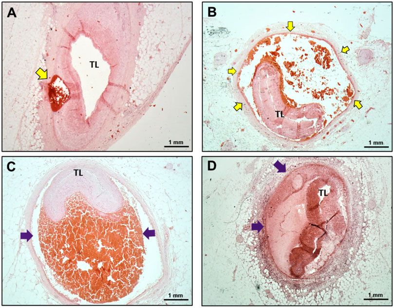
4                                                                                                                                                                                         M. Margaritis et al.

transverse sectional orientation were also examined for the presence of
                                                                                          Table 1 Demographics and cardiovascular risk factors of
irregular fibrils (fibrils with irregular edges) in 8 to 10 different collagen            autopsy cases and UKSCAD Cohort
bundles distributed in 4 to 5 areas of the copper grid. An average of 8
images of elastin and fibroblasts per subject were also taken and the wid-                                                                      Autopsy                    UKSCAD                       P-value
                                                                                                                                                cases                      cohort
est diameter in each image, as well as for the diameters of irregular fibrils,
                                                                                                                                                n 5 36                     n 5 359
was measured by post-analysis of the images with ImageJ. Elastin images                     ......................................................................................................
were qualitatively analysed for the presence of features reported in he-                    Female, n (%)                                       29 (81%)                   342 (95%)                     0.0004
reditary connective tissue disorders such as frayed edges, moth-eaten                       Post-partum,a n (%)                                 3 (10%)                    30 (9%)                       0.7223

                                                                                                                                                                                                                             Downloaded from https://academic.oup.com/cardiovascres/advance-article/doi/10.1093/cvr/cvab183/6287653 by guest on 06 September 2021
edges, calcified microcavities, dense internal spots, thick surface coat,                   Age (years)                                         49.4 ± 2.5                 47.0 ± 0.5                    0.3491
and indentations.14,15 A percentage was then calculated for each feature                    Body mass index (kg/m2)                             29.6 ± 1.5                 26.2 ± 0.3                    0.0290
observed in each subject. All analysis was conducted blinded to SCAD                        Active smoking, n (%)                               5 (14%)                    14 (4%)                       0.0014
and HV status.                                                                              Hypertension, n (%)                                 7 (19%)                    85 (24%)                      0.6311
                                                                                            Dyslipidaemia, n (%)                                7 (19%)                    33 (9%)                       0.0114
2.3 Statistical analysis                                                                    Diabetes mellitus, n (%)                            1 (3%)                     7 (2%)                        0.4433
Summary statistics are provided for all variables, including means ±                         a
                                                                                               Females only. Data presented as N (%) or mean ± standard error.
standard errors for continuous variables and counts and percentages
for categorical variables. Continuous variables were tested for nor-
mality using Kolmogorov–Smirnov test, and by visual inspection of                ..
                                                                                  .. 3.2 Macroscopic examination
the data.                                                                          ..
   When comparing the UKSCAD cohort with the autopsy cases,                         .. All autopsy cases involved a single coronary artery (example of typical
                                                                                     .. macroscopic findings in Supplementary material online, Figure S2).
as well as in the histology and IHC experiment analyses, differences                  ..
in continuous and categorical variables were tested using independent                  .. Localization of SCAD in the autopsy series was compared with angio-
                                                                                        .. graphic data from the SCAD-survivor cohort (Table 2). There was a sig-
sample t-test or Mann–Whitney U-test and Fisher exact tests,                             ..
respectively.
                                                                                          .. nificantly higher proportion of SCAD autopsy cases involving the LMS or
                                                                                           .. proximal coronary artery segments compared to SCAD survivors (au-
   In the electron microscopy experiments, univariate and multivariable                     ..
                                                                                             .. topsy—20/36, 56% vs. survivors 64/359, 18%; P < 0.0001). These in-
regression analyses were carried out to test the impact of the most rele-                     ..
vant clinical data (group SCAD vs. HV; Beighton score >4 vs.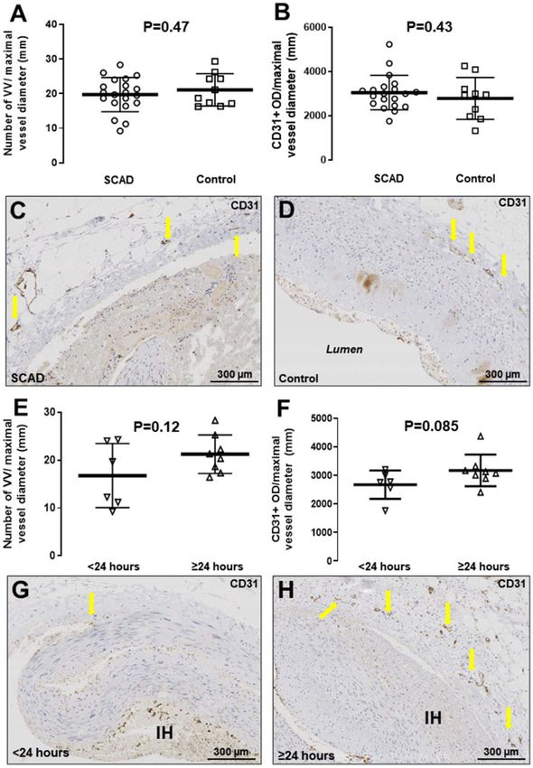
Micro- and ultrastructural features of SCAD 5
..
Table 2 Anatomic localization of culprit lesions in autopsy
.. number of peri-adventitial inflammatory cells surrounding the dissected
.. segment was significantly higher compared to the non-dissected seg-
cases and UKSCAD Cohort ..
.. ment, adjusted for the percentage of vessel circumference affected by
Autopsy UKSCAD P-value .. the dissection (Figure 3B, P < 0.0001). These results were replicated
cases cohort
..
.. when examining the number of CD68þ macrophages (Figure 3D,
(n 5 36) (n 5 359)
......................................................................................................
.. P < 0.0001) and CD3þ T-lymphocytes (Figure 3E, P = 0.016) corre-
..
LMS (n, % total) 6 (17%) 16 (4%) 0.0095 .. sponding to the dissected vs. non-dissected segment in IHC analysis.
..
LAD (n, % total) 14 (33%) 234 (65%) 0.0033 ..
Downloaded from https://academic.oup.com/cardiovascres/advance-article/doi/10.1093/cvr/cvab183/6287653 by guest on 06 September 2021
Proximal (n, % LAD) 9 (75%) 29 (13%) 0.001 .. 3.3.2 Vasa vasorum
..
Mid-distal (n, % LAD) 3 (25%) 192 (87%) .. After adjusting for vessel diameter, no significant differences in the
LCx (n, % total) 6 (17%) 104 (29%) 0.1706 .. density of VV were found between SCAD sections and controls
..
Proximal (n, % LCx) 2 (33%) 13 (14%) .. (Figure 4A). Total CD31 staining (PECAM-1, expressed on the surface
Mid-distal (n, % LCx) 4 (67%) 81 (86%)
.. of endothelial cells) in the media and adventitia also did not differ be-
..
RCA (n, % total) 12 (33%) 68 (19%) 0.0501 .. tween the two groups (Figure 4B–D). When comparing SCAD au-
Proximal (n, % RCA) 3 (25%) 6 (9%)
.. topsy cases with rapid- vs. delayed-onset death, we observed a trend
..
Mid-distal (n, % RCA) 9 (75%) 61 (91%) .. towards denser VV (Figure 4E) and more abundant medial and adven-
Multi-vessel (n, % total) 0 33 (9%)
.. titial CD31 staining (Figure 4F–H), although the association did not
..
Triple vessel (n, %) 6 (1.7%) .. reach statistical significance. The distribution of CD34 staining was
.. similar, confirming the structures stained as VV (Supplementary mate-
LMS cases include cases where extension into the LAD was noted. LAD, LCx, ..
and RCA cases include all cases where the origin of SCAD lesion was noted
.. rial online, Figure S3).
..
within the vessel, including multi-vessel cases. ..
LAD, left anterior descending artery; LCx, left circumflex artery; LMS, left main .. 3.3.3 Medial dissection and intramural haematoma
stem; RCA, right coronary artery. ..
.. In most cases (n = 31, 86.1%), the dissection event occurred near the
.. outer media, close to the adventitial border. In the remaining cases, the
..
.. medial intramural haematoma was localized close to the internal elastic
To compare the SCAD inflammatory cell infiltrate with the estab- .. lamina (IEL).
..
lished histopathology of medium- and large-vessel arteritides, we per- .. The proportion of the total coronary circumference affected varied
formed IHC for CD68 (surface marker of macrophages) and CD3 ..
.. widely both within and between cases. Some sections displayed a small
(surface marker of T-lymphocytes) and compared with age- and sex- .. intramural haematoma accounting for less than 10% of the vessel medial
matched control cases. There was, as expected, significantly higher infil- ..
.. area (e.g. Figure 5A); on the other hand, more proximal sections belong-
tration of CD68þ and CD3þ cells in the peri-adventitial tissues sur- .. ing to the same case displayed a false lumen enveloping almost the en-
rounding an SCAD section compared to control cases (Figure 1A and B
..
.. tirety of the coronary circumference (Figure 5B).
and microphotographs Figure 1C–H). In SCAD cases with significant in- .. The appearance and constituents of the false lumen were heteroge-
flammation, there was abundant CD68þ staining throughout the adven-
..
.. neous. Some cases displayed dense red clot with trapped white cell nu-
titial infiltrate, extending into the perivascular adipose tissue and in the .. clei and minimal fibrin formation (Figure 5C). Other cases had varying
..
media surrounding the dissection plane and haematoma (Figure 1D). .. degrees of mixed layers of fibrin formation, with almost distinct ‘com-
Similarly, there was significant, albeit less pronounced staining for CD3, .. partmentalization’ of different segments of the intramural haematoma,
..
which appeared to be more spatially localized over the adventitial bor- .. giving a ‘trabeculated’ appearance, akin to the so-called lines of Zahn
der of the vascular wall, as well as the outer rim of the media and adven- .. seen in fresh thrombus (Figure 5D).
..
titial inflammatory infiltrate (Figure 1E). ..
We next assessed the association between time elapsed from symp-
..
.. 3.3.4 Intimal features and atherosclerotic changes
tom onset to death and degree of inflammatory infiltrate. Two research- .. More than half (n = 28, 78%) of the autopsy cases studied displayed
ers, both blinded to clinical details, independently analysed n = 27 cases
..
.. some intimal changes. These ranged from mild to moderate thickening (a
for whom symptom-to-death time was available, semi-quantifying the .. recognized feature of ageing) but also included changes consistent with
..
degree of inflammatory cell infiltrate as ‘high’ or ‘low/absent’. There was .. underlying atherosclerosis. Approximately half of the cases (n = 17, 47%)
95% observer concordance. The degree of peri-adventitial inflammatory .. had mild to moderate atherosclerotic changes in both culprit and non-
..
cell infiltration was significantly associated with a longer time period .. culprit coronary arteries. These changes were mostly limited to early
from symptom onset to death (Figure 2A, P = 0.006; e.g. Figures 2C vs. 2G). .. neo-intima formation, with proliferation of vascular smooth muscle cells
..
Similarly, IHC staining for CD68þ (Figure 2B, E & H) and CD3þ (Figure .. and sometimes the formation of foam cells in the intima. Only one case
2B, F & I) showed a similar significant link between abundant cellular
..
.. displayed significant atheroma (80% stenosis) on macroscopic examina-
staining and longer time interval from symptom onset to death. .. tion, but in a non-dissected coronary vessel.
..
In addition to this temporal association, we sought to establish a spa- .. No recognized histological features of fibromuscular dysplasia (FMD)
tial association between inflammation and the dissected media. In the .. were identified. FMD is characterized by thickening and proliferation of
..
n = 18 autopsy cases that exhibited high inflammatory infiltrate, there .. the intimal layer and obliteration of the medial layer through extensive,
was a significantly larger surface area of peri-adventitial reactive tissue ad- .. dense, deeply stained collagen deposition, as well as fragmentation of the
..
jacent to dissected vs. non-dissected segments of the coronary sections .. internal and/or external elastic lamina. These features are prominent in
examined, after adjusting for the percentage of total vascular circumfer- .. the example provided in Supplementary material online, Figure S4, which
..
ence affected (Figure 3A, P < 0.001). Furthermore, in H&E sections, the . displays two internal mammary arteries from our archives, showing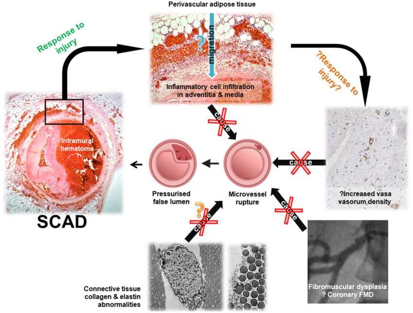
6                                                                                                                                                          M. Margaritis et al.

                                                                                                                                                                                  Downloaded from https://academic.oup.com/cardiovascres/advance-article/doi/10.1093/cvr/cvab183/6287653 by guest on 06 September 2021
    Figure 1 Composition of peri-adventitial inflammatory cell infiltrate in SCAD. Compared to age- and sex-matched control cases (n = 10), SCAD autopsy
    cases (n = 20) showed significantly higher infiltration with CD68þ macrophages (A, P < 0.001) and CD3þ T cells (B, P < 0.001). This infiltrate comprising
    lymphocytes, macrophages, and eosinophils was visualized in H&E staining (C) and spatially analysed with IHC. CD68þ cells were abundant throughout (D),
    whereas CD3þ T cells were less numerous and localized to the adventitial border (E). Control sections did not feature these findings (F–H). All compari-
    sons between groups were made using unpaired t-test on log-transformed values.

                                                                                ..
typical FMD features. These features were absent in the SCAD autopsy             ..             this mature endothelial cell marker showed the normal presence of en-
cases studied: Figure 6 shows a typical SCAD case, displaying a minor de-         ..            dothelial cells in the intima of all histological sections studied (e.g. Figure
                                                                                   ..
gree of intimal thickening and collagen deposition, with intact tunica me-          ..          6C). EVG staining also did not reveal evidence of excessive collagen de-
dia in the non-dissected segment and without fragmentation of the                    ..         position in the intima or the media, which is distinct from the mild fibro-
                                                                                      ..
elastic laminae. Specifically, none of the cases examined demonstrated                 ..       elastic intimal thickening seen in some cases (e.g. Figure 6B). In addition,
the extensive, dense, deeply stained collagen deposition seen in FMD
                                                                                        ..      IHC for a-smooth muscle actin showed a normal pattern of staining
                                                                                         ..
(e.g. Figure 6A and B). To further confirm the presence of intact endothe-                ..    across the non-dissected segments of the media in SCAD sections (e.g.
lial cells, we performed IHC staining for CD31 (PECAM-1). Staining for
                                                                                           ..   Figure 6D).
Micro- and ultrastructural features of SCAD                                                                                                                              7

                                                                                                                                                                              Downloaded from https://academic.oup.com/cardiovascres/advance-article/doi/10.1093/cvr/cvab183/6287653 by guest on 06 September 2021
  Figure 2 Association between the degree of peri-adventitial inflammatory infiltrate and time interval from SCAD symptom onset to death. Increased de-
  gree of peri-adventitial inflammatory cell infiltration was significantly associated with increased time from symptom onset to death (A, n = 27, P = 0.006, v2
  test). CD68þ and CD3þ staining showing a higher macrophage (B, CD68þ, P < 0.001, n = 17), and T-cell infiltrate (B, CD3þ, P = 0.03, n = 17) in cases with
  more than 24 h delay between onset of SCAD-related symptoms and death. Comparisons made using an unpaired t-test on log-transformed values. Panels
  (C–I) provide examples of autopsy cases belonging to the delayed-onset (C–F) vs. rapid-onset (G–I) death groups.

  We did not observe evidence of IEL degradation or fragmentation in            ..
                                                                                 .. 3.4 Electron microscopy
any of the sections studied or differences when compared to control               .. Dermal connective tissue from 31 patients and 16 HV was assessed by
cases (e.g. Figure 6B).
                                                                                   ..
                                                                                    .. trasmission electron microscopy . Demographic characteristics and car-
                                                                                     .. diovascular risk factors can be found in Table 3. No significant differences
                                                                                      ..
                                                                                       .. were found in the size of the major constituents of extracellular matrix
3.3.5 Extra-coronary arterial findings                                                  .. (Figure 7); fibroblasts and their subcellular synthetic organelles
No FMD in extra-coronary arteries was reported on the autopsy
                                                                                         ..
                                                                                          .. (Supplementary material online, Figure S5); or features of elastin damage
reports of the SCAD cases. Complete non-coronary arterial material                         .. (Supplementary material online, Figure S6) between SCAD cases and HV.
                                                                                            ..
was, however, not available for examination. Non-coronary arteries                           ..        Univariate and multivariable analyses are presented in Supplementary
were examined from 19 autopsy cases. No FMD was identified from 12                            .. material online, Tables S1 and S2. This demonstrated a significant effect
                                                                                               ..
renal arteries, 7 renal arterioles, 1 splenic artery, 1 vertebral artery, 1                     .. of age on minimum collagen fibril diameter (P = 0.0011), the number of
aorta, and 1 cerebral artery examined.
                                                                                                 .. irregular fibrils (P = 0.015), and elastin calcified microcavities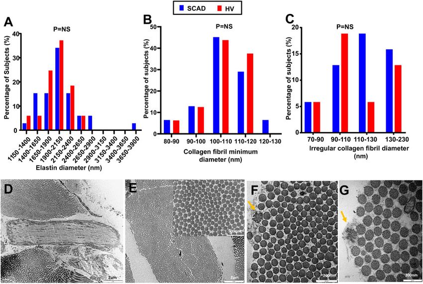
8                                                                                                                                                               M. Margaritis et al.

                                                                                                                                                                                       Downloaded from https://academic.oup.com/cardiovascres/advance-article/doi/10.1093/cvr/cvab183/6287653 by guest on 06 September 2021
    Figure 3 Association between the degree of peri-adventitial inflammatory infiltrate and proximity to dissected portion of the medial layer. In SCAD cases,
    we observed significantly higher inflammatory area (A, P < 0.0001; n = 18) and denser peri-adventitial inflammatory cell infiltrate (B, P < 0.0001, n = 18) adja-
    cent to dissected segments vs. non-dissected coronary segments. In a typical SCAD section (C), there is denser reactive adventitial tissue (green arrows) sur-
    rounding the intramural haematoma (IH) vs. areas adjacent to healthy, non-dissected portions of the medial layer (black arrows). Similarly, IHC showed that
    the number of CD68þ macrophages (D, P < 0.0001, n = 8) and CD3þ T-lymphocytes (E, P = 0.016, n = 8) was higher in the adventitia surrounding the dis-
    sected vs. non-dissected coronary circumference. All comparisons between dissected and non-dissected segments were made using paired t-test.

(P = 0.0031). The number of elastic calcified microcavities was also signif-        ..                   a majority of autopsy cases suggesting a rapid or arrhythmic death.
                                                                                     ..
icantly different (p = 0.0046) between those with low and high beighton               ..                 Secondly, inflammatory infiltration develops over time and likely consti-
score. In addition, significant differences between SCAD and HV were
                                                                                       ..                tutes a healing response to injury. Thirdly, we find no evidence of endo-
                                                                                        ..
found for elastin thick surface coat (P = 0.0285) and elastin calcified                  ...             thelial/intimal injury, no coronary histological features of FMD, and no
microcavities (P = 0.0491).                                                                ..            evidence of an increased VV density in SCAD. Finally, we show no ultra-
                                                                                            ..
                                                                                             ..          structural differences in dermal collagen and no evidence of changes in
                                                                                              ..         the cellular activity of skin fibroblasts in SCAD. Nevertheless, some fea-
4. Discussion                                                                                  ..
                                                                                                ..       tures of elastin damage do appear to significantly differ between the two
We present the largest study to date of SCAD coronary histopathology                             ..      groups.
                                                                                                  ..
and the first systematic assessment of dermal collagen ultrastructure in                           ..       The study findings have important implication for the autopsy assess-
SCAD survivors. We report, firstly, that myocardial necrosis is absent in
                                                                                                    ..   ment of SCD. The presence of more proximal dissections when
Micro- and ultrastructural features of SCAD 9
Downloaded from https://academic.oup.com/cardiovascres/advance-article/doi/10.1093/cvr/cvab183/6287653 by guest on 06 September 2021
Figure 4 VV density in SCAD. VV in SCAD coronary sections (n = 20) vs. control cases (n = 10) (A, P = 0.47) or total CD31þ optical density in the vascu-
lar media adjusted for maximal vessel diameter (B, P = 0.43). Panels (C and D) are representative SCAD and control microphotographs of CD31-stained sec-
tions, respectively. VV density comparing rapid-onset (24 h) SCAD fatalities (E and F, G and H
representative microphotographs for 24 h (n = 8) groups, respectively. P = NS). All comparisons between groups were made using an un-
paired t-test on log-transformed values.
compared to patients surviving to angiography is consistent with higher-
.. finding that P-SCAD was associated with more extensive myocardial in-
..
risk cases leading to fatality. However, the lack of myocardial infarction .. farction requires validation but is consistent with a number of studies
in a majority of cases suggests many deaths are arrhythmic and an ab-
.. reporting P-SCAD is a more extreme phenotype.16,17 This, coupled with
..
sence of myocardial necrosis cannot rule out this diagnosis. These find- .. the recent demonstration that post-menopausal women with SCAD
..
ings also suggest some patients with shorter, more distal SCAD .. may have a more benign phenotype than pre-menopausal women,18 sug-
presenting with arrhythmic death may be missed at autopsy unless this .. gests changes in female sex hormones may have a role in determining
..
diagnosis is carefully excluded by systematic assessment of the entire .. the severity of SCAD at presentation, although the mechanism remains
coronary tree, as suggested by a previous smaller case series.3 The
.. unclear.10 M. Margaritis et al.
Downloaded from https://academic.oup.com/cardiovascres/advance-article/doi/10.1093/cvr/cvab183/6287653 by guest on 06 September 2021
Figure 5 Intramural haematoma features from the SCAD autopsy case series. (A and B) Sequential H&E sections from the same autopsy case. Distally
only a small proportion of the total vessel circumference is affected (A), leaving the true lumen (TL) relatively patent, whereas proximally the false lumen
envelops almost the entire vessel causing significant luminal compression (B). (C) Intramural haematoma (purple arrow) displaying dense red clot with mini-
mal fibrin formation. (D) Intramural haematoma (purple arrows) showing varying degrees of maturation with fibrin formation, with almost distinct ‘compart-
mentalization’ of segments within. Yellow arrows: intramural haematoma; TL, true lumen.
A number of hypotheses as to the arterial vulnerability and patho-
.. coronary arterial tone leading to speculation of an additional poten-
..
physiological mechanism underlying SCAD have been proposed, to .. tial element to the outside-in pathophysiological hypothesis of
..
which our data provide novel insights. Although it was not possible .. SCAD.20
to serially section the entire coronary tree of affected patients, no .. A significant proportion of patients with SCAD have been shown to
..
structural abnormalities of the coronary intima or the IEL were dem- .. have co-existent remote arteriopathies, particularly the ‘string-of-beads’
onstrated, as might be expected for a spontaneous tear to develop .. sign of radiological FMD.1,2 The exact proportion of SCAD cases with
..
(as implied from the inside-out hypothesis). Intracoronary imaging has .. extra-coronary arteriopathies is unclear due to variations in the defini-
provided evidence of false lumen pressurization prior to the develop-
.. tions and imaging modalities used in different studies.1,21 One recent
..
ment of fenestrations between false and true lumens and angio- .. study even reported 100% of SCAD cases had radiological arterial ‘ab-
..
graphic findings have shown these fenestrations occur after the .. normalities’ of some sort.22 This has led to speculation that SCAD arises
development of intramural haematoma and not as a pre-requisite for .. primarily as a complication of pre-existent coronary histological FMD.23
..
SCAD.6,19 These findings taken together are supportive of the out- .. In this study, the typical histopathological features of FMD were not seen
side-in hypothesis as the predominant mechanism for SCAD. .. in the coronary artery sections studied, suggesting that changes of local-
..
Additionally, coronary microvessels have been proposed as the po- .. ized coronary histological FMD are not a pre-requisite to SCAD in many
tential source of intramural bleeding in SCAD. A previous intracoro-
.. cases. It is therefore likely that other, more subtle changes in the coro-
..
nary imaging study reported an increase in VV density in SCAD .. nary vessel wall, such as differences in cell–cell adhesion or extracellular
..
coronaries5 although this was not confirmed in a subsequent larger .. matrix function, are responsible for the vulnerability to SCAD.
series.6 This histological study confirms no evidence for increased VV .. Histological FMD was not found on a limited review of available non-cor-
..
density suggesting that absolute vessel density may be less important .. onary arterial material from the autopsy cases. This may represent in-
than the vulnerability of traversing microvessels and the disrupting .. complete sampling but it remains to be confirmed that the ‘string-of-
..
forces to which they are subjected. It has been demonstrated that .. beads’ appearance of radiological FMD seen in SCAD invariably corre-
adventitial vasoactive factors play an important role in regulating
.. sponds to histological changes consistent with the pathologicalMicro- and ultrastructural features of SCAD 11
Downloaded from https://academic.oup.com/cardiovascres/advance-article/doi/10.1093/cvr/cvab183/6287653 by guest on 06 September 2021
Figure 6 Intimal and medial layer features in SCAD. (A) H&E staining of SCAD lesion with medial dissection and intramural haematoma (IH) leading to
compression true lumen (TL) compression (black arrows). Moderate fibro-elastic thickening of the endothelial layer (red arrows) but no abnormalities in
structure or orientation of medial smooth muscle cells (yellow arrows). (B) EVG staining. Fibro-elastic intimal thickening is well-delineated (red arrows); the
IEL clearly visualized and smooth muscle cell medial layer distinguished from intima and adventitia (yellow arrows). (C) CD31 staining shows mature endo-
thelial cells on the intimal surface (green arrows). (D) aSMA showing normal pattern of staining in the non-dissected segment of the media (blue arrows).
..
definitions of FMD. The only reported post-mortem case presenting the .. across the SCAD case series as described in eosinophilic coronary peri-
gross pathological appearances of a renal artery ‘string-of-beads’, does .. arteritis.28 Our findings are consistent with the fact that, although inflam-
..
not describe the histological findings of this artery.24 In this study, we are .. matory disorders are often reported as a predisposing condition in
unable to definitively address the question as to whether the coronary .. SCAD,11 rates of inflammatory diseases are probably similar to the gen-
..
histology of patients with SCAD and extra-coronary arteriopathies (in- .. eral population.29,30
cluding the radiological string-of-beads) differs from SCAD cases with-
.. SCAD is associated with hereditary connective tissue disorders in a
..
out such arteriopathies. A future prospective series with systematic .. small proportion of cases.9,31,32 Features of hypermobility have also
serial sectioning of relevant arterial beds will be required to address
.. been described in a subgroup of SCAD survivors. This has led to specu-
..
these questions definitively. The low rates of atherosclerotic changes .. lation that even without a monogenetic cause, abnormalities of connec-
seen in the autopsy cases may reflect the low-risk profile of this predom-
.. tive tissue might be a common mechanism underlying SCAD.1,2
..
inantly female population but are also consistent with recent findings .. Abnormalities of dermal collagen ultrastructure have been shown in a
..
suggesting an opposing influence of common genetic variants on SCAD .. range of established connective tissue disorders.15 We found no general-
vs. ischaemic heart disease risk.25,26 .. izable difference in a range of connective tissue ultrastructural features
..
Previous histopathological case reports have described SCAD as a .. on blinded analysis. Importantly, previously reported effects of age33 on
mono-arteritis because of the density of the reported associated inflam- .. collagen fibril size and the number of irregular fibrils were confirmed,
..
matory infiltrate.7,8 Our study presents the most comprehensive evi- .. providing effectively a positive control within the analysis. Some features
dence so far supportive that coronary inflammation in SCAD is a time ... of elastin damage were different between HV and SCAD survivors, sug-
dependent and localized healing response to the injury rather than a .. gesting a possible underlying predisposition towards more unstable elas-
..
causal vasculitic process. This inflammatory infiltrate is distinct from that .. tin in these patients. These findings are hypothesis-generating and will
of medium- and large-vessel arteritides: There were scarcely any giant .. require further validation but are in keeping with recent genetic stud-
..
cells noted, a predominant feature in giant cell arteritis (GCA).27 The .. ies9,34 showing causal connective tissue disorder variants in SCAD affect
predominant cell type was CD68þ macrophages, as opposed to GCA .. only12 M. Margaritis et al.
..
Table 3 Demographics, cardiovascular risk factors, and .. 4.1 Limitations
SCAD event details of SCAD cases and HV recruited in the .. SCAD leading to SCD is rare, thus making a prospective unbiased design
..
electron microscopy studies .. logistically impossible. As a retrospective observational study, we cannot
.. conclude that the associations demonstrated are causative. All SCAD
SCAD cases (n 5 31) HV (n 5 16) ..
...................................................................................................... .. autopsy cases were initially referred for clinical autopsy and as such, it
.. was impossible to employ a uniform methodology or sequential section-
Age at biopsy (years) 45.8 ± 1.34 44.0 ± 1.49 ..
Body mass index (kg/m2) 27.15 ± 1.13 26.37 ± 01.82 .. ing of the entire length of the coronary tree. Our ethical permissions did
.. not permit genetic analysis of autopsy cases. As most autopsies are ini-
Active smoking, n (%) 1 (3.1%) 0 ..
Downloaded from https://academic.oup.com/cardiovascres/advance-article/doi/10.1093/cvr/cvab183/6287653 by guest on 06 September 2021
Hypertension, n (%) 8 (25%) 0 .. tially performed by non-cardiovascular pathologists and the heart (only)
.. is retained for later examination by a cardiovascular pathologist, limited
Dyslipidaemia, n (%) 1 (3.1%) 0 ..
Diabetes mellitus, n (%) 0 0 .. non-coronary arteries were available for assessment. Interpretation of
.. the frequency of non-coronary arteriopathies is therefore limited by in-
P-SCAD, n (%) 5 (15.6%) N/A ..
Age at SCAD event (years) 42.4 ± 1.39 N/A .. complete sampling. The numbers of included patients will impact on
.. power meaning small effects in the measured indices may not be demon-
Multi-vessel SCAD, n (%) 5 (15.6%) N/A ..
Recurrent SCAD, n (%) 4 (12.5%) N/A .. strated. Dermal connective ultrastructure was used as a surrogate for
.. coronary connective tissue as trasmission electron microscopy could
..
Continuous variable values presented as mean ± SEM. .. not be undertaken on the dissected coronaries, given that fresh tissue is
P-SCAD, pregnancy-associated SCAD; SCAD, spontaneous coronary artery .. required.
dissection. ..
..
..
.
Figure 7 Ultrastructural analysis of the main ECM components in SCAD patients vs. HV. Elastin diameter (A), collagen fibril minimum diameter (B) and ir-
regular collagen fibril diameter (C) distributions in SCAD (n = 31) vs. HV (n = 16). Representative images of elastin (D), collagen fibrils (E), and irregular fibrils
(F and G). T-test between the averages, standard deviations, minimum and maximum values, and ranges of these parameters was performed between SCAD
and HV. No significant differences were observed.Micro- and ultrastructural features of SCAD 13
..
5. Conclusions .. References
.. 1. Adlam D, Alfonso F, Maas A, Vrints C; Writing Committee. European Society of
Care is required during autopsy for SCD to exclude SCAD, particularly
.. Cardiology, acute cardiovascular care association, SCAD study group: a position pa-
..
when affecting more distal coronary locations and presenting without .. 2. per on spontaneous coronary artery dissection. Eur Heart J 2018;39:3353–3368.
myocardial necrosis. This study found no supporting evidence for a
.. Hayes SN, Kim ESH, Saw J, Adlam D, Arslanian-Engoren C, Economy KE, Ganesh SK,
.. Gulati R, Lindsay ME, Mieres JH, Naderi S, Shah S, Thaler DE, Tweet MS, Wood MJ;
causal role for peri-coronary inflammation, which is more likely an evolv- .. American Heart Association Council on Peripheral Vascular Disease; Council on
.. Clinical Cardiology; Council on Cardiovascular and Stroke Nursing; Council on
ing response to injury than a coronary mono-arteritis triggering an .. Genomic and Precision Medicine; and Stroke Council. Spontaneous coronary artery
SCAD event. We also found no evidence to support the inside-out hy- .. dissection: current state of the science: a scientific statement from the American
..
Downloaded from https://academic.oup.com/cardiovascres/advance-article/doi/10.1093/cvr/cvab183/6287653 by guest on 06 September 2021
pothesis, no features of coronary FMD, and no evidence for an increased .. Heart Association. Circulation 2018;137:e523–e557.
VV density as the primary source for intramural bleeding. Finally, we find .. 3. Desai S, Sheppard MN. Sudden cardiac death: look closely at the coronaries for
.. spontaneous dissection which can be missed. A study of 9 cases. Am J Forensic Med
no generalized changes in dermal collagen ultrastructure, suggesting .. Pathol 2012;33:26–29.
these changes may not be a prime pathophysiological driver in most .. 4. Vrints CJ. Spontaneous coronary artery dissection. Heart 2010;96:801–808.
.. 5. Kwon TG, Gulati R, Matsuzawa Y, Aoki T, Guddeti RR, Herrmann J, Lennon RJ,
patients. .. Ritman EL, Lerman LO, Lerman A. Proliferation of coronary adventitial vasa vasorum
..
.. in patients with spontaneous coronary artery dissection. JACC Cardiovasc Imaging
.. 2016;9:891–892.
Supplementary material .. 6. Jackson R, Al-Hussaini A, Joseph S, van Soest G, Wood A, Macaya F, Gonzalo N,
.. Cade J, Caixeta A, Hlinomaz O, Leinveber P, O’Kane P, Garcı́a-Guimaraes M,
.. Cortese B, Samani NJ, Escaned J, Alfonso F, Johnson T, Adlam D. Spontaneous coro-
Supplementary material is available at Cardiovascular Research online. .. nary artery dissection: pathophysiological insights from optical coherence tomogra-
..
.. phy. JACC Cardiovasc Imaging 2019;12:2475–2488.
.. 7. Carreon CK, Esposito MJ. Eosinophilic coronary monoarteritis. Arch Pathol Lab Med
Authors’ contribution .. 2014;138:979–981.
.. 8. Mandal R, Brooks EG, Corliss RF. Eosinophilic coronary periarteritis with arterial dis-
..
All authors contributed to the design of the work, interpretation of the .. 9. section: the mast cell hypothesis. J Forensic Sci 2015;60:1088–1092.
Henkin S, Negrotto SM, Tweet MS, Kirmani S, Deyle DR, Gulati R, Olson TM, Hayes
data and the final writing of the manuscript. M.M., F.S., and A.A.B.-C. addi- ..
.. SN. Spontaneous coronary artery dissection and its association with heritable con-
tionally contributed to the acquisition and analysis of the data and C.B. to .. nective tissue disorders. Heart 2016;102:876–881.
the statistical analysis of the data. D.A. and M.N.S. are accountable for all .. 10. Buja LM, Butany J. Cardiovascular Pathology. 4th ed. Amsterdam, Netherlands: Elsevier;
.. 2016.
aspects of the work and undertake to ensure that questions related to .. 11. Saw J, Starovoytov A, Humphries K, Sheth T, So D, Minhas K, Brass N, Lavoie A,
the accuracy or integrity of any part of the work are appropriately inves- .. Bishop H, Lavi S, Pearce C, Renner S, Madan M, Welsh RC, Lutchmedial S,
.. Vijayaraghavan R, Aymong E, Har B, Ibrahim R, Gornik HL, Ganesh S, Buller C,
tigated and resolved. ..
.. Matteau A, Martucci G, Ko D, Mancini GBJ. Canadian spontaneous coronary artery
.. dissection cohort study: in-hospital and 30-day outcomes. Eur Heart J 2019;40:
.. 1188–1197.
Acknowledgements .. 12. Billaud M, Donnenberg VS, Ellis BW, Meyer EM, Donnenberg AD, Hill JC, Richards
.. TD, Gleason TG, Phillippi JA. Classification and functional characterization of vasa
The authors are grateful for the support of SCAD survivors, the families .. vasorum-associated perivascular progenitor cells in human aorta. Stem Cell Reports
of deceased SCAD victims, and our non-SCAD and healthy controls. ..
.. 2017;9:292–303.
They thank our clinical and pathology colleagues who have referred .. 13. Schneider CA, Rasband WS, Eliceiri KW. NIH Image to ImageJ: 25 years of image
SCAD cases to our research study. They specifically acknowledge the .. analysis. Nat Methods 2012;9:671–675.
.. 14. Kobayasi T. Dermal elastic fibres in the inherited hypermobile disorders. J Dermatol
support of Jenny Middleton, Jane Plume, Donna Alexander, Sue Sterland, ..
Daniel Lawday, Emma Beeston, Tara Maitland, and Andrea Marshall for .. 15. Sci 2006;41:175–185.
Hermanns-Le T, Pierard GE. Ultrastructural alterations of elastic fibers and other
..
all their support for SCAD research. They acknowledge the leadership .. dermal components in Ehlers-Danlos syndrome of the hypermobile type. Am J
of the ESC-ACCA SCAD Study Group. Finally we acknowledge Kees .. Dermatopathol 2007;29:370–373.
.. 16. Tweet MS, Hayes SN, Codsi E, Gulati R, Rose CH, Best PJM. Spontaneous coronary
Straatman for designing the macro used in the EM analysis. .. artery dissection associated with pregnancy. J Am Coll Cardiol 2017;70:426–435.
.. 17. Havakuk O, Goland S, Mehra A, Elkayam U. Pregnancy and the risk of spontaneous
Conflict of interest: D.A. has received funding to support a clinical re- .. coronary artery dissection: an analysis of 120 contemporary cases. Circ Cardiovasc
..
search fellow from Abbott vascular. He has also received funding from .. 18. Interv 2017;10:e004941.
Astra Zeneca, Inc. for unrelated research and has conducted unrelated .. Diez-Villanueva P, Garcia-Guimaraes MM, Macaya F, Masotti M, Nogales JM, Jimenez-
.. Kockar M, Velazquez M, Lozano I, Moreu J, Avanzas P, Salamanca J, Alfonso F.
consultancy for GE, Inc. All other authors have reported that they have .. Spontaneous coronary artery dissection and menopause. Am J Cardiol 2021;148:
no relationships relevant to the contents of this paper to disclose. .. 53–59.
.. 19. Waterbury TM, Tweet MS, Hayes SN, Eleid MF, Bell MR, Lerman A, Singh M, Best
.. PJM, Lewis BR, Rihal CS, Gersh BJ, Gulati R. Early natural history of spontaneous cor-
..
Funding .. onary artery dissection. Circ Cardiovasc Interv 2018;11:e006772.
.. 20. Meyer MR, Barton M. Role of perivascular adipose tissue for sex differences in coro-
This work was supported by BeatSCAD, the British Heart Foundation (BHF) .. nary artery disease and spontaneous coronary artery dissection (SCAD). Endocr
[PG/13/96/3060], the National Institute for Health Research (NIHR) rare dis-
.. Metab Sci 2021;2:100068.
.. 21. Gornik HL, Persu A, Adlam D, Aparicio LS, Azizi M, Boulanger M, Bruno RM, de
ease translational collaboration, and the Leicester NIHR Biomedical Research .. Leeuw P, Fendrikova-Mahlay N, Froehlich J, Ganesh SK, Gray BH, Jamison C,
Centre. The authors also acknowledge Cardiac Risk in the Young (CRY) UK, ..
.. Januszewicz A, Jeunemaitre X, Kadian-Dodov D, Kim ES, Kovacic JC, Mace P,
the charity that supports MNS’ laboratory. .. Morganti A, Sharma A, Southerland AM, Touze E, van der Niepen P, Wang J,
.. Weinberg I, Wilson S, Olin JW, Plouin PF. First International Consensus on the diag-
.. nosis and management of fibromuscular dysplasia. Vasc Med 2019;24:164–189.
.. 22. Sharma S, Kaadan MI, Duran JM, Ponzini F, Mishra S, Tsiaras SV, Scott NS, Weinberg
Data availability .. I, Ghoshhajra B, Lindsay M, Gibson CM, Chi G, Michalak N, Wood MJ. Risk factors,
..
.. imaging findings, and sex differences in spontaneous coronary artery dissection. Am J
The data underlying this article will be shared on reasonable request to
.. Cardiol 2019;123:1783–1787.
.. 23. Saw J, Bezerra H, Gornik HL, Machan L, Mancini GB. Angiographic and intracoronary
the corresponding author. . manifestations of coronary fibromuscular dysplasia. Circulation 2016;133:1548–1559.14 M. Margaritis et al.
24. Moulson N, Kelly J, Iqbal MB, Saw J. Histopathology of coronary fibromuscular dys-
.. of coronary arteritis: report of seven autopsy cases and a review of the literature.
..
plasia causing spontaneous coronary artery dissection. JACC Cardiovasc Interv 2018;11: .. Virchows Arch 2013;462:239–248.
909–910. .. 29. Kronzer VL, Tarabochia AD, Lobo Romero AS, Tan NY, O’Byrne TJ, Crowson CS,
25. Adlam D, Olson TM, Combaret N, Kovacic JC, Iismaa SE, Al-Hussaini A, O’Byrne .. Turley TN, Myasoedova E, Davis JM 3rd, Raphael CE, Gulati R, Hayes SN, Tweet MS.
MM, Bouajila S, Georges A, Mishra K, Braund PS, d’Escamard V, Huang S, Margaritis .. Lack of association of spontaneous coronary artery dissection with autoimmune dis-
M, Nelson CP, de Andrade M, Kadian-Dodov D, Welch CA, Mazurkiewicz S, .. ease. J Am Coll Cardiol 2020;76:2226–2234.
Jeunemaitre X, Consortium D, Wong CMY, Giannoulatou E, Sweeting M, Muller D, .. 30. Adlam D, Cortese B, Kadziela J. Autoimmune disease and spontaneous coronary
Wood A, McGrath-Cadell L, Fatkin D, Dunwoodie SL, Harvey R, Holloway C,
.. artery dissection: causation versus coexistence. J Am Coll Cardiol 2020;76:
..
Empana JP, Jouven X, Olin JW, Gulati R, Tweet MS, Hayes SN, Samani NJ, Graham .. 2235–2237.
RM, Motreff P, Bouatia-Naji N; CARDIoGRAMPlusC4D Study Group. Association of .. 31. Kadian-Dodov D, Gornik HL, Gu X, Froehlich J, Bacharach JM, Chi YW, Gray BH,
the PHACTR1/EDN1 genetic locus with spontaneous coronary artery dissection. J .. Jaff MR, Kim ES, Mace P, Sharma A, Kline-Rogers E, White C, Olin JW. Dissection
Downloaded from https://academic.oup.com/cardiovascres/advance-article/doi/10.1093/cvr/cvab183/6287653 by guest on 06 September 2021
Am Coll Cardiol 2019;73:58–66. .. and aneurysm in patients with fibromuscular dysplasia: findings from the U.S. Registry
26. Saw J, Yang M-L, Trinder M, Tcheandjieu C, Xu C, Starovoytov A, Birt I, Mathis MR, .. for FMD. J Am Coll Cardiol 2016;68:176–185.
Hunker KL, Schmidt EM, Jackson L, Fendrikova-Mahlay N, Zawistowski M, Brummett .. 32. Carss KJ, Baranowska AA, Armisen J, Webb TR, Hamby SE, Premawardhana D,
CM, Zoellner S, Katz A, Coleman DM, Swan K, O’Donnell CJ, Assimes TL,
.. Al-Hussaini A, Wood A, Wang Q, Deevi SVV, Vitsios D, Lewis SH, Kotecha D,
O’Donnell CJ, Zhou X, Li JZ, Gornik HL, Assimes TL, Stanley JC, Brunham LR,
.. Bouatia-Naji N, Hesselson S, Iismaa SE, Tarr I, McGrath-Cadell L, Muller DW,
..
Ganesh SK, Million Veteran P; Million Veteran Program. Chromosome 1q21.2 and .. Dunwoodie SL, Fatkin D, Graham RM, Giannoulatou E, Samani NJ, Petrovski S,
additional loci influence risk of spontaneous coronary artery dissection and myocar- .. Haefliger C, Adlam D. Spontaneous coronary artery dissection: insights on rare
dial infarction. Nat Commun 2020;11:4432. .. genetic variation from genome sequencing. Circ Genom Precis Med 2020;13:
27. Kurata A, Saito A, Hashimoto H, Fujita K, Ohno SI, Kamma H, Nagao T, Kobayashi S, .. e003030.
Yamashina A, Kuroda M. Difference in immunohistochemical characteristics between .. 33. Breiteneder-Geleff S, Mallinger R, Böck P. Quantitation of collagen fibril cross-section
Takayasu arteritis and giant cell arteritis: it may be better to distinguish them in the .. profiles in aging human veins. Hum Pathol 1990;21:1031–1035.
same age. Mod Rheumatol 2019;29:992–1001.
.. 34. Kaadan MI, MacDonald C, Ponzini F, Duran J, Newell K, Pitler L, Lin A, Weinberg I,
..
28. Kajihara H, Tachiyama Y, Hirose T, Takada A, Takata A, Saito K, Murai T, Yasui W. .. Wood MJ, Lindsay ME. Prospective cardiovascular genetics evaluation in spontaneous
Eosinophilic coronary periarteritis (vasospastic angina and sudden death), a new type .. coronary artery dissection. Circ Genom Precis Med 2018;11:e001933.
.
Translational Perspective
Spontaneous coronary artery dissection (SCAD), especially of distal coronary territories should be carefully assessed at post-mortem in SCD cases,
even where there are no signs of myocardial infarction. The immediate cause of SCAD is likely to be the development of a spontaneous intramural
haematoma rather than an intimal disruption or ‘tear’. This does not seem to be directly related to increased VV density, coronary fibromuscular
dysplasia, or local inflammation (except as a response to injury). Although SCAD is rarely associated with hereditary connective tissue disorders,
there does not seem to be a more generalizable global connective tissue ultrastructural abnormality in most cases.You can also read