Detection of Fungal Toxins Produced by Dermatophytes by using Thin Layer Chromatography - Open Journal Systems
←
→
Page content transcription
If your browser does not render page correctly, please read the page content below
2434    Indian Journal of Forensic Medicine & Toxicology, January-March 2021, Vol. 15, No. 1

       Detection of Fungal Toxins Produced by Dermatophytes by
                   using Thin Layer Chromatography

             Abeer Mohammed Ali Al-garawyi1, Adnan H. Al-Hamadani2, Adnan waheed AL- Bederi3
  1
  Post graduate Biology Department, Education for Pure Science College, Al-Muthanna University, Iraq, 2 Prof.
Department Microbiology/ College of Medicine / Al-Qadisiyah University, 3Prof. Department Anatomy/ College of
                                     Medicine / Al-Qadisiyah University

                                                                          Abstract
       Dermatophyte fungi include a wide range of filamentous fungi that are pathogenic to humans including
       three superficial cutaneous genera such as ; Epidermophyton, Microsporum and Trichophyton. However,
       the aim of this study isolation and identification of fungi responsible of dermatophytosis and detection of
       the dermatophyte ability to produce mycotoxins. The results of this study showed that 49(69.01%) out of
       71 specimens were gave positive results , while 22 (30.98%) were gave negative results by examination
       on 10%KOH and cultured on Sabouraud dextrose agar with cyclohexmaide. Out of 49 culture positive
       isolates, 31(63.3%) T. rubrum isolates was the most frequent etiological agent followed by 11( 22.4%)
       T.mentagrophytes ; 5(14.3%) M. canis and 2

       (4.1%) Epidermophyton floccosum isolates. The use of the TLC method to detect of mycotoxins in chloroform
       extracts for 49 dermatophyte mycelia. It was found all dermatophyte isolates are able to produce fluorescent
       stains with different colors under UV light at 365 nm , also these stains may be act as a non-enzymatic
       virulence indicator ( mycotoxins).

       Keywords: E. floccosum , Dermatophytosis, TLC , Mycotoxins.

                            Introduction                                            lipase , elastase, keratinase, phospholipase and protease
                                                                                    depending on the specificity of the different substrate ,
     Dermatophytes are anamorphic genera which
                                                                                    the pathogenicity of host tissues occurs (2) . Keratinase
includes      Epidermophyton,        Microsporum     and
                                                                                    enzyme produced by Bacillus species (3),(4) Streptomyces
Trichophyton , all of these genera cause superficial                                (5) and also dermatophytes fungi (6) . The outer layer of
fungal infection called dermatophytosis. Dermatophytes
                                                                                    the epidermis characterized by being rich in keratinous
are also characterized as keratinophilic fungi, as they
                                                                                    substance such as the skin, hair and nails , which is
infect the skin, hair, and nails of humans and animals.
                                                                                    considered a favorite for the dermatophytes fungi. In
Usually, dermatophyte infections are limited only
                                                                                    addition to keratinase, protease is also regarded one of the
to the outer layer of the epidermis and also unable to
                                                                                    most important enzymes produced by dermatophytes as
penetrate the deep tissues of a healthy individual (1) .
                                                                                    a virulence factor (7) . There are non-enzymatic virulence
Dermatophytes are included Anthropophiles ( infect
                                                                                    factor that are produced by dermatophytes fungi such as
human) , zoophiles ( infect animals) and geophiles (soil
                                                                                    ; xanthomegnin is mycotoxin produced by T. rubrum,
dwelling), all of these subdivisions according to their
                                                                                    which is also produced from Penicillium and Aspergillus
natural habitat. Dermatophytes are characterized by the
                                                                                    , whether in vitro or in vivo causing nephropathy and
secretion of large quantities of analyzed enzymes such as
                                                                                    death in animals. The T. rubrum culture characterized by
                                                                                    red pigmentation on the reverse side , this is explained
Corresponding author:                                                               by the production of xanthomegnin which is also noted
Abeer Mohammed Ali Al-garawyi                                                       in infected skin and nail specimens (8). There are limited
E-mail: aliabeer297@gmail.com                                                       species of dermatophytes that have the ability to produce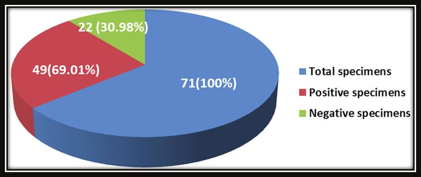
Indian Journal of Forensic Medicine & Toxicology, January-March 2021, Vol. 15, No. 1   2435

melanin or melanin-like compounds , whether in vitro               Detection of mycotoxin by using Thin Layer
or in vivo , which play a similar role in pathogenesis          Chromatography technique (TLC):
of dermatophytic diseases (9) . Aim of this study;
                                                                     This experiment was carried out in a toxicology
isolation and identification of fungi responsible of
                                                                laboratory at the College of Applied Medical Sciences
dermatophytosis and detection of dermatophyte ability to
                                                                / Karbala University , where used Thin Layer
produce mycotoxins such as Aflatoxin - like compounds
                                                                Chromatography plates (TLC) with dimensions of 20 *
chemically and separation of these mycotoxins by Thin
                                                                20 cm, after activated in the electric oven at a temperature
Layer Chromatography (TLC).
                                                                of 105 0C for an hour before use (11) . The separation
              Materials and Methods                             system used consists of chloroform : methanol 98:2 and
                                                                15µl of standard mycotoxin was taken by capillary tube
    Isolation and Identification of fungal isolates:
                                                                , and put on the line a distance of 2 cm from left edge of
71samples were collected from patients with
                                                                the plate and at a distance of 2 cm between the spot of
dermatomycosis infections during period from February
                                                                standard mycotoxin put an amount equal to the standard
2018 to January 2019. All the specimens were divided
                                                                mycotoxin from the first fungal isolate extract and thus to
into two portions, one portion for direct examination
                                                                the rest isolates extract. After that, the plate was placed
under light microscope and other was cultured on
                                                                in the separation tank containing the separation system
sabouraud dextrose agar.
                                                                consisting of a mixture of chloroform and methanol,
   Detection of dermatophytes ability to produce                at a ratio of 98: 2 v/v. The separation solution was
mycotoxins:                                                     monitored until it reached a distance of approximately
                                                                2 cm from the top end of the plate, then plates removed
    Isolation and purification of mycotoxins :                  and dried under laboratory conditions for a period of
                                                                5min (12) . Then compare the RF value and colour for the
     We used nutrient broth ( NB) to detection
                                                                mycotoxins spots with standard.
of mycotoxins produced by dermatophyte fungi.
Subsequently , prepared 25 flask with volume 500ml                    Determination of UV absorbance:
then each flask added to it 250 ml NB, after sterilization
and cooling, the culture media was inoculated with agar                The partially purified mycotoxin compounds which
blocks of 5 mm of pure isolates grown on sabouraud              produced by dermatophytes fungi subjected to UV
dextrose agar SDA for 7 days at a rate of one disk              radiation at a wavelength of 365 nm absorption spectrum
per flask, except one flask left without inoculation as         on TLC to distinguish the absorption bands in the sample
                                                                (12) .
a control for comparison , and incubated at 28 °C for
three weeks. After incubation, the entire contents were
                                                                                    Results and Discussion
filtered with a sterile, clean gauze and then chloroform
was added to the broth (1:1) in a separation funnel. The            The result of this study showed 49(69.01%) out
mixture was shaken for a few minutes then separated             of 71 specimens were gave positive results , while 22
an upper layer containing spores and mycelia, and a             (30.98%) were gave negative results by examination on
lower layer containing chloroform and mycotoxins . The          10%KOH and cultured on Sabouraud dextrose agar with
bottom layer filtered through a Whatman No. 1 filter            cyclohexmaide (SDAC) (Fig-1) .
paper then concentrated by using reflective condenser to
approximately 1 ml in dark bottles (10) .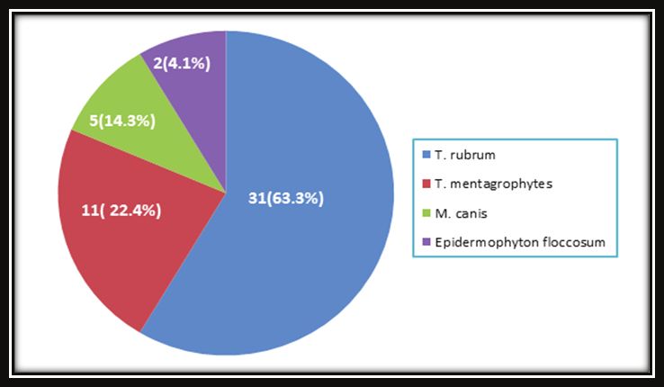
2436    Indian Journal of Forensic Medicine & Toxicology, January-March 2021, Vol. 15, No. 1

       Fig(1):Numbers and percentages of clinical samples with dermatomycosis infections (P value 0.001) .

    The results of this study showed that there is a statistical significant (P
Indian Journal of Forensic Medicine & Toxicology, January-March 2021, Vol. 15, No. 1 2437
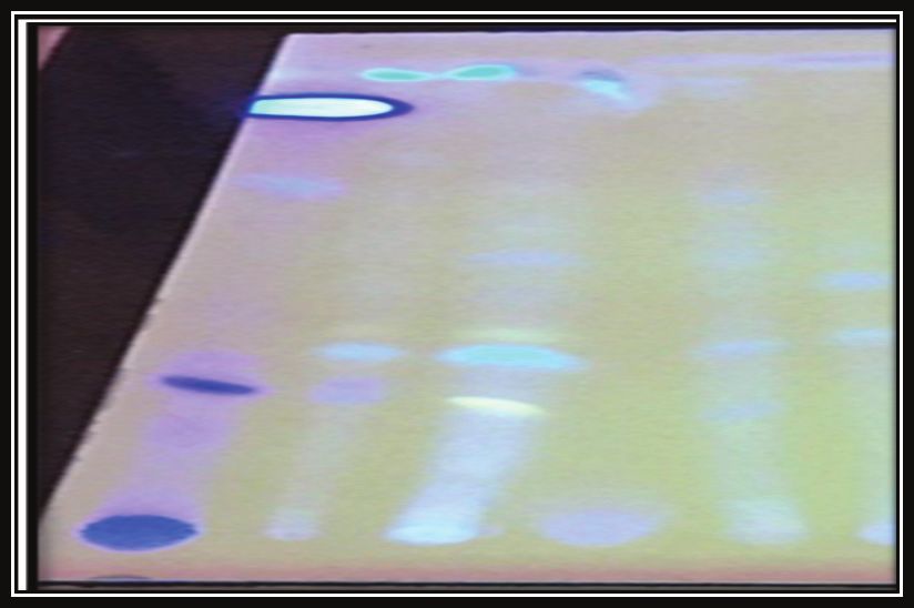
Fig(3): Detection of Mycotoxins by using TLC technique; A: Standard mycotoxin, 1: T. rubrum,2: T.
mentagrophytes, 3: M. canis and 4: E. floccosum extracts grown on the NB after18 days of incubation at28 ◦
C.
The results of current study also showed that some References
of these spots had Rf. an identical to the standard 1. Weitzman I and Summerbell R C .
Rf., while the other spots had Rf. different from the The dermatophytes. Clinical microbiology
standard Rf. Although there are no previous studies and reviews,1995; 8(2): 240-259.
scarcity of references about this topic for the purpose of
comparison, the precise diagnosis for these compounds 2. Chinnapun D. Virulence factors involved in
is focus of the current studies, in our lab. pathogenicity of dermatophytes. Walailak Journal of
Science and Technology (WJST),2015; 12(7): 573-580.
Conclusion
3. Jeevana Lakshmi P, Kumari Chitturi, C M
The use of the TLC method to detect of mycotoxins
and Lakshmi V V. Efficient degradation of feather by
in chloroform extracts for 49 dermatophyte mycelia. It
keratinase producing Bacillus sp. International journal
was found all dermatophyte isolates are able to produce
of microbiology,2013; 2013: 608-321.
fluorescent stains with different colors under UV light at
365 nm , also these stains may be act as a non-enzymatic 4. Kainoor P S and Naik G R. Production and
virulence indicator ( mycotoxins). characterization of feather degrading keratinase from
Bacillus sp.JB,2010; 99( 9):384-90.
Ethical Clearance: The Research Ethical
Committee at scientific research by ethical approval of 5. Syed D G, Lee J C, Li W J, Kim C J and Agasar
both MOH and MOHSER in Iraq D. Production, characterization and application of
keratinase from Streptomyces gulbargensis. Bioresource
Conflict of Interest: None
technology,2009; 100(5): 1868-1871.
Funding: Self-funding 6. Sharma A., Chandra S and Sharma M. Difference
in keratinase activity of dermatophytes at different
environmental conditions is an attribute of2438 Indian Journal of Forensic Medicine & Toxicology, January-March 2021, Vol. 15, No. 1
adaptation to parasitism. Mycoses,2012: 55(5): 12. Al- Khafji N A. Molecular properties of A.niger
410-415. isolates producing toxins and contaminating feed
7. Elavarashi E., Kindo A. J. and Rangarajan S. grain and its laboratory control. M. Sc. Thesis.,
Enzymatic and non-enzymatic virulence activities College of science,2017 Al-Qadisiyah Univ., Iraq.
of dermatophytes on solid media. Journal of clinical 13. Lafta A K . Biosynthesis of nanoparticales and
and diagnostic research: JCDR,2017; 11(2): 23- evaluation of antifungal activity against pathogenic
25. fungi causing Onychomycosis . M. Sc. Thesis.,2019
8. Gupta A K, Ahmad I, Borst I and Summerbell R C. College of science, Al- Mustansiriyah Univ., Iraq.
Detection of xanthomegnin in epidermal materials 14. Maluki A H and Al-Hulli A S. The Frequency of
infected with Trichophyton rubrum. Journal of Nail Changes and Disorders in Iraqi People above 50
investigative dermatology,2000; 115(5): 901-905. Years Old. Journal of Cosmetics, Dermatological
9. Youngchim S, Pornsuwan S, Nosanchuk J D, Sciences and Applications ,2016; 6(04): 124.
Dankai W and Vanittanakom N. Melanogenesis 15. AL-Shamei S K. Study of some genes encoded
in dermatophyte species in vitro and during to azole antifungal resistance in Dermatophytes.
infection. Microbiology, 2011; 157(8): 2348-56. M. Sc. Thesis.,2019 Education College of Pure
10. Mohamed M A, Emeish W F, Braeuning A and Science, Thi-Qar University, Iraq.
Hammad S. Supplementary data to: Detection 16. Hindy N A and Abiess A A. Isolation and Identification
of Afla-toxin producing fungi isolated from nile of Dermatophytes Causing Dermatophytosis in
tilapia and fish feed . Excli J.,2017; 16:1308-1318. Hilla City, Iraq. Indian Journal of Public Health
11. Al-Jumaili S A and Al-Mousawi. Isolation and Research & Development,2019; 10(10): 2225-
diagnosis of fungi associated with imported apple 2230.
and study of toxicological effects of A. terreus in 17. Anupama A. Isolation and Identification of
male white rat. Al-Kufa University Journal for Dermatophytes from Clinical Samples – One Year
biology, 2011. Study. Int.J.Curr.Microbiol.App.Sci.,2017; 6(11):
1276-1281.You can also read