Differential expansion of circulating human MDSC subsets in patients with cancer, infection and inflammation
←
→
Page content transcription
If your browser does not render page correctly, please read the page content below

Open access Original research
J Immunother Cancer: first published as 10.1136/jitc-2020-001223 on 8 September 2020. Downloaded from http://jitc.bmj.com/ on January 29, 2022 by guest. Protected by copyright.
Differential expansion of circulating
human MDSC subsets in patients with
cancer, infection and inflammation
Luca Cassetta,1 Kirsten Bruderek,2 Joanna Skrzeczynska-Moncznik,3
Oktawia Osiecka,3 Xiaoying Hu,4,5 Ida Marie Rundgren,6 Ang Lin,7,8
Kim Santegoets,9 Utku Horzum,10 Ana Godinho-Santos,11 Gennadiy Zelinskyy,12
Thalia Garcia-Tellez,13 Sunčica Bjelica,14 Bartłomiej Taciak,15,16
Astrid Olsnes Kittang,17 Benedikt Höing,2 Stephan Lang,2 Michael Dixon,18
Verena Müller,4,5 Jochen Sven Utikal,5,19 Derya Karakoç,20,21 Kerim Bora Yilmaz,20,22
Emilia Górka,15,16 Lubomir Bodnar,23,24 Olympia Evdoxia Anastasiou,12
Christine Bourgeois,25 Robert Badura,11,26 Monika Kapinska-Mrowiecka ,27
Mirjana Gotic,28 Mark ter Laan,29 Esther Kers-Rebel,9 Magdalena Król,15,16
Juan Francisco Santibañez,14,30 Michaela Müller-Trutwin,13 Ulf Dittmer,12
Ana Espada de Sousa,11 Güneş Esendağlı,10,20 Gosse Adema ,31 Karin Loré,7,8
Elisabeth Ersvær,6 Viktor Umansky,5,19 Jeffrey W Pollard,1 Joanna Cichy,3
Sven Brandau 2,32
To cite: Cassetta L, Bruderek K, ABSTRACT Programmed death-ligand 1 was primarily expressed
Skrzeczynska-Moncznik J, Background Myeloid-derived suppressor cells (MDSC) in M-MDSC and e-MDSC and was not upregulated as a
et al. Differential expansion are a functional myeloid cell subset that includes myeloid consequence of disease. LOX-1 expression was confined
of circulating human MDSC
cells with immune suppressive properties. The presence to PMN-MDSC.
subsets in patients with cancer,
of MDSC has been reported in the peripheral blood of Conclusions This study provides improved technical
infection and inflammation.
Journal for ImmunoTherapy patients with several malignant and non-malignant protocols and workflows for the multi-center analysis of
of Cancer 2020;8:e001223. diseases. So far, direct comparison of MDSC across circulating human MDSC subsets. Application of these
doi:10.1136/jitc-2020-001223 different diseases and Centers is hindered by technical workflows revealed a predominant expansion of PMN-
pitfalls and a lack of standardized methodology. To MDSC in solid tumors that exceeds expansion in chronic
►► Additional material is overcome this issue, we formed a network through the infection and inflammation.
published online only. To view COST Action Mye-EUNITER (www.mye-euniter.eu) with the
please visit the journal online goal to standardize and facilitate the comparative analysis
(http://dx.doi.org/10.1136/jitc- of human circulating MDSC in cancer, inflammation and
2020-001223). BACKGROUND
infection. In this manuscript, we present the results of
Myeloid-derived suppressor cells (MDSCs)
the multicenter study Mye-EUNITER MDSC Monitoring
Accepted 24 July 2020 can expand in the peripheral blood of patients
Initiative, that involved 13 laboratories and compared
circulating MDSC subsets across multiple diseases, using with several malignant and non- malignant
a common protocol for the isolation, identification and diseases as a consequence of altered myelo-
characterization of these cells. poiesis. MDSCs represent a still relatively
Methods We developed, tested, executed and optimized new functional myeloid cell subset and there
a standard operating procedure for the isolation and are few methods available to compare this
immunophenotyping of MDSC using blood from healthy subset across different diseases.1 2 MDSCs
donors. We applied this procedure to the blood of almost are commonly subdivided into two main
400 patients and controls with different solid tumors and subtypes, based on their morphology, density
© Author(s) (or their non-malignant diseases. The latter included viral infections and cell surface markers: polymorphonuclear
employer(s)) 2020. Re-use such as HIV and hepatitis B virus, but also psoriasis and MDSC (PMN-MDSC) and monocytic MDSC
permitted under CC BY-NC. No cardiovascular disorders.
commercial re-use. See rights
(M-MDSC). An additional subtype, which
Results We observed that the frequency of MDSC in
and permissions. Published by lacks macrophage and granulocyte markers,
healthy donors varied substantially between centers
BMJ.
and was influenced by technical aspects such as the is called early-
stage MDSC (e- MDSC) and
For numbered affiliations see anticoagulant and separation method used. Expansion of it has been shown to accumulate in several
end of article.
polymorphonuclear (PMN)-MDSC exceeded the expansion disease settings.3 4 The main biological func-
Correspondence to of monocytic MDSC (M-MDSC) in five out of six solid tion attributed to MDSC is immune suppres-
Dr Sven Brandau; tumors. PMN-MDSC expansion was more pronounced sion of T cells through several mechanisms
sven.brandau@uk-essen.d e in cancer compared with infection and inflammation. such as production of arginase 1, inducible
Cassetta L, et al. J Immunother Cancer 2020;8:e001223. doi:10.1136/jitc-2020-001223 1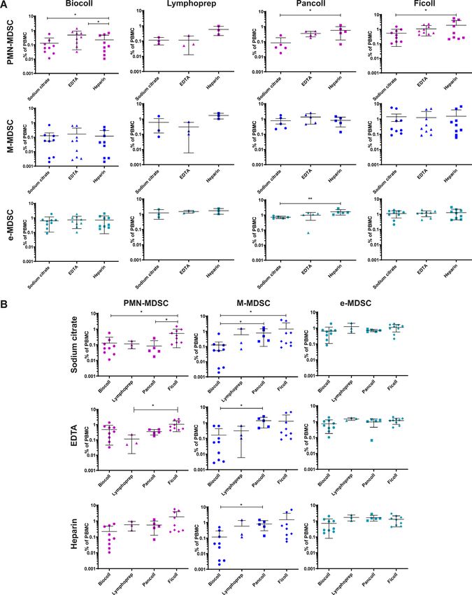
Open access
J Immunother Cancer: first published as 10.1136/jitc-2020-001223 on 8 September 2020. Downloaded from http://jitc.bmj.com/ on January 29, 2022 by guest. Protected by copyright.
nitric oxide synthase, indoleamine dioxygenase, cycloox- patients recruited were chemotherapy and radiotherapy
ygenase and reactive oxygen species.5 naive before collection. All control samples are age
Bronte et al published in 2016 an extensive paper matched and sex matched to the patient group.
containing recommendations for the study of human and
mouse MDSC; the paper provided a useful list of markers, Lab 3 (Nijmegen, The Netherlands)
gating strategies and functional tests to be used for MDSC Peripheral blood samples were collected from healthy
study.4 This was definitely one of the first solid milestones individuals and glioma patients undergoing neurosur-
in MDSC study standardization. However, still there is the gical resection or biopsy for intracranial tumors at the
need to further refine the process by comparing MDSC Radboud University Medical Center (Radboudumc).
frequencies and functions across multiple diseases. A Glioma patients receive dexamethasone as part of
significant problem in the field is the lack of robust and their treatment. All patients had histologically proven
reproducible consensus protocols and markers that would brain tumors diagnosed by neuropathologists of the
allow the comparative analysis of MDSC in different Radboudumc. The tumors were classified according
disease settings and multicenter trials. to WHO 2016 Classification of tumors of the Central
In order to overcome these limitations, the authors of Nervous System, and encompassed low- grade diffuse
this paper formed a study group within the COST Action astrocytoma, isocitrate dehydrogenase (IDH)- mutant
Mye-EUNITER (www.mye-euniter.eu). The main goal of (WHO grade II), oligodendrogliomas, IDH-mutant and
Mye-EUNITER was to further standardize and facilitate 1 p/19q-codeleted (WHO grade II, WHO grade III), and
the study of human MDSC in multiple human diseases. glioblastomas (grade IV). The healthy donors were anon-
Within this network, we developed an international ymous and not age and sex matched.
research program (Mye- EUNITER MDSC Monitoring
Initiative, Mye-MMI), which involved 13 European labs Lab 4 (Heidelberg, Germany)
and performed the first pan-European cross comparative Peripheral blood samples (30 mL) were obtained from
study on human MDSC, with almost 400 patients analyzed. 21 melanoma patients of stage I–IV who were seen at the
This study uses a consensus standard operating procedure Skin Cancer Center (University Medical Center Mann-
(SOP)-like protocol for the isolation and phenotyping of heim, Germany) from February 2017 to March 2018.
human circulating MDSC. Analysis of peripheral blood Tumor stages were determined according to the eighth
mononuclear cells (PBMC) from healthy donors showed edition of the American Joint Committee on Cancer
that technical variables, such as the anticoagulant and (AJCC) classification. All the patients recruited were not
separation method used, cause artifactual variabilities in treated within the last 6 months before blood collection.
the frequency of MDSC. When using an optimized and For control samples, mononuclear cells were isolated
standardized protocol and comparing results to healthy from peripheral blood obtained from the Institute of
control subjects, we observed a significant expansion of Transfusion Medicine and Immunology, Medical Faculty
PMN-MDSC in solid cancers that was less pronounced Mannheim, Heidelberg University, German Red Cross
in non-malignant chronic infectious and inflammatory Blood Service Baden Württemberg–Hessen (Mannheim,
disease settings. Expression of programmed death-ligand Germany) after informed consent. All control samples
1 (PD-L1) was mainly observed in M-MDSC and e-MDSC, are age matched to the patient group.
while PMN- MDSC express Lectin- like oxidized low- Lab 5 (Belgrade, Serbia)
density lipoprotein receptor-1 (LOX-1). No blood samples from lab five are presented in this
manuscript.
METHODS Lab 6 (Ankara, Turkey)
Human blood collection and description of patient cohorts Peripheral blood samples (~10 mL) were obtained from
Lab 1 (Essen, Germany) healthy volunteers, and newly diagnosed, treatment-naïve
Peripheral blood was prospectively collected from colorectal cancer patients. All control samples are age
treatment- naïve patients with primary head and neck matched and sex matched to the patient group.
cancer (HNC) and from healthy donors. Patients with
synchronous carcinoma in another location or concomi- Lab 7 (Warsaw, Poland)
tant systemic infectious disease were excluded. All control For control samples, mononuclear cells were isolated from
samples are age matched to the patient group. peripheral blood obtained from female healthy individ-
uals from Military Institute of Medicine. Peripheral blood
Lab 2 (Edinburgh, UK) (10 mL) was obtained from ovarian cancer patients from
Peripheral blood (10 mL) was obtained from breast Military Institute of Medicine (Warsaw, Poland). The
cancer patients from National Health Service, Edinburgh, exclusion criteria for all patients with cancer at baseline
Scotland, UK. The exclusion criteria for all patients with included systemic metastatic disease, any inflammatory
cancer at baseline included systemic metastatic disease, disorder and active infection or immunocompromised
any inflammatory disorder and active infection or immu- status not related to cancer. All the patients recruited were
nocompromised status not related to cancer. All the chemotherapy and radiotherapy naive before collection.
2 Cassetta L, et al. J Immunother Cancer 2020;8:e001223. doi:10.1136/jitc-2020-001223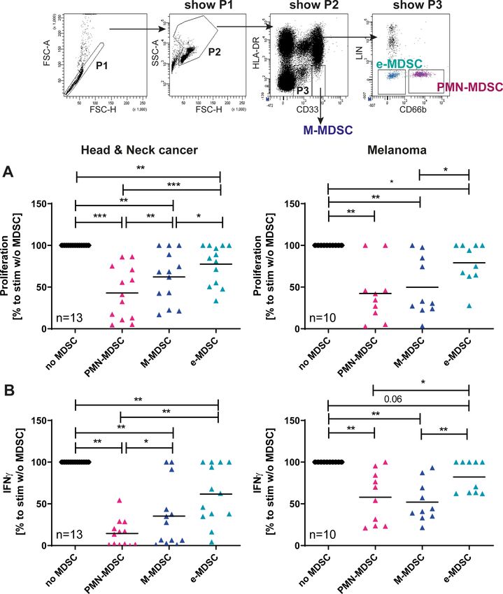
Open access
J Immunother Cancer: first published as 10.1136/jitc-2020-001223 on 8 September 2020. Downloaded from http://jitc.bmj.com/ on January 29, 2022 by guest. Protected by copyright.
from Dept. of Dermatology, Zeromski Hospital, Krakow,
Poland. The severity of the psoriatic skin lesions was
assessed according to the Psoriasis Area Severity Index
score. Patients on UV therapy, systemic or local corticoste-
roid treatment were excluded from the studies. Healthy
control subjects had no clinical signs of dermatologic
or inflammatory diseases. All control samples are age
Figure 1 Schematic representation of the Mye-MMI study. matched to the patient group.
See manuscript text for details on the different phases and
steps. The SOP-like protocol for MDSC immunophenotyping Lab 12 (Stockholm, Sweden)
that was generated in the context of this study is available Healthy adults were intramuscularly vaccinated with one
from the corresponding author on reasonable request. dose of the yellow fever vaccine (Stamaril, Sanofi) or the
MDSC, Myeloid-derived suppressor cells; Mye-MMI, Mye-
tick-
borne encephalitis vaccine (Encepur, GlaxoSmith-
EUNITER MDSC Monitoring Initiative; PD-L1, programmed
death-ligand 1; SOP, standard operating procedure.
Kline). Peripheral venous blood was collected at the day
prior to vaccination and at day 2 after vaccination. Prevac-
cination controls and postvaccination samples from the
All control samples are age matched and sex matched to same individuals were compared.
the patient group.
Lab 13 (Bergen, Norway)
Lab 8 (Essen, Germany) Peripheral blood was collected from patients with acute
For control samples, mononuclear cells were isolated chest pain admitted at Haukeland University Hospital
from the peripheral blood obtained from healthy volun- (Bergen, Norway). The patients were included consec-
teers at the Institute of Virology with their agreement to utively in two cohorts. We included subjects above 18
use the blood for the Mye-MMI study. Peripheral blood years of age who were hospitalized due to recent-onset
(10 mL) was obtained from non- treated patients with chest pain of suspected cardiac origin and who received
hepatitis B virus (HBV) infection from Clinic for Gastro- acetylsalicylic acid during ambulance transport or when
enterology and Hepatology, University Hospital Essen. All arriving at the hospital, on suspicion of cardiovascular
control samples are age matched to the patient group. disease (CVD). Patients who were on immunomodu-
lating medications, for example, corticosteroids, or with
Lab 9 (Paris, France)
severe infections, were excluded from the study. Patients
For the study on HIV-1 infection, peripheral blood was
in whom CVD was confirmed were defined as acute chest
collected on heparin from eleven antiretroviral treated,
pain patients with CVD. The patients where no CVD could
aviremic individuals living with HIV and from eleven
be detected were defined as acute chest pain patients
healthy donors. The inclusion criteria for the people living
without CVD.
with HIV-1 were as follows: plasma HIV RNA levels of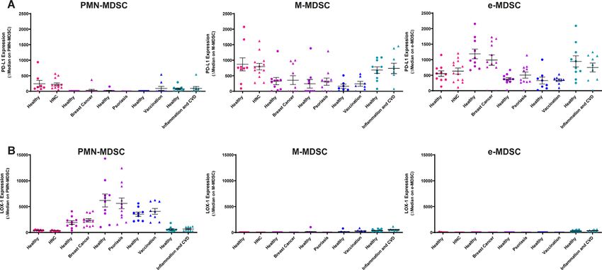
Open access
J Immunother Cancer: first published as 10.1136/jitc-2020-001223 on 8 September 2020. Downloaded from http://jitc.bmj.com/ on January 29, 2022 by guest. Protected by copyright.
were washed by filling the tube up with PBS followed by CD28.2 Beckman coulter) in L- lysine and L-arginine free
centrifugation at 300xg for 8 min at room temperature RPMI medium (Thermo Fisher Scientific) supplemented with
until the supernatant was clear. After the last washing 150 µM L-Arginine, 0.218 mM L-Lysine hydrochloride (both
step, cells were resuspended in Roswell Park Memorial Sigma-Aldrich, Taufkirchen, Germany), 100 IU/mL penicillin,
Institute (RPMI) 1640 culture medium supplemented 100 mg/mL streptomycin and 10% (v/v) heat-inactivated FCS.
with 100 IU/mL penicillin, 100 mg/mL streptomycin Autologous MDSC-subsets were added in a T-cell: MDSC ratio
and 10% (v/v) heat- inactivated FCS for counting. To of 2.5:1. CPDye405 intensity was analyzed by flow cytometry
study the influence of the anticoagulants all participants after 4 days of coculture and proliferation. Supernatants of
received the same batch of heparin, trisodium citrate or the coculture were collected and IFN-γ measured by ELISA
EDTA collection tubes and Safety-Multifly-needle 21G (all according to manufacturer’s protocol.
Sarstedt, Nümbrecht, Germany) that were shipped from
the core lab (lab 1) to the participating centers (phase Patient and public involvement
IIA, figure 1). Patients were not involved in the design of the study and the
In phase III of the study, the samples were collected in analysis and discussion of the data. The background of this
a harmonized fashion using trisodium citrate as anticoag- study and the overall concept of Mye-EUNITER Initiative
ulant and using Biocoll as the separation medium. are shared with the general public by two publicly available
videos. Videos are currently available at www.Mye-EUNITER.
Immunophenotyping of MDSC subsets by flow cytometry eu.
For characterization of MDSC subsets in phase I and II of Analysis and statistics
the study, all participants used the same antibody clones with Data from all participants were collected, uploaded to a
free choice of fluorochromes. In brief, 1×10⁶ cells in a total protected data server and centrally analyzed in the core lab
volume of 50 µL were stained with CD15 clone HI98, CD14 (Essen, Germany). GraphPad Prism Software (GraphPad Soft-
clone MOP9, CD33 clone WM53, CD11b clone ICRF44, ware, La Jolla, California, USA) was used for statistical analysis
HLA-DR clone G46-6n and lineage cocktail including CD3 and significance was assessed with Mann-Whitney, Wilcoxon
clone SK7, CD19 clone H1B19, CD20 clone 2H7 (not manda- matched-pairs signed rank or Kruskal-Wallis test. Results were
tory) and CD56 clone NCAM16.2 in the presence of FcR considered significant at *p≤0.05, **p≤0.01 and ***p≤0.001.
block in Fluorescence-activated cell sorting (FACS) buffer
(PBS/2% bovie serum albumine (BSA)) for 20 min at 4°C. RESULTS
FMO control was used for CD11b and HLA-DR. Cells were Development of consensus protocols and execution of training
washed with FACS buffer, centrifuged at 400xg for 5 min and schools
resuspended in 200 µL FACS buffer for acquisition. Within this network, we developed an international research
In phase III, participants included the functional marker program (Mye- MMI), which involved 13 European labs.
PD-L1 clone 29E.2A3 and LOX-1 clone 15C4. Isotypes were The Mye-MMI study was divided into three phases: during
used as negative controls. Isolation of PBMC, MDSC anti- phase I participants designed and agreed to use a SOP for
body labeling and data acquisition was locally performed in the immunophenotyping and analysis of MDSC in the blood
the participating centers. Because of technical constraints of human patients and controls (Phase IA, figure 1) The Mye-
with multiparameter options at local flow cytometers and MMI network organized a training school, where scientists
because of limited choices of commercially available fluo- from all participating labs executed the SOP and trained
rochromes for LOX-1, CD11b could not be included in the together in the core lab (Essen, Germany) using the same
second step. Locally acquired data files were uploaded to a samples; the outputs of the training school indicated that the
protected data server and centrally analyzed in the core lab SOP was reproducible and the variance among users minimal
(Essen, Germany) to ensure uniform conditions for gating (phase IB, figure 1). The training school also provided all
for all samples from all centers. participants with practical and hands-on experience with the
SOP. T cell suppression is a hallmark of MDSC function. We
T cell suppression assay used a previously published protocol to assess the suppres-
For validation of MDSC suppressive capacity, we used a previ- sion of cancer patient-derived T cells by autologous circu-
ously published protocol of flow cytometry MDSC sorting lating MDSC.6 This protocol was trained in the Essen core
and suppression of polyclonally stimulated autologous cancer lab with selected participants and subsequently used for func-
patient-derived T cells.6 In brief, CD3 depleted PBMC from tional validation using patients with HNC and patients with
cancer patients were labeled with CD66b clone 80H3, CD33 melanoma. All these steps enabled the network to perform
and HLA-DR and sorted on lowest flow rate for HLA-DR-/ the study in different labs and geographical locations using
CD33high (M-MDSC), HLA-DR-/CD33dim/CD66b+ (PMN- a commonly agreed SOP for immunophenotyping and
MDSC), HLA-DR-/CD33dim/CD66b- (e-MDSC). common assays validating MDSC function.
The positively selected CD3 T Lymphocytes were labeled with
10 µM Proliferation Dye eFluor 450 (CPDye405) according to Expansion of circulating MDSC subsets in cancer exceeds
manufacturer instructions (eBioscience, Frankfurt am Main, expansion in infectious and inflammatory diseases
Germany). T cells were stimulated with coated CD3 (1 µg/ After development of an initial consensus protocol for the
mL, clone OKT-3, eBioscience) and CD28 (2 µg/mL, clone immunophenotyping of circulating MDSC and execution
4 Cassetta L, et al. J Immunother Cancer 2020;8:e001223. doi:10.1136/jitc-2020-001223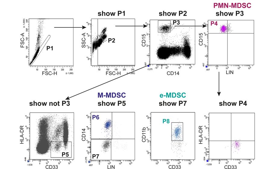
Open access
J Immunother Cancer: first published as 10.1136/jitc-2020-001223 on 8 September 2020. Downloaded from http://jitc.bmj.com/ on January 29, 2022 by guest. Protected by copyright.
Figure 2 Flow cytometry gating of the three major subsets of human circulating MDSC. PBMCs were isolated from the
peripheral blood of patients and healthy donor controls by density gradient centrifugation in 12 different participating centers
and according to a harmonized protocol. Use of predefined clones was mandatory. In a centralized analysis, MDSC subsets
were classified as CD15+/CD14-/CD33dim/HLA-DRneg=PMN-MDSC (P4), CD15-/CD14+/CD33pos/HLA-DRneg=M-MDSC (P6)
and CD15-/CD14-/CD11b+/CD33dim/HLA-DRneg as e-MDSC (P8).4 An example for the gating strategy is shown (head and neck
cancer patient). Note that all MDSC subsets are negative for the lymphocyte lineage markers CD3, CD19 and CD56 (CD20 not
mandatory). MDSC, myeloid-derived suppressor cells; PBMCs, peripheral blood mononuclear cells; PMN, polymorphonuclear.
of a training school, the trained researchers performed In most cancer types, we observed a substantial induction of
MDSC immunophenotyping in their own Centers (phase PMN-MDSC frequency over healthy controls (figure 3A). No
IC, figure 1). In each center, a particular cancer type or statistically significant increase of PMN-MDSC was observed in
infection or inflammatory disease was investigated. Data patients with infectious and inflammatory diseases, although
files (.fcs format) from all patients and control subjects were a tendency for higher frequency of PMN-MDSC was observed
uploaded to a protected server at the coordinating Center in some diseases, in agreement with previous reports.7 In
(Essen) and centrally analyzed. Figure 2 shows an example of patients with cancer, the induction of the PMN-MDSC subset
the employed gating strategy. Based on previous experience exceeded expansion of other MDSC subsets (figure 3A).
in the participating centers, we derived a sequential gating When compared with healthy donors the PMN- MDSC
strategy that identifies the three major human circulating frequency was induced in 5/6 cancer types (figure 3B). In
MDSC subsets with no overlap between subsets. In a first step, contrast, M-MDSC frequency was only significantly induced
we determined the frequency of PMN-MDSC, M-MDSC and in glioma or was even reduced in breast cancer. A particular
e-MDSC in six different types of cancer, major viral infections, induction of M-MDSC was observed in patients with inflam-
psoriasis, a mixed cohort of patients with inflammatory CVDs matory CVD and after vaccination confirming previous find-
and in patients that received the yellow fever or the tick-borne ings,8 but did not change in any other conditions. With the
encephalitis vaccine. In each center, an independent group exception of glioma and viremic HIV-1, e-MDSC frequencies
healthy donor controls was analyzed (see online supplemen- were not significantly altered between healthy donors and
tary table S1 for patient and healthy donor characteristics). patients.
At this stage, the anticoagulant and the blood separation Although our study was focused on the immunomon-
medium were free of choice to the lab. itoring and immunophenotpying of MDSC in different
Cassetta L, et al. J Immunother Cancer 2020;8:e001223. doi:10.1136/jitc-2020-001223 5Open access
J Immunother Cancer: first published as 10.1136/jitc-2020-001223 on 8 September 2020. Downloaded from http://jitc.bmj.com/ on January 29, 2022 by guest. Protected by copyright.
Figure 3 Frequency of the three MDSC subsets in malignant and non-malignant disease. For each center the frequency of the
three MDSC subsets was determined for patients and the respective local healthy controls. Staining procedure and gating were
performed according to figure 1. (A) Median frequency of MDSC compared with healthy donor’s controls (set as value 1) and
(B) total frequency within the PBMC are shown. Mean values with SD are shown. Mann-Whitney U test was used for statistical
analysis. Results were considered significant at *p≤0.05, **p≤0.001 and ***p≤0.0001. CVD, cardiovascular disease; HBV,
hepatitis B virus; MDSC, Myeloid-derived suppressor cells; PBMCs, peripheral blood mononuclear cells.
diseases, we sought to test and confirm the T cell suppressive and e-MDSC in suppressing the function of polyclonally stim-
activity of the MDSC subsets under investigation. Employing ulated autologous T cells (figure 4).
previously published protocols6 we tested the immunosup-
pressive capacity of MDSC isolated from patients with HNC Identification of technical and experimental variables that
and melanoma using identical protocols after execution of influence immunomonitoring of MDSC
a training school. In line with earlier findings,6 and in both Intercenter analysis of data presented in figure 3 revealed
tumor entities tested, PMN-MDSC were superior to M-MDSC an interesting variability in the frequency of PMN-MDSC
6 Cassetta L, et al. J Immunother Cancer 2020;8:e001223. doi:10.1136/jitc-2020-001223Open access
J Immunother Cancer: first published as 10.1136/jitc-2020-001223 on 8 September 2020. Downloaded from http://jitc.bmj.com/ on January 29, 2022 by guest. Protected by copyright.
Figure 4 Suppressive activity of MDSC subsets. Suppressive activity of the MDSC subsets was confirmed in two independent
laboratories using a shared and previously published protocol.6 (A) Cell proliferation dye labeled Responder T cells from patients
were stimulated with plate-bound CD3 and CD28 mAb in the presence or absence of autologous MDSC (T cell: MDSC ratio of
2.5:1). Lymphocyte proliferation was measured at day 4 and analyzed centrally. Relative proliferation to stimulated T cell without
MDSC (set as 100%) and mean values are shown. (B) Levels of IFNγ were determined in supernatants of cocultures consisting
of T cells activated by plate-bound CD3/CD28 with and without additions of MDSC. IFNγ was determined after 4 days by
ELISA. Relative release was calculated to stimulated T cells without MDSC. Mean values are shown. Wilcoxon signed-rank test
was used for statistical analysis. Results were considered significant at *p≤0.05, **p≤0.001 and ***p≤0.0001. Gating strategy
data are from a patient with head and neck cancer. IFNγ, interferon-γ; M-MDSC, monocytic myeloid-derived suppressor cells;
PMN, polymorphonuclear.
Cassetta L, et al. J Immunother Cancer 2020;8:e001223. doi:10.1136/jitc-2020-001223 7Open access
J Immunother Cancer: first published as 10.1136/jitc-2020-001223 on 8 September 2020. Downloaded from http://jitc.bmj.com/ on January 29, 2022 by guest. Protected by copyright.
in healthy donors (figure 3B, upper panels). Frequency PD-L1 is primarily expressed in mononuclear MDSC and is not
of this MDSC subset in healthy donors varied between upregulated as a consequence of disease
0.08% (lab 1) and 1,8% (lab 3) in the cancer Centers and LOX-1 has been suggested as a marker associated with
between 0.3% (lab 11) and 1.9% (lab 12) in the infection suppressive activity of PMN-MDSC.9 PD-L1 is expressed on
& inflammation Centers. We considered it unlikely that myeloid cells and considered as an important biomarker
this difference is based on biological differences between and functional target in current immunotherapies.10
healthy donors in the respective Centers. A reanalysis of In the final step of our study (phase IIIB, figure 1), we
the underlying data and protocols further revealed that, tested the expression of both markers in five participating
despite standardized immunolabelling and analysis, the centers.
participating centers used different anticoagulants and Quantitative analysis of expression levels showed that
separation media for isolation of MDSC from venous PD-L1 is primarily expressed on M-MDSC and e-MDSC
blood of healthy donors and patients. In order to test for (figure 7 and online supplementary figure S1). In contrast,
a potential effect of the anticoagulant (sodium citrate, PD-L1 is absent or expressed at very low levels on PMN-
EDTA, heparin), we purchased the three types of blood MDSC. As expected, LOX-1 is expressed on PMN-MDSC
collection tubes at the coordinating Center (lab 1) and and absent on M-MDSC and e-MDSC (figure 7B, online
shipped them to eight participating Centers for further supplementary figure S2). It is important to note that in
use (phase IIA, figure 1). Centers obtained venous blood all tested diseases and conditions both markers were not
from healthy donors into three different collection tubes upregulated in patients over healthy controls.
and used their preferred separation medium for MDSC
isolation. MDSC labeling was performed according to the
standardized consensus protocol and data files were sent DISCUSSION
to the coordination Center for centralized analysis. Anal- Since their initial identification, MDSCs have received substan-
ysis of MDSC frequencies showed that the type of antico- tial consideration from immunologists, who performed
agulant had little influence on the frequency of M-MDSC several studies to elucidate their immunopathological role
and e-MDSC. In contrast, a clear increase in PMN-MDSC in cancer, inflammation and infectious diseases.11–14 In addi-
frequency was observed when heparin was compared with tion, MDSCs are considered as major cellular mediators of
sodium citrate (figure 5A). This increase was observed resistance to cancer immunotherapy and may serve as poten-
for all types of separation medium. In order to test for a tial future biomarkers to predict response to conventional
potential effect of the separation medium, we reanalyzed and immune-based cancer therapy.15 Thus, precise and reli-
the data of healthy donors with the separation medium able immunomonitoring of MDSC, even in intercenter anal-
being the variable factor (figure 5B). Results revealed yses, is of utmost importance.
an increased frequency of PMN-MDSC when Ficoll was Human circulating MDSCs are normally isolated from
whole blood after density gradient centrifugation and subdi-
used as the separation medium (figure 5B, left column).
vided into at least three different subsets (granulocytic PMN-
Samples prepared via Biocoll showed the lowest frequency
MDSC, monocytic M- MDSC and myeloid precursor- like
of M-MDSC in healthy donors. For PMN-MDSC Biocoll
e-MDSC) using flow cytometry.6
and Lymphoprep showed the lowest frequencies. Based
The main limitation of the current studies on human
on these data we concluded that a combination of Biocoll
MDSC lies in the extreme variability in the protocols used to
and sodium citrate would provide lowest frequencies of
extract, identify and phenotype these cells. Variable isolation
MDSC in healthy donor blood samples; these data were,
procedures, antibody panels and gating strategies are used
therefore, used to refine the SOP by including precise
to isolate and characterize human MDSC. Previous reports
indications on the separation medium and anticoagulant
already highlighted the need for marker and gating harmo-
to be used (phase IIB, figure 1).
nization4 16 and outlined technical variables that could affect
MDSC immunomonitoring.17 A recommendation paper
Validation of the refined SOP and comparison with previous was published in order to suggest minimal characterization
dataset standards when researching MDSC.4 However, also in those
In order to confirm the utility of standardization of the reports no consensus strategy for MDSC preparation, isola-
isolation procedure, five participating Centers volun- tion and immunophenotyping is provided and an accepted
teered to repeat the analysis shown in figure 3 using the gating strategy for enumeration of non-overlapping human
refined SOP (phase IIIA, figure 1). Figure 6 shows the MDSC subsets does not exist until now. These obstacles make
level of variance in healthy donors for the original data it nearly impossible to directly compare MDSC data sets
(figure 3, phase IC) and the variance of data with the between published studies.
harmonized isolation procedure (phase IIIA). Data show In this study, we developed a consensus protocol for the
that choice and standardization of blood collection tubes isolation, identification and immunophenotypic charac-
and separation medium reduced PMN-MDSC frequency terization and analysis of human MDSC. After centralized
in healthy donors, further improved intercenter compara- hands-on training, this protocol was executed in the partic-
bility of results and reduced differences in MDSC counts ipating Centers, followed by a centralized flow cytometry
between healthy control cohorts from different Centers. analysis that utilized a uniform gating strategy. Our initial
8 Cassetta L, et al. J Immunother Cancer 2020;8:e001223. doi:10.1136/jitc-2020-001223Open access Figure 5 Identification of further technical variables in MDSC immunomonitoring. Participating centers stained for MDSC J Immunother Cancer: first published as 10.1136/jitc-2020-001223 on 8 September 2020. Downloaded from http://jitc.bmj.com/ on January 29, 2022 by guest. Protected by copyright. subsets in healthy donor controls. Beforehand every participant received the identical batch of blood collection tubes. From the same donor sodium citrate-, EDTA and heparin blood was collected and PBMC were stained for MDSC subsets in PBMC. Lymphocyte separation medium was free of choice. MDSC frequency was determined in the core lab as described for figure 2. Influence of anticoagulants (A) and lymphocyte separation medium (B) on total frequency of putative ‘MDSC subsets’ in healthy individuals with mean and SD is shown. Kruskal-Wallis was used for statistical analysis. Results were considered significant at *p≤0.05, **p≤0.001 and ***p≤0.0001. M-MDSC, monocytic myeloid-derived suppressor cell; PBMC, peripheral blood mononuclear cell; PMN, polymorphonuclear. analysis on malignant and non- malignant infectious/ per cancer type, and consistent to what is already reported inflammatory disease patients revealed that PMN- MDSC in the literature.6 18–22 No statistically significant induc- are significantly expanded in malignant disease with 5/6 tion of PMN-MDSC frequency was found in melanoma. cancer types (ie, Glioma as well as head and neck, breast, Interestingly, and in contrast to cancer, this induction of colorectal and ovarian cancer) showing statistically signifi- PMN-MDSC was not observed in patients with infection cant upregulation even with our relatively small sample size and inflammation. For M-MDSC and e-MDSC we did not Cassetta L, et al. J Immunother Cancer 2020;8:e001223. doi:10.1136/jitc-2020-001223 9
Open access
J Immunother Cancer: first published as 10.1136/jitc-2020-001223 on 8 September 2020. Downloaded from http://jitc.bmj.com/ on January 29, 2022 by guest. Protected by copyright.
Figure 6 Standardization of anticoagulant and separation medium reduces intercenter variability of PMN-MDSC frequencies.
All participants used sodium citrate as anticoagulant and Biocoll as separation medium and determined the frequency of
PMN-MDSC in healthy donor controls. Data were compared with data obtained for figure 2. (A) Frequency of PMN-MDSC in
Step1 (figure 2, free choice of anticoagulant and separation medium) and step 2 (sodium citrate and Biocoll) from all healthy
blood donors in the five centers is shown (plus mean and SD). (B) The mean frequency was determined for each of the five
participating centers and (C) % cv was calculated. In all panels the comparison to step 1 (data from figure 2) is shown. F test
was used for statistical analysis. Results were considered significant at *p≤0.05 and ****p≤0.0001. PBMC; peripheral blood
mononuclear cell.
find such significant changes in most disease entities. We MDSC subset in infectious and inflammatory diseases and
acknowledge that the sample size could be responsible for also exceeds the relative expansion of M-MDSC in cancer.
the lack of statistically significant expansion of MDSC in These data partially challenge previously published data
some types of disease. Nevertheless, the comparative pan- that reported robust MDSC expansion in infections and
disease analysis in our study unequivocally shows that PMN- non-malignant inflammatory diseases such as psoriasis,
MDSC expansion in solid cancers exceeds expansion of this HIV, HBV and CVDs.23–27
Figure 7 Expression of molecules associated with T cell suppression expression of PD-L1 and LOX-1 on MDSC subsets in
patients and healthy donor controls was determined using the harmonized flow cytometry labeling protocol, Biocoll as standard
separation medium and sodium citrate as standard anticoagulant. Flow cytometry analysis was performed in the core lab to
ensure standardized gating. Staining intensity of PD-L1 (A) and LOX-1 (B) on MDSC-subsets was determined in five different
disease settings. Delta median (median signal intensity of antibody minus median signal intensity of isotype control) is shown.
Data are depicted as mean and SD is shown. CVD, cardiovascular disease; M-MDSC, monocytic myeloid-derived suppressor
cells; PD-L1, programmed death-ligand 1; PMN, polymorphonuclear.
10 Cassetta L, et al. J Immunother Cancer 2020;8:e001223. doi:10.1136/jitc-2020-001223Open access
J Immunother Cancer: first published as 10.1136/jitc-2020-001223 on 8 September 2020. Downloaded from http://jitc.bmj.com/ on January 29, 2022 by guest. Protected by copyright.
In this context, it is interesting to note the very divergent PD-L1 is a promising target for checkpoint blockade
levels of MDSC expansion reported in the literature. The use immunotherapy. From our data we may assume that this
of different methodologies and techniques to isolate MDSC type of immunotherapy will primarily co-target M-MDSC
could potentially lead to technical, rather than biologically in cancer patients. Most likely, additional and distinct ther-
caused differences among labs. It is a particular strength of apeutic interventions will be required to target human
our study that, by using a SOP-like protocol, harmonized PMN-MDSC. In the context of these considerations, we
reagents and antibodies, core Center analysis and a uniform are convinced that the use of standardized protocols for
gating strategy, we were able to facilitate a comparative anal- immunomonitoring of MDSC subsets and their associ-
ysis of human MDSC expansion across many different disease ated functional molecules will aid future patient selection
types. and stratification for treatment.
Our study also revealed the importance of further tech-
nical variables during blood collection and separation.
These findings were triggered by initial observations that CONCLUSION
showed substantial differences in the frequency of MDSC In summary, our optimized study design allows us to
in healthy donors across the different Centers (see PMN- conclude that expansion of PMN-MDSC exceeds expan-
MDSC frequencies in healthy donors in figure 3B). These sion of other MDSC subsets in cancer and is more
differences suggested that additional technical variables, pronounced in solid tumors as opposed to various
not standardized in the original SOP, could affect the infectious and inflammatory diseases. In addition, we
results. We then decided to analyze the anticoagulant and report several technical and analytical aspects that will
gradient separation medium variables, by performing guide the future analysis of MDSC, an important poten-
additional experiments; results indicated that different tial cellular resistance mechanism in current cancer
anticoagulants and separation media can alter the base- immunotherapies.
line frequency of MDSC in healthy donors. In particular
the combination of sodium citrate as anticoagulant and Author affiliations
1
MRC Centre for Reproductive Health, The University of Edinburgh The Queen's
Biocoll as stratification solution allowed for the lowest
Medical Research Institute, Edinburgh, UK
MDSC frequencies in healthy donors. 2
Department of Otorhinolaryngology, University Hospital Essen, Essen, Germany
Anticoagulants can affect the frequency of MDSC in 3
Department of Immunology, Faculty of Biochemistry, Biophysics and Biotechnology,
human blood as already reported by Apodaca et al; the Jagiellonian University, Krakow, Małopolska, Poland
4
authors compared the total number of M-MDSC in blood Clinical Cooperation Unit Dermato-Oncology, DKFZ, Heidelberg, Baden-
Württemberg, Germany
of healthy donors collected using EDTA or Heparin 5
Department of Dermatology, Venereology and Allergology, University Medical
and observed a significant difference between the two Centre Mannheim, Mannheim, Baden-Württemberg, Germany
anticoagulants.17 6
Department of Biomedical Laboratory Scientist Education and Chemical
We included these two additional standardized variables Engineering, Faculty of Engineering and Natural Sciences, Western Norway
in the SOP and repeated the experiments in a selected University of Applied Sciences, Bergen, Hordaland, Norway
7
Division of Immunology and Allergy, Department of Medicine Solna, Karolinska
number of centers, showing a reduced degree of variance
Institute, Stockholm, Stockholm, Sweden
among healthy donors across centers. It is worth noting 8
Center for Molecular Medicine, Karolinska Institute, Stockholm, Stockholm,
that by this approach we obtained PMN-MDSC frequen- Sweden
cies well below 1% in four out of five Centers. Again, a 9
Medical Center, Radiotherapy & OncoImmunology Laboratory, Department of
great variability in MDSC, and in particular PMN-MDSC Radiation Oncology, Radboud University, Nijmegen, Gelderland, The Netherlands
10
Department of Basic Oncology, Cancer Institute, Hacettepe University, Ankara,
frequency is reported in the literature.4 17 28
Ankara, Turkey
In the final phase of our study, we also evaluated the 11
Instituto de Medicina Molecular João Lobo Antunes, Faculdade de Medicina,
expression of PDL1 and LOX-1, two surrogate suppressive University of Lisbon, Lisboa, Lisboa, Portugal
functional markers associated with M-MDSC and PMN- 12
Institute for Virology, University Hospital Essen, Essen, Nordrhein-Westfalen,
MDSC, respectively. Our data confirmed the restricted Germany
13
HIV Inflammation and Persistence, Pasteur Institute, Paris, Île-de-France, France
expression of LOX-1 on PMN- MDSC as previously 14
Department of Molecular Oncology, Institute for Medical Research, University of
reported for circulating PMN-MDSC9 and PMN-MDSC in Belgrade, Beograd, Beograd, Serbia
cancer tissues.29 On a per cell basis the expression level of 15
Department of Cancer Biology, Institute of Biology, Warsaw University of Life
LOX1 was not different between PMN-MDSC extracted Sciences, Warszawa, Poland
16
from healthy donor controls and cancer patients. It is, Cellis AG, Zurich, Switzerland
17
Department of Clinical Science, University of Bergen, Bergen, Hordaland, Norway
however, important to note that LOX1- positive PMN- 18
Edinburgh Breast Unit and Breast Cancer Now Research Unit, The University of
MDSC were substantially expanded in cancer patients Edinburgh, Edinburgh, UK
over healthy controls. This is also in line with previous 19
Clinical Cooperation Unit Dermato-Oncology, German Cancer Research Centre,
studies9 that showed an expansion of this LOX-1-positive Heidelberg, Baden-Württemberg, Germany
20
subset in the unseparated blood of cancer patients. In Department of Medical and Surgical Research, Institute of Health Sciences,
Hacettepe University, Ankara, Ankara, Turkey
contrast, strong expression of PD- L1 was restricted to 21
Department of General Surgery, Faculty of Medicine, Hacettepe University, Ankara,
M-MDSC and e-MDSC and similar to LOX-1, on a per Ankara, Turkey
cell basis, the expression was not substantially induced in 22
Department of General Surgery, Gulhane Egitim ve Arastirma Hastanesi, Ankara,
patients with cancer. Ankara, Turkey
Cassetta L, et al. J Immunother Cancer 2020;8:e001223. doi:10.1136/jitc-2020-001223 11Open access
J Immunother Cancer: first published as 10.1136/jitc-2020-001223 on 8 September 2020. Downloaded from http://jitc.bmj.com/ on January 29, 2022 by guest. Protected by copyright.
23
Department of Oncology and Immunooncology, Hospital Ministry of the Interior from the Serbian Ministry of Education, Science and Technological Development
and Administration & Warmia and Masuria Oncology Centre, Olsztyn, Poland (451-03-68/2020-14/200015)'. Lab6: This work was partially supported by The
24
Department of Oncology, University of Warmia and Mazury in Olsztyn, Olsztyn, Scientific and Technological Research Council of Turkey, (TÜBİTAK; project no.
Poland 115S636) and covered under the European Cooperation in Science and Technology
25
Center for Immunology of Viral Infections and Autoimmune Diseases, IDMIT (COST-EU) Action BM1404 (Mye-EUNITER). Lab7: This result is part of a project
Department, IBFJ, CEA, Université Paris-Sud, Saint-Aubin, Île-de-France, France that has received funding from the European Research Council (ERC) under the
26
Serviço de Doenças Infecciosas, Northern Lisbon University Hospital Centre, European Union’s Horizon 2020 research and innovation programmeprogram (Grant
Lisboa, Lisboa, Portugal agreement No. 715048). Lab9: TG-T was recipient of a fellowship from the Pasteur
27
Department of Dermatology, Specialised Hospital of Stefan Zeromski in Krakow, Paris University International PhD program and and Institut Carnot Microbes et
Krakow, Poland Santé. This work was supported by Sidaction. Lab10: This work was funded by
28 the following grants: PTDC/MED-IMU/30474/2017 – project cofunded by FEDER
Clinic of Hematology, Clinical Center of Serbia, Beograd, Beograd, Serbia
29 LISBOA-01–0145-FEDER-030474, through Programa Operacional Regional de
Medical Center, Department of Neurosurgery, Radboud University, Nijmegen,
Lisboa, do PORTUGAL 2020, and Fundação para a Ciencia e a Tecnologia and by
Gelderland, The Netherlands
30 Gilead Genesis to AES. Lab 11: This work was supported by grants from Polish
Centro Integrativo de Biología y Química Aplicada (CIBQA), Universidad Bernardo
National Science Center UMO-2011/02/A/NZ5/00337 and UMO-2017/25/B/
O'Higgins, Santiago, Chile NZ6/01003 (to JC). Lab12: This study was funded by the Swedish Research Council
31
Department of Radiation Oncology, Radboud University Radboud Institute for (2019–01036). Lab13: This work was supported by grants given by Faculty of
Molecular Life Sciences, Nijmegen, The Netherlands Engineering and Natural Sciences, Western Norway University of Applied Sciences,
32
German Cancer Consortium, Partner Site Essen-Düsseldorf, Germany Norway.
Competing interests None declared.
Twitter Lubomir Bodnar @lbodnar
Patient consent for publication Not required.
Acknowledgements Lab1: We would like to thank Delia Cosgrove for her
enthusiastic organizational input and for support of this project. We also thank Ethics approval Lab1: The collection and use of peripheral blood cells was
the staff of the ORL department for their support during blood sample collection. approved by the ethics committee of the medical faculty of the University of
Lab2: The work was supported extensively by the Edinburgh Breast Unit team and Duisburg–Essen. Lab2: All study protocols were approved by The University of
particularly by Lorna Renshaw and Jane Keys in this unit. We would like to thank Edinburgh (Edinburgh, UK) ethics committees as appropriate. Lab3: Experiments
the CIR blood resource (AMREC #15-HV-013) for the recruitment of blood from were performed in accordance to the Helsinki Declaration and approved by the local
normal controls and the CIR flow facility (Shonna Johnston, Will Ramsay and Mari Medical Ethics Committee of the Radboudumc (registration number 2011/307).
Pattinson). Lab3: We would like to thank Sandra Bossmann en/of Fleur Brienen for Lab4: The study protocol was approved by the Institution's ethics committee (2010–
excellent support. Lab4: We thank the staff of the Core Facility Live Cell Imaging 318N-MA). Lab6: The study was approved by Hacettepe University and University
Mannheim and S. Uhlig from FlowCore Mannheim for help with the cell sorting. of Health Sciences - Diskapi Yildirim Beyazit Research and Training Hospital local
Lab6: We would like to thank all patients and nurses who contributed to the study, ethics committees and conducted in agreement with guiding principles of the
especially Nuraydın Sahin for collecting blood samples. Lab9: We would like to declaration of Helsinki and the good clinical practice. Lab7: All study protocols were
thank Beatrice Jacquelin for her contribution in supervising the PhD student and approved by Military Institute of Medicine (Warsaw, Poland) ethics committees as
obtention of financial support. We would like to thank all the members of the appropriate. Lab8: All study protocols were approved by University Hospital Essen's
Kremlin-Bicêtre hospital who helped for this study. Lab10: We would like to thank Ethics Committee (Approval Number 15–6495-BO) as appropriate. Lab9: Peripheral
the Infectious Diseases Department of Hospital de Santa Maria/CHULN/Lisboa blood samples from non-HIV-infected blood donors were obtained from the French
Portugal and all the patients for their collaboration, as well as the team members of blood bank (Etablissement Français du Sang) as part of an agreement with the
Flow Cytometry Unit at Instituto de Medicina Molecular Joao Lobo Antunes (Lisboa, Institut Pasteur (C CPSL UNT, number 15/EFS/023). Lab10: All study protocols were
Portugal) for technical assistance. Lab11: We would like to thank lab members approved by the Faculdade de Medicina da Universidade de Lisboa and Centro
that enrolled in this study as healthy donors. Lab12: We would like to thank Tyler Hospitalar Universitário Lisboa Norte (Lisbon, Portugal) ethics committees. Lab11:
Sandberg for the help in the recruitment of the healthy volunteers. Lab13: We would All study protocols were approved by the Jagiellonian University Institutional
like to thank all patients who contributed to the study. We would like to thank Aud Bioethics Committee. Lab12: The study was approved by the Stockholm Local
Valle Hansen at the Western Norway University of Applied Sciences for collecting Ethical Committee and was performed according to the Declaration of Helsinki
blood samples and Professor Øystein Bruserud at Department of Clinical Science, principles. Lab13: Both clinical protocols (phase I and II) were approved by The
University of Bergen, for kindly letting us use the BD FACS Verse flow cytometer Norwegian Regional Committee for Medical and Health Research Ethics (REK Vest
at the Leukemia Research Laboratory. Participants of this study also thank all 2017/49).
members of the Mye-EUNITER network for their scientific input and for fruitful Provenance and peer review Not commissioned; externally peer reviewed.
discussions.
Data availability statement Data are available on reasonable request.
Contributors Lab1 (SB, KB, BH and SL) coordinated the study, designed the Experimental data and protocols are available on reasonable request via the
SOP, participated to Phase I, II and III of the study and performed centralized data corresponding author. Reuse requires citation of this manuscript. Identifiable
analysis. Lab2 (LC, MD and JWP) coordinated the study, designed the SOP and patient information and data are not available, as patient material was used
participated to phase I, II and III of the study. Lab3 (GA, KS, EK-R and MtL) designed in an anonymous manner. The SOP-like protocol for MDSC immunomonitoring
the SOP and participated to phase I and II of the study. Lab4 (VU and XH) designed generated in the context of this study is available from the corresponding author on
the SOP and participated to phase I and II of the study. Lab5 (SB, MG and JFS) reasonable request.
contributed to phase I of the study.Lab6 (GE and UH) participated to phase I of the
study. Lab7 (BT, LB, EG and MK) participated to phase I of the study. Lab8 (GZ, Open access This is an open access article distributed in accordance with the
OEA and UD) participated to Phase I and II of the study. Lab9 (TG-T, B and MM-T) Creative Commons Attribution Non Commercial (CC BY-NC 4.0) license, which
participated to phase I of the study. Lab 10 (AG-S, RB and AEdS) participated in permits others to distribute, remix, adapt, build upon this work non-commercially,
Phase I and II of the study. Lab11 (JC, JS-M, OO and MK-M) participated to phase and license their derivative works on different terms, provided the original work is
I, II and III of the study. Lab12 (AL and KL) participated to phase I, II and III of the properly cited, appropriate credit is given, any changes made indicated, and the use
study. Lab13 (IMR, AOK and EE) participated to phase I, II and III of the study. is non-commercial. See http://creativecommons.org/licenses/by-nc/4.0/.
Funding Lab1: This research was supported by European Cooperation in ORCID iDs
Science and Technology (COST) Action Mye-EUNITER (BM1404) and by a grant Monika Kapinska-Mrowiecka http://orcid.org/0000-0001-8184-9265
from the Deutsche Forschungsgemeinschaft to SB (DFG, BR 2278/6–1). Lab2: Gosse Adema http://orcid.org/0000-0002-6750-1665
This research was supported by Wellcome Trust (101067/Z/13/Z), MRC Centre\ Sven Brandau http://orcid.org/0000-0002-2702-4163
grant MR/N022556/1 to JWP. Lab3: This work was supported by grants from the
STOPbraintumors Foundation, COST (European Union) and grants from the Dutch
Cancer Society awarded to GA and CBüll (KUN2015-7604) and GA, KS and P
Wesseling (KWF11266). Lab4: The work was supported by the grant from German
Research Council RTG2099/2 (JSU, VU). Lab5: Our work is supported by a grant
12 Cassetta L, et al. J Immunother Cancer 2020;8:e001223. doi:10.1136/jitc-2020-001223Open access
J Immunother Cancer: first published as 10.1136/jitc-2020-001223 on 8 September 2020. Downloaded from http://jitc.bmj.com/ on January 29, 2022 by guest. Protected by copyright.
REFERENCES 16 Mandruzzato S, Brandau S, Britten CM, et al. Toward harmonized
1 Talmadge JE, Gabrilovich DI. History of myeloid-derived suppressor phenotyping of human myeloid-derived suppressor cells by
cells. Nat Rev Cancer 2013;13:739–52. flow cytometry: results from an interim study. Cancer Immunol
2 Tesi RJ. Mdsc; the most important cell you have never heard of. Immunother 2016;65:161–9.
Trends Pharmacol Sci 2019;40:4–7. 17 Apodaca MC, Wright AE, Riggins AM, et al. Characterization of a
3 Cassetta L, Baekkevold ES, Brandau S, et al. Deciphering myeloid- whole blood assay for quantifying myeloid-derived suppressor cells.
derived suppressor cells: isolation and markers in humans, mice and J Immunother Cancer 2019;7:230.
18 Markowitz J, Wesolowski R, Papenfuss T, et al. Myeloid-derived
non-human primates. Cancer Immunol Immunother
suppressor cells in breast cancer. Breast Cancer Res Treat
2019;68:687–97.
2013;140:13–21.
4 Bronte V, Brandau S, Chen S-H, et al. Recommendations for
19 Walankiewicz M, Grywalska E, Polak G, et al. Myeloid-derived
myeloid-derived suppressor cell nomenclature and characterization suppressor cells in ovarian cancer: friend or foe? Cent Eur J Immunol
standards. Nat Commun 2016;7:12150. 2017;42:383–9.
5 Umansky V, Adema GJ, Baran J, et al. Interactions among 20 Weber R, Fleming V, Hu X, et al. Myeloid-derived suppressor cells
myeloid regulatory cells in cancer. Cancer Immunol Immunother hinder the anti-cancer activity of immune checkpoint inhibitors. Front
2019;68:645–60. Immunol 2018;9:1310.
6 Lang S, Bruderek K, Kaspar C, et al. Clinical relevance and 21 Karakasheva TA, Dominguez GA, Hashimoto A, et al. CD38+ M-
suppressive capacity of human myeloid-derived suppressor cell MDSC expansion characterizes a subset of advanced colorectal
subsets. Clin Cancer Res 2018;24:4834–44. cancer patients. JCI Insight 2018;3. doi:10.1172/jci.insight.97022.
7 Skrzeczynska-Moncznik J, Zabieglo K, Osiecka O, et al. Differences [Epub ahead of print: 22 Mar 2018].
in staining for neutrophil elastase and its controlling inhibitor SLPI 22 Ding AS, Routkevitch D, Jackson C, et al. Targeting myeloid cells in
reveal heterogeneity among neutrophils in psoriasis. J Invest combination treatments for glioma and other tumors. Front Immunol
Dermatol 2020;140:1371–8. 2019;10:10.
8 Lin A, Liang F, Thompson EA, et al. Rhesus macaque myeloid- 23 Greifenberg V, Ribechini E, Rössner S, et al. Myeloid-derived
derived suppressor cells demonstrate T cell inhibitory functions suppressor cell activation by combined LPS and IFN-gamma
and are transiently increased after vaccination. J Immunol treatment impairs DC development. Eur J Immunol
2018;200:286–94. 2009;39:2865–76.
9 Condamine T, Dominguez GA, Youn J-I, et al. Lectin-type 24 Ilkovitch D, Ferris LK. Myeloid-derived suppressor cells are elevated
oxidized LDL receptor-1 distinguishes population of human in patients with psoriasis and produce various molecules. Mol Med
polymorphonuclear myeloid-derived suppressor cells in cancer Rep 2016;14:3935–40.
patients. Sci Immunol 2016;1. doi:10.1126/sciimmunol.aaf8943. 25 Zhang Z-N, Yi N, Zhang T-W, et al. Myeloid-derived suppressor
[Epub ahead of print: 05 Aug 2016]. cells associated with disease progression in primary HIV infection:
10 Wu Y, Chen W, Xu ZP, et al. Pd-L1 distribution and perspective for PD-L1 blockade attenuates inhibition. J Acquir Immune Defic Syndr
cancer Immunotherapy-Blockade, knockdown, or inhibition. Front 2017;76:200–8.
26 Zhou L, Miao K, Yin B, et al. Cardioprotective role of myeloid-derived
Immunol 2019;10:2022.
suppressor cells in heart failure. Circulation
11 Gabrilovich DI, Bronte V, Chen S-H, et al. The terminology issue for
2018;138:181–97.
myeloid-derived suppressor cells. Cancer Res
27 Pal S, Nandi M, Dey D, et al. Myeloid-derived suppressor cells
2007;67:425. induce regulatory T cells in chronically HBV infected patients with
12 Gabrilovich DI, Nagaraj S. Myeloid-derived suppressor cells as high levels of hepatitis B surface antigen and persist after antiviral
regulators of the immune system. Nat Rev Immunol therapy. Aliment Pharmacol Ther 2019;49:1346–59.
2009;9:162–74. 28 Jiang J, Guo W, Liang X, Phenotypes LX. Phenotypes, accumulation,
13 Gabrilovich DI. Myeloid-derived suppressor cells. Cancer Immunol and functions of myeloid-derived suppressor cells and
Res 2017;5:3–8. associated treatment strategies in cancer patients. Hum Immunol
14 Veglia F, Perego M, Gabrilovich D. Myeloid-derived suppressor cells 2014;75:1128–37.
coming of age. Nat Immunol 2018;19:108–119. 29 Si Y, Merz SF, Jansen P, et al. Multidimensional imaging provides
15 Fleming V, Hu X, Weber R, et al. Targeting myeloid-derived evidence for down-regulation of T cell effector function by MDSC in
suppressor cells to bypass tumor-induced immunosuppression. human cancer tissue. Sci Immunol 2019;4. doi:10.1126/sciimmunol.
Front Immunol 2018;9:398. aaw9159. [Epub ahead of print: 18 Oct 2019].
Cassetta L, et al. J Immunother Cancer 2020;8:e001223. doi:10.1136/jitc-2020-001223 13You can also read