Retinoic Acid Receptors and the Control of Positional Information in the Regenerating Axolotl Limb - MDPI

Page created by Benjamin Logan
 
CONTINUE READING
Retinoic Acid Receptors and the Control of Positional Information in the Regenerating Axolotl Limb - MDPI
cells
Article
Retinoic Acid Receptors and the Control of Positional
Information in the Regenerating Axolotl Limb
Trey Polvadore and Malcolm Maden *

                                          Department of Biology & UF Genetics Institute, University of Florida, Gainesville, FL 32610, USA;
                                          polvadore@ufl.edu
                                          * Correspondence: malcmaden@ufl.edu

                                          Abstract: We know little about the control of positional information (PI) during axolotl limb regener-
                                          ation, which ensures that the limb regenerates exactly what was amputated, and the work reported
                                          here investigates this phenomenon. Retinoic acid administration changes the PI in a proximal direc-
                                          tion so that a complete limb can be regenerated from a hand. Rather than identifying all the genes
                                          altered by RA treatment of the limb, we have eliminated many off-target effects by using retinoic acid
                                          receptor selective agonists. We firstly identify the receptor involved in this respecification process as
                                          RARα and secondly, identify the genes involved by RNA sequencing of the RARα-treated blastemal
                                          mesenchyme. We find 1177 upregulated genes and 1403 downregulated genes, which could be
                                          identified using the axolotl genome. These include several genes known to be involved in retinoic
                                          acid metabolism and in patterning. Since positional information is thought to be a property of the
                                          cell surface of blastemal cells when we examine our dataset with an emphasis on this aspect, we find
                                          the top canonical pathway is integrin signaling. In the extracellular matrix compartment, we find
                                          a MMP and several collagens are upregulated; several cell membrane genes and secretory factors
                                          are also upregulated. This provides data for future testing of the function of these candidates in the
                                          control of PI during limb regeneration.
         
         
                                          Keywords: retinoic acid; retinoic acid receptor; RARα; positional information; axolotl; limb regeneration
Citation: Polvadore, T.; Maden, M.
Retinoic Acid Receptors and the
Control of Positional Information in
the Regenerating Axolotl Limb. Cells      1. Introduction
2021, 10, 2174. https://doi.org/
                                                The axolotl (Ambystoma mexicanum), a salamander native to Mexico, is capable of
10.3390/cells10092174
                                          extraordinary feats of regeneration including complete regeneration of severed limbs.
                                          Regardless of where the amputation occurs along the proximal–distal (PD; shoulder to
Received: 20 July 2021
Accepted: 19 August 2021
                                          hand) limb axis, only the missing tissue is regrown. This indicates that cells in the limb
Published: 24 August 2021
                                          blastema, a conical assembly of proliferating cells that will form the regenerated tissue,
                                          are somehow aware of their position along the PD axis. This property is reflected in
Publisher’s Note: MDPI stays neutral
                                          the rule of distal transformation, which states that regeneration can only occur in the
with regard to jurisdictional claims in
                                          proximal-to-distal direction, as blastema cells can only create cells with an identity more
published maps and institutional affil-   distal than their own. An amputation between the elbow and wrist, for example, is unable
iations.                                  to regenerate a second elbow as those tissues would require more proximal identities.
                                          While this phenomenon has been widely studied, the molecular mechanisms that govern
                                          the establishment, maintenance, and interpretation of positional information (PI) are still
                                          poorly understood.
Copyright: © 2021 by the authors.
                                                Unlike development, where concentration gradients of extracellular morphogens are
Licensee MDPI, Basel, Switzerland.
                                          considered to be the means by which positional information is encoded, in regeneration,
This article is an open access article
                                          the cell surface of connective tissue blastemal cells has become the focus of attention. For
distributed under the terms and           example, when a wrist blastema is grafted proximally to the upper arm level and the limb
conditions of the Creative Commons        is then amputated through the upper arm, the grafted blastema will move distally as the
Attribution (CC BY) license (https://     limb regenerates and then cease moving and integrate at the wrist level from which it
creativecommons.org/licenses/by/          originated [1]. When a blastema is cut off from the limb, rotated 180 degrees around the
4.0/).                                    circumferential axis, and placed back on the limb stump, it will frequently de-rotate and

Cells 2021, 10, 2174. https://doi.org/10.3390/cells10092174                                                      https://www.mdpi.com/journal/cells
Retinoic Acid Receptors and the Control of Positional Information in the Regenerating Axolotl Limb - MDPI
Cells 2021, 10, x FOR PEER REVIEW                                                                                                                        2 of 17

Cells 2021, 10, 2174                 limb regenerates and then cease moving and integrate at the wrist level from which                                  2 of 17it
                                     originated [1]. When a blastema is cut off from the limb, rotated 180 degrees around the
                                     circumferential axis, and placed back on the limb stump, it will frequently de-rotate and
                                     end up in the same position at which it started [2]. When proximal and distal blastemas
                                    end up in the same position at which it started [2]. When proximal and distal blastemas
                                     are cultured in vitro, proximal blastemas engulf distal blastemas, while blastemas from
                                    are cultured in vitro, proximal blastemas engulf distal blastemas, while blastemas from
                                     similar positions simply fuse with each other [3], suggesting there is a gradient of cell
                                    similar positions simply fuse with each other [3], suggesting there is a gradient of cell
                                     adhesion along the PD axis, which is used to interpret positional information through cell
                                    adhesion along the PD axis, which is used to interpret positional information through cell
                                     surface interactions.
                                    surface interactions.
                                           An exception
                                           An    exception to to the
                                                                  the rule
                                                                        rule ofofdistal
                                                                                  distaltransformation
                                                                                           transformation was     was found
                                                                                                                        foundwhen whendistal
                                                                                                                                         distalblastemas
                                                                                                                                                  blastemas
                                     were   treated   with   retinoic    acid   (RA),    a derivative     of vitamin
                                    were treated with retinoic acid (RA), a derivative of vitamin A, and regenerated an  A,   and  regenerated      anentire
                                                                                                                                                         entire
                                     arm   composed      of  all three    limb   segments      (upper    arm,    forearm,
                                    arm composed of all three limb segments (upper arm, forearm, and hand). This respeci-     and  hand).    This   respecifi-
                                     cation ofofdistal
                                    fication       distalidentity
                                                           identity  totoa proximal
                                                                            a proximal   state  byby
                                                                                            state   RARA was    originally
                                                                                                              was   originally seen  in axolotl
                                                                                                                                   seen           limbs
                                                                                                                                         in axolotl         and
                                                                                                                                                        limbs
                                     found
                                    and       to betodependent
                                          found        be dependent  on both on the
                                                                                  bothconcentration
                                                                                          the concentrationof RA of and RA length   of treatment
                                                                                                                              and length              (Figure
                                                                                                                                              of treatment
                                     1) [4,5].  In an  attempt     to  understand        the  molecular      basis   of  respecification,
                                    (Figure 1) [4,5]. In an attempt to understand the molecular basis of respecification, a                   a subtractive
                                     screen was screen
                                    subtractive      performed       between untreated
                                                             was performed                       and RA-treated
                                                                                    between untreated                   distal blastemas
                                                                                                               and RA-treated                  in the newt
                                                                                                                                      distal blastemas        in
                                    the newt (Notophtalmus viridescens). In accordance with the suggested locationthis
                                     (Notophtalmus      viridescens).      In accordance        with   the   suggested       location   of  PI,  of PI,screen
                                                                                                                                                           this
                                     identified
                                    screen         prod1, aprod1,
                                              identified      gene aencoding       a cell surface
                                                                        gene encoding                  protein, protein,
                                                                                               a cell surface      which was       upregulated
                                                                                                                               which                 approxi-
                                                                                                                                       was upregulated
                                     mately 15-fold 15-fold
                                    approximately         after RA     treatment
                                                                    after              [6]. Further
                                                                            RA treatment                experimentation
                                                                                                [6]. Further      experimentationshowedshowed
                                                                                                                                            that removing
                                                                                                                                                      that re-
                                     Prod1 from
                                    moving      Prod1thefrom
                                                          membrane         or treating
                                                                 the membrane           orcells  with cells
                                                                                           treating     a blocking
                                                                                                               with aantibody
                                                                                                                         blockingchanged
                                                                                                                                      antibodythe     engulf-
                                                                                                                                                    changed
                                    the engulfment behavior of distal cells, and over-expression of prod1 in the blastematrans-
                                     ment    behavior     of  distal   cells,  and    over-expression         of  prod1    in  the  blastema    cells     cells
                                     formed distal
                                    transformed         cellscells
                                                     distal    intointo
                                                                      more moreproximal
                                                                                  proximal   cells [7].
                                                                                                cells [7].However,
                                                                                                           However,the    theaxolotl
                                                                                                                                axolotl homolog
                                                                                                                                        homolog to newt   newt
                                     Prod1 is
                                    Prod1    is not
                                                 not membrane-bound
                                                      membrane-bound and        and does
                                                                                      does notnot appear
                                                                                                   appear to  to have
                                                                                                                  have direct
                                                                                                                         direct effects
                                                                                                                                  effectson
                                                                                                                                          onPI PI[8],
                                                                                                                                                   [8],so
                                                                                                                                                        sothethe
                                     searchfor
                                    search    foraapositional
                                                     positionalidentity
                                                                    identitygenegene ininthethe  axolotl
                                                                                              axolotl      continues.
                                                                                                        continues.    RecentRecent   studies
                                                                                                                                 studies   havehave    identi-
                                                                                                                                                  identified
                                     fied differential
                                    differential    gene gene     expression
                                                           expression      in thein   the axolotl
                                                                                   axolotl           limb blastema
                                                                                              limb blastema       followingfollowing    RA treatment
                                                                                                                                 RA treatment      [9], but[9],it
                                     but proven
                                    has   it has proven
                                                    difficultdifficult
                                                               to parsetopositionally
                                                                              parse positionally
                                                                                             relevantrelevant
                                                                                                         genes from genes thefrom
                                                                                                                                manythe   many
                                                                                                                                       genes        genes in-
                                                                                                                                               involved       in
                                     volved
                                    RA         in RA signaling.
                                         signaling.

                                    Figure      Retinoicacid
                                     Figure 1. Retinoic      acidinduces
                                                                    inducesthethe  proximalization
                                                                                proximalization           of regenerating
                                                                                                      of regenerating         tissue
                                                                                                                         tissue   andand     duplicates
                                                                                                                                         duplicates  limblimb
                                                                                                                                                           ele-
                                     ments along
                                    elements   alongthethe
                                                         PD PD axis.
                                                                 axis.(A) Following
                                                                        (A) Followingamputation
                                                                                         amputationofofthe theaxolotl
                                                                                                                axolotlforelimb
                                                                                                                        forelimbat  at the
                                                                                                                                        the mid-radius/ulna,
                                                                                                                                            mid-radius/ulna,
                                     theregenerate
                                    the  regenerate(gray)
                                                      (gray)isisidentical
                                                                  identicaltotothe
                                                                                 the  tissue
                                                                                   tissue    lost.
                                                                                           lost. If If
                                                                                                    thethe  regenerating
                                                                                                         regenerating   limblimb   is treated
                                                                                                                               is treated      with
                                                                                                                                            with     retinoic
                                                                                                                                                 retinoic acid,
                                     acid, more  tissue   is  regenerated    and  limb   elements    are  duplicated   (red).   Regenerates
                                    more tissue is regenerated and limb elements are duplicated (red). Regenerates 1 through 5 depict           1 through   5
                                     depict five possibilities, each showing higher levels of duplication caused by increasing concentra-
                                    five possibilities, each showing higher levels of duplication caused by increasing concentrations of
                                     tions of RA (blue gradient, increasing from left to right). Regenerate 5 shows complete duplication
                                    RA (blue gradient, increasing from left to right). Regenerate 5 shows complete duplication of the
                                     of the humerus, a result indicative of proximalization to the shoulder level; (B) Images of reference
                                    humerus,    a resultscored
                                     limb regenerates      indicative
                                                                   fromofnormal
                                                                            proximalization
                                                                                    (score 0) totocomplete
                                                                                                    the shoulder
                                                                                                               limb level;   (B) Images
                                                                                                                    duplication      (scoreof5),
                                                                                                                                              reference   limb
                                                                                                                                                 reproduced
                                    regenerates
                                     from [5].    scored    from  normal    (score 0)  to complete     limb  duplication   (score   5), reproduced    from  [5].

                                         We sought to perform another differential expression analysis that could better subset
                                          We sought to perform another differential expression analysis that could better sub-
                                    positionally relevant genes from the overall response to RA treatment of the limb blastema.
                                    set positionally relevant genes from the overall response to RA treatment of the limb blas-
                                    In the early 1990s, synthetic RA agonists were designed to specifically bind to only one
                                    tema. In the early 1990s, synthetic RA agonists were designed to specifically bind to only
                                    of the three known retinoic acid receptors (RARs) when administered at low concentra-
                                    one of[10,11].
                                    tions   the three
                                                   The known
                                                        three retinoic
                                                               agonistsacid receptors
                                                                        we have  used(RARs)  when
                                                                                      here are Am580administered  at low
                                                                                                        (also known       concen-
                                                                                                                      as CD336),
                                    trations  [10,11]. The three agonists we  have used here are Am580   (also known
                                    CD2019, and CD437, which selectively bind to RARα, RARβ, and RARγ, respectively. Weas CD336),
                                    treated axolotl forelimbs amputated mid-radius/ulna with each RAR agonist and com-
                                    pared their effects on limb duplication that follows PI respecification. Our results indicate
                                    that all three agonists can induce complete limb duplication at 250 nM treatments, but
Retinoic Acid Receptors and the Control of Positional Information in the Regenerating Axolotl Limb - MDPI
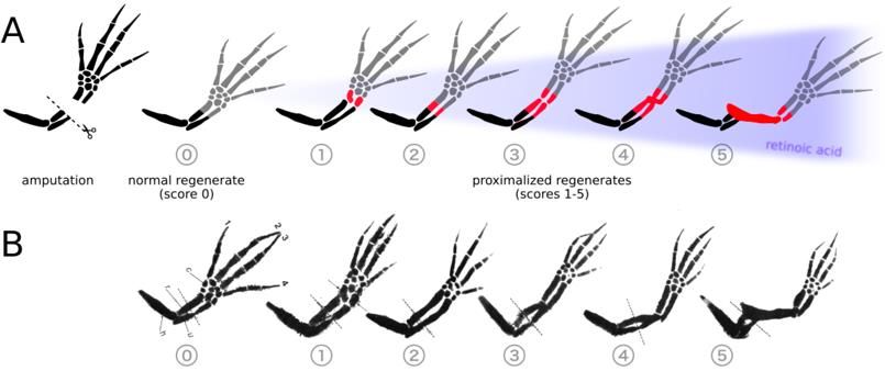
Cells 2021, 10, 2174                                                                                        3 of 17

                       only activation of RARα by Am580 consistently results in complete PD duplication at
                       25 nM treatment.
                             Following treatment of axolotl blastemas with 25 nM Am580, and thereby minimizing
                       off-target gene expression induced by RARβ and RARγ, we extracted total RNA from only
                       the blastema mesenchyme where the PI is known to reside. Am580 treatment induced
                       significant differential expression in 3637 genes by RNA-sequencing, of which 2580 could
                       be matched to an available reference sequence. A subset of 505 annotated genes was
                       analyzed and revealed several cell adhesion, cell membrane, matrix, and secretory proteins.
                       We also identified 23 unknown genes with homology limited to the genus Ambystoma
                       and differential expression of fourfold or greater, which may represent candidates for
                       future studies.

                       2. Materials and Methods
                       2.1. Axolotl Care
                       2.1.1. Axolotl Procurement and Husbandry
                           Albino and wild type axolotls were purchased from the Ambystoma Genetic Stock
                       Center at the University of Kentucky at sizes ranging from hatchlings (1 cm) to large
                       juveniles (10–13 cm). All axolotls were housed individually for the duration of the ex-
                       periments to prevent cannibalistic limb loss. Each animal was kept in room temperature,
                       40% Holtfreter’s solution (HS). Axolotls were fed a mixture of live brine shrimp, frozen
                       bloodworms, and fish pellets. All axolotl experimentation was performed in compliance
                       with the University of Florida’s Institutional Animal Care and Use Committee (protocol
                       number 201810351, approved 25 September 2018).

                       2.1.2. Limb Amputations
                            Limb amputations were conducted with a single cut to the mid-zeugopod following
                       systemic anesthetization in tricaine-s (Sigma-Aldrich, St Louis, MO, USA) dissolved in HS.
                       The concentration of tricaine-s used was dependent on axolotl length and ranged from 300
                       to 1000 mg/L.

                       2.1.3. Systemic RAR Agonist Treatment
                            Following limb amputation, the axolotls were placed in HS for 24 h to recover. Ani-
                       mals in the treatment groups were then placed in the working RAR agonist solution for
                       3–5 days post amputation and the animals in the control group were placed in HS with
                       DMSO (volume equal to amount of agonist solution used in treatment animals). The
                       solutions were changed every 24 to 48 h. All animals were then returned to HS until limb
                       regeneration was complete or until tissue was harvested.

                       2.2. Solution Preparation
                       2.2.1. Victoria Blue Stain
                            Victoria Blue stain was prepared by dissolving 1% w/v Victoria Blue dye in
                       acid alcohol.

                       2.2.2. RAR Agonist Solutions
                           Stock solutions of all-trans-RA, Am580 (Sigma-Aldrich, St. Louis, MO, USA), CD271,
                       CD2019, and CD437 (Tocris Bioscience, Bio-Techne Corp, Minneapolis, MN, USA) were
                       prepared to 1 mM by dissolving in DMSO and stored at −20 ◦ C. Working solutions were
                       prepared by dilution with HS to achieve the desired treatment concentrations ranging from
                       10 nM to 250 nM.

                       2.3. Limb Analysis
                       2.3.1. Limb Collection and Staining
                            Regenerated limbs were collected and fixed in 10% neutral buffered formalin (NBF)
                       for at least 24 h at 4 ◦ C. The limbs were bleached overnight at room temperature in 30%
Retinoic Acid Receptors and the Control of Positional Information in the Regenerating Axolotl Limb - MDPI
Cells 2021, 10, 2174                                                                                           4 of 17

                       hydrogen peroxide. Each limb was dehydrated in 50% ethanol 1 h, treated with 1% HCl in
                       70% ethanol for 2 h, and then stained with Victoria blue for 45 min. After dehydrating to
                       100% ethanol, the tissue was cleared in methyl salicylate.

                       2.3.2. Duplication Scoring
                            Following collection of the limbs and staining with Victoria blue, the regenerates
                       were photographed and visually evaluated for proximodistal respecification. Duplication
                       scores (Figure 1) were assigned ranging from 0 (normal regenerate with no duplication) to
                       5 (complete duplication from stylopod to autopod) [4]. Intermediate scores are assigned
                       according to the degree of duplication.

                       2.4. RNA Sequencing
                       2.4.1. RNA Extraction
                            For the sequencing experiment, mesenchyme tissue was collected only from the right
                       forelimb blastemas, which had the epidermis removed, of both Am580 and DMSO-treated
                       axolotls. The left contralateral limb was allowed to continue regeneration and served as
                       confirmation that respecification had taken place, since contralateral pairs of limbs from any
                       one animal always behaved identically. The right mesenchymes were immediately placed
                       in RNALater, incubated at 4 ◦ C overnight, and then stored at −20 ◦ C. Total RNA was
                       extracted using a RNeasy Fibrous Tissue Mini Kit (QIAGEN 74704, Qiagen, Germantown,
                       MD, USA) according to the manufacturer’s protocol. A total of 675–2889 ng of RNA was
                       extracted from each sample (means of 1350 ng (s = 574 ng) and 2168 ng (s = 563 ng) for
                       Am580 and DMSO-treated tissue, respectively), as quantified using a Qubit fluorometer.
                       Agilent TapeStation analysis assigned RINs ranging from 9.5 to 10.0 (mean RIN of 9.9,
                       s = 0.14). From each treatment group of ten axolotls, RNA extracts were used for RNA-
                       sequence library construction (n = 6).

                       2.4.2. Library Construction and RNA Sequencing
                            An Illumina TruSeq stranded library was constructed via poly-A selection for each
                       individual blastema sample for a total of 12 libraries (six from the Am580-treated group
                       and six from the DMSO-control group). The libraries were sequenced on the Illumina
                       NovaSeq6000 platform to yield approximately 642 million paired-end, 150 bp reads (mean
                       of 53.5 M read pairs (s = 6.5 M read pairs) per library). Library construction and sequencing
                       were performed by HudsonAlpha Discovery, Huntsville, AL, USA.

                       2.4.3. Read Processing and Quality Control
                            All reads were evaluated with FastQC [12] for base sequence quality, adapter content,
                       and duplication level to identify any library-level errors. Adapter sequences and low-
                       quality bases were trimmed from each read using Trimmomatic [13] and then re-evaluated
                       with FastQC. Surviving paired-end reads were not merged, and single-end reads were
                       excluded from downstream analyses.

                       2.4.4. Data Processing and Visualization
                           Data processing was performed in R [14] using RStudio [15]. Data visualization was
                       accomplished with the R graphics package ggplot2 [16].

                       2.5. RNA-Seq Analysis
                       2.5.1. Transcriptome Assembly
                           Reads in each fastq file were aligned to the AmexG_v3.0.0 axolotl genome [17] using
                       HISAT2 [hisat2/2.1.0, -q -dta] [18]. The resulting SAM files were converted to BAM files
                       using samtools [samtools/1.8, samtools view -S], sorted [samtools sort], and indexed
                       [samtools index -c] [19]. Due to the large size of the individual chromosome arms, it was
                       necessary to create a CSI index instead of the standard BAI index.
Cells 2021, 10, 2174                                                                                          5 of 17

                             A combined transcriptome for treated and control blastemas was constructed using
                       StringTie [stringtie/1.3.4d] [20,21] and gffread [gffread/0.9.8c] [22]. The GenePred annota-
                       tion file provided with the genome was downloaded (https://www.axolotl-omics.org/dl/
                       AmexG_3.0.0_FinalGeneSet.gp) and used to generate new transcript names that retained
                       the reference names when possible and utilized the “MSTRG” name from StringTie for
                       novel transcripts. Custom R [14] scripts were used in RStudio [15] to manipulate the
                       reference GTF and StringTie output.

                       2.5.2. Differential Expression Analysis
                             Transcript counts were produced with featureCounts [subread/1.6.2, featureCounts -s
                       2 -p -t exon -g gene_id] [23] using the transcriptome annotation file generated by StringTie.
                       Custom R scripts were used to clean the featureCounts output and differential expression
                       analysis was conducted with edgeR [24]. Low expressed reads were removed from the
                       analysis by filtering for transcripts that did not have at least ten counts in at least six
                       samples. The differential expression model fit a quasi-likelihood method and significance
                       was determined with the [edgeR/3.26.8, glmQLFit, adjust.method = “BH”, p value = 0.05].
                       A total of 3454 genes were identified as differentially expressed at a log2 fold change
                       greater than zero, with 1650 genes upregulated and 1804 downregulated. An additional
                       18,693 genes were present but did not exhibit significant differential expression.

                       2.5.3. Transcript Annotation
                            All differentially expressed transcripts identified by edgeR were annotated with
                       BLASTx [ncbi_blast, blastx -db swissprot -outfmt 10 -max_target_seqs 1] against the Swis-
                       sProt database, supplementing the existing genome annotation. The top hit for each gene
                       was retained for downstream analyses.

                       2.5.4. Pathway Analysis
                             Differentially expressed genes with human Entrez designations were loaded into
                       Ingenuity Pathway Analysis (IPA; QIAGEN) and subjected to pathway analyses with
                       various fold change cutoffs. Using only genes with at least a twofold change, only
                       275 analysis-ready molecules were available, whereas decreasing the foldchange cutoff to
                       1.5 increased the analyzed genes to 485. IPA was used primarily for the identification of
                       significantly activated or inhibited canonical pathways.

                       2.6. Data
                           The data are currently being submitted to NCBI and the accession numbers will be
                       provided during review.

                       3. Results
                       3.1. Human and Axolotl RARs Are Homologous
                            As the agonists were designed to selectively bind to human RARs [10,11], we sought
                       to determine if agonist specificity would likely be retained when paired with the axolotl
                       RAR homologs. Three amino acid residues within the ligand-binding domain of the human
                       RARs are responsible for the binding specificity of each agonist [25]. Sequence similarities
                       between human and axolotl RARs are high (87%, 90%, and 83% for RARα, RARβ, and
                       RARγ, respectively) and axolotl RARs contain the same three specificity-granting amino
                       acid residues, indicating that the agonists show similar selectivity with axolotl RARs
                       (Figure 2).
Cells 2021, 10, x FOR PEER REVIEW                                                                                                           6 of 17

Cells 2021, 10, 2174                and RARγ, respectively) and axolotl RARs contain the same three specificity-granting
                                                                                                                  6 of 17
                                    amino acid residues, indicating that the agonists show similar selectivity with axolotl
                                    RARs (Figure 2).

                                     Figure2.2. The
                                    Figure       The three
                                                      three retinoic
                                                             retinoicacid
                                                                       acidreceptors
                                                                            receptors(RARα,
                                                                                       (RARα,RARβ,
                                                                                               RARβ,and andRARγ)
                                                                                                             RARγ)are arehomologous,
                                                                                                                          homologous,asasseenseen in
                                    in the alignments of select regions of the ligand-binding domain of both human (top) and axolotl(bot-
                                     the alignments    of  select regions of the ligand-binding   domain   of both  human    (top) and  axolotl
                                     tom). Three
                                    (bottom).       amino
                                                 Three      acidacid
                                                        amino     substitutions, depicted
                                                                      substitutions,      in red,
                                                                                     depicted     grant
                                                                                              in red,    binding
                                                                                                       grant       specificity
                                                                                                              binding          to the
                                                                                                                       specificity  to RAR  agonists
                                                                                                                                       the RAR
                                    agonists Am580, CD2019, and CD437. The protein sequences were obtained from the EST database at
                                     Am580,    CD2019,    and  CD437.   The protein  sequences were   obtained   from  the EST   database  hosted
                                     Sal-Siteat[26].
                                    hosted      Sal-Site [26].

                                    3.2.
                                     3.2.Effects
                                          EffectsofofSelective
                                                      SelectiveRAR
                                                                RARAgonists
                                                                      Agonists ononLimb
                                                                                      LimbRegeneration
                                                                                             Regeneration
                                          As   previous    studies have  shown    that  individual
                                           As previous studies have shown that individual            RARs   are responsible
                                                                                                          RARs                 for thefor
                                                                                                                  are responsible       forma-
                                                                                                                                           the for-
                                    tion  of specific   phenotypes,   we  hypothesized      that positional  information   can
                                     mation of specific phenotypes, we hypothesized that positional information can be prox-     be proximal-
                                    ized  by thebyactivation
                                     imalized                   of a single
                                                      the activation         RAR [27–29].
                                                                       of a single             Forelimb
                                                                                      RAR [27–29].        stumps,
                                                                                                       Forelimb     24 h post-amputation,
                                                                                                                  stumps,    24 h post-amputa-
                                    were
                                     tion, were treated with synthetic agonists Am580 (RARα agonist), CD2019 agonist),
                                           treated    with   synthetic agonists    Am580     (RARα    agonist),  CD2019    (RARβ     (RARβ ago-
                                    CD271 (RARα and RARβ dual-agonist), CD437 (RARγ agonist), or all-trans-RA. Systemic
                                     nist), CD271 (RARα and RARβ dual-agonist), CD437 (RARγ agonist), or all-trans-RA. Sys-
                                    treatment of 3–5 cm-long axolotls at a concentration of 250 nM agonist resulted in varying
                                     temic treatment of 3–5 cm-long axolotls at a concentration of 250 nM agonist resulted in
                                    degrees of proximal–distal limb duplication following mid-zeugopod amputation of both
                                     varying degrees of proximal–distal limb duplication following mid-zeugopod amputa-
                                    forelimbs. Qualitative, visual scoring of Victoria blue-stained regenerates were used to
                                     tion of both forelimbs. Qualitative, visual scoring of Victoria blue-stained regenerates
                                    compare the proximal–distal (PD) duplication of each treatment (Figure 3). The limbs
                                     were used
                                    treated   withto250compare
                                                           nM RAthe   proximal–distal
                                                                    received   a mean score (PD)ofduplication
                                                                                                   2.5 out of 5,ofwhile
                                                                                                                    each the
                                                                                                                          treatment
                                                                                                                               control(Figure
                                                                                                                                         limbs 3).
                                     The limbs
                                    treated   withtreated    with 250ofnM
                                                    equal volumes       theRA    received
                                                                             solvent   DMSO  a mean   score
                                                                                               received      of 2.5
                                                                                                         a score of out  of 5,
                                                                                                                     0 with nowhile    the control
                                                                                                                                indication   of
                                     limbs   treated   with   equal  volumes    of the  solvent   DMSO    received    a score
                                    PD duplication. While all four agonist-treated groups had at least one limb with complete  of 0 with   no indi-
                                     cation of PDonly
                                    duplication,      duplication.   While all
                                                            Am580 (RARα)         four
                                                                               and      agonist-treated
                                                                                     CD2019               groupsgroups
                                                                                               (RARβ)-treated       had atwere
                                                                                                                            leastcomprised
                                                                                                                                   one limb with
                                     complete
                                    of  completeduplication,
                                                     duplication only
                                                                   in allAm580
                                                                          treated (RARα)
                                                                                    limbs. The andRARγ
                                                                                                     CD2019    (RARβ)-treated
                                                                                                          agonist,  CD437, had the  groups    were
                                                                                                                                         great-
                                     comprised
                                    est  variationofwithcomplete
                                                             a meanduplication
                                                                     score of 2.7,inandall the
                                                                                           treated
                                                                                               RARβγlimbs.  The RARγCD271
                                                                                                         dual-agonist     agonist,   CD437,
                                                                                                                                  resulted   in had
                                    athe  greatest
                                       mean    scorevariation    withresults
                                                       of 3.8. These    a meandemonstrate
                                                                                  score of 2.7,successful
                                                                                                  and the RARβγ        dual-agonist
                                                                                                             respecification            CD271 re-
                                                                                                                                of positional
                                    identity   to a  more    proximal  state  in all  treatment   groups   as they  all contain
                                     sulted in a mean score of 3.8. These results demonstrate successful respecification of posi- duplicated
                                    proximal    elements.
                                     tional identity     to a more proximal state in all treatment groups as they all contain dupli-
                                     cated proximal elements.
                                    3.3. Systemic Treatment of 25 nM Am580 Consistently Respecifies Positional Information
                                         While the 250 nM agonist treatments suggest that activation of both RARα and RARβ
                                    lead to the highest degree of PD respecification, the possibility remained that only a single
                                    receptor is truly responsible for proximalization. As previous studies [10] have shown that
                                    the specificity of each agonist for a single RAR is highly dependent on agonist concentration
                                    and that, at any one concentration, the agonists elicit comparable binding [11], we repeated
                                    the systemic treatments at 25 nM on slightly larger axolotls (6–8 cm). When the agonists
                                    are more selective at this lower concentration of 25 nM, only activation of RARα by Am580
                                    consistently results in PD duplication with a mean score of 4.9, compared to the CD2019
                                    and CD437 treatment groups with mean scores of 0.1 and 0.3, respectively (Figure 4A).
                                    Comparing the 250 nM (Figure 3) and 25 nM (Figure 4A) treatments reveals that the 10X
                                    dilution results in a decrease in limb duplication following CD2019 and CD437 treatments,
                                    while there is minimal difference between the duplication scores of Am580-treated limbs.
                                    Representative Victoria blue-stained limbs from each treatment group at both 250 nM and
                                    25 nM are compared in Figure 4B.
CellsCells
      2021,  10, x FOR PEER REVIEW
           2021, 10, 2174                                                                                                                                7 of 17
                                                                                                                                                                   7 of 17

                                       Figure 3. Duplication of forelimb regenerates from juvenile axolotls (3–5 cm) treated systemically
                                       with 250 nM Am580 (n = 20 limbs), CD2019 (n = 10), CD271 (n = 10), or CD437 (n = 15). Retinoic acid
                                       (n = 8) and DMSO (n = 8) served as the positive and negative controls, respectively. A score of 0–5
                                       was assigned to each limb based on the degree of duplication (Figure 1). Limbs treated with Am580
                                       and CD2019 were all completely duplicated, while CD271 and CD437 induced a range of duplica-
                                       tion. All treatments have greater duplication than the DMSO control (DMSO vs. RA, p = 6.08 × 10⁻⁴;
                                       DMSO vs. CD271, p = 9.1 × 10⁻⁶; DMSO vs. CD437, p = 4.5 × 10⁻⁴). Am580 and CD2019 are greater
                                       than RA (RA vs. Am580 or CD2019, p = 1.9 × 10⁻³; RA vs. CD271). CD271 is just significantly more
                                       effective than RA (p = 0.016) and CD437 is not significantly more effective than RA (p = 0.438).

                                       3.3. Systemic Treatment of 25 nM Am580 Consistently Respecifies Positional Information
                                               While the 250 nM agonist treatments suggest that activation of both RARα and RARβ
                                        lead to the highest degree of PD respecification, the possibility remained that only a single
                                        receptor is truly responsible for proximalization. As previous studies [10] have shown that
                                        the specificity of each agonist for a single RAR is highly dependent on agonist concentra-
                                        tion and
                                       Figure        that, at anyofone
                                                3. Duplication               concentration,
                                                                        forelimb     regeneratesthefromagonists   elicit
                                                                                                           juvenile      comparable
                                                                                                                     axolotls  (3–5 cm) binding       [11], we
                                                                                                                                          treated systemically
                                     Figure
                                        repeated3. Duplication
                                                      the  systemic   of forelimb
                                                                        treatments      regenerates
                                                                                         at 25 nM    on from   juvenile
                                                                                                         slightly larger   axolotls
                                                                                                                            axolotls
                                       with 250 nM Am580 (n = 20 limbs), CD2019 (n = 10), CD271 (n = 10), or CD437 (n = 15). Retinoic (3–5
                                                                                                                                      (6–8   cm)
                                                                                                                                             cm). treated
                                                                                                                                                   When      systemically
                                                                                                                                                            the
                                                                                                                                                              acid
                                     with   250
                                        agonists  nM areAm580
                                                          more     (n  = 20
                                                                 selective    limbs),
                                                                               at  this  CD2019
                                                                                         lower      (n  = 10),
                                                                                                 concentration CD271
                                                                                                                   of 25(n  =
                                                                                                                          nM, 10), or
                                                                                                                                only   CD437
                                                                                                                                       activation
                                       (n = 8) and DMSO (n = 8) served as the positive and negative controls, respectively. A score of 0–5       (n  =
                                                                                                                                                     of15).
                                                                                                                                                        RARα Retinoic acid
                                     (nwas
                                         = 8)assigned
                                        by     and DMSO
                                            Am580               (n
                                                        consistently=  8)  served
                                                                          results     as
                                                                                     in   the
                                                                                         PD    positive
                                                                                             duplication   and  negative
                                                                                                              with  a mean   controls,
                                                                                                                               score  of respectively.
                                                                                                                                          4.9,
                                                         to each limb based on the degree of duplication (Figure 1). Limbs treated with Am580
                                                                                                                                               compared    A  score
                                                                                                                                                             to     of 0–5
                                     wastheassigned
                                              CD2019 to   and   CD437
                                                             each   limb   treatment
                                                                            based     on   groups
                                                                                          the       with
                                                                                               degree    of mean   scores (Figure
                                                                                                            duplication      of 0.1 and
                                                                                                                                      1).  0.3, respectively
                                                                                                                                          Limbs    treated   with  Am580
                                       and CD2019 were all completely duplicated, while CD271 and CD437 induced a range of duplication.
                                     and(Figure
                                           CD2019  4A).were
                                                          Comparing        the 250 duplicated,
                                                                all completely         nM (Figure 3)      and CD271
                                                                                                      while    25 nM (Figure
                                                                                                                        and CD437 4A) treatments
                                                                                                                                        induced a reveals
                                                                                                                                                       range of duplica-
                                       All treatments have greater duplication than the DMSO control (DMSO vs. RA, p = 6.08 × 10⁻⁴ ;
                                        that
                                     tion.  Allthe   10X
                                                 treatments dilution
                                                                 have   results
                                                                          greater
                                                                               − 6  in  a  decrease
                                                                                      duplication      in
                                                                                                      than limb
                                                                                                             the duplication
                                                                                                                 DMSO   − 4      following
                                                                                                                            control  (DMSO      CD2019
                                                                                                                                                 vs.  RA,  and
                                                                                                                                                           p = 6.08 × 10⁻⁴;
                                       DMSO vs. CD271, p = 9.1 × 10 ; DMSO vs. CD437, p = 4.5 × 10 ). Am580 and CD2019 are greater
                                        CD437      treatments,      while    there
                                     DMSO vs. CD271, p = 9.1 × 10 ; DMSO vs. CD437,
                                                                                  −6  is  minimal    differencep = 4.5 × 10 ). Am580 and CD2019ofare greater
                                                                                                                  between      the
                                                                                                                              −4    duplication      scores
                                       than RA (RA vs. Am580 or CD2019, p = 1.9 × 10⁻³ ; RA vs. CD271). CD271 is just significantly more
                                        Am580-treated
                                     than   RA (RA vs. Am580  limbs. Representative
                                                                         or CD2019, p = Victoria1.9 × 10 blue-stained
                                                                                                          −3; RA vs. CD271).limbsCD271
                                                                                                                                    from eachis justtreatment
                                                                                                                                                      significantly more
                                       effective than RA (p = 0.016) and CD437 is not significantly more effective than RA (p = 0.438).
                                        group at
                                     effective       both
                                                  than    RA250(pnM    and 25
                                                                  = 0.016)     and nM   are compared
                                                                                      CD437                in Figure 4B.
                                                                                               is not significantly     more effective than RA (p = 0.438).

                                     3.3. Systemic Treatment of 25 nM Am580 Consistently Respecifies Positional Information
                                           While the 250 nM agonist treatments suggest that activation of both RARα and RARβ
                                    lead to the highest degree of PD respecification, the possibility remained that only a single
                                    receptor is truly responsible for proximalization. As previous studies [10] have shown that
                                    the specificity of each agonist for a single RAR is highly dependent on agonist concentra-
                                    tion and that, at any one concentration, the agonists elicit comparable binding [11], we
                                    repeated the systemic treatments at 25 nM on slightly larger axolotls (6–8 cm). When the
                                    agonists are more selective at this lower concentration of 25 nM, only activation of RARα
                                    by Am580 consistently results in PD duplication with a mean score of 4.9, compared to
                                    the CD2019 and CD437 treatment groups with mean scores of 0.1 and 0.3, respectively
                                    (Figure 4A). Comparing the 250 nM (Figure 3) and 25 nM (Figure 4A) treatments reveals
         Figure 4. Comparison of PD that   theduplication
                                        limb    10X dilution
                                                           at 250 results
                                                                  nM and 25 innM a decrease     in limb
                                                                                    systemic agonist        duplication
                                                                                                        treatment.          following
                                                                                                                   (A) Scoring           CD2019 and
                                                                                                                                 following
                                    CD437 treatments,
         Victoria blue staining of mid-zeugopod               while treated
                                                   limb regenerates    there with
                                                                               is minimal     difference
                                                                                     25 nM agonist    reveals between    the
                                                                                                              a decrease in   duplication
                                                                                                                            duplication to scores of
                                                                    −10 ) and CD437 (n = 12; p = 5.1 × 10⁻⁴ ). There is no significant
         nearly zero in both CD2019 Am580-treated
                                       (n = 14 limbs; p =limbs.   Representative
                                                           9.4 × 10                     Victoria blue-stained limbs from each treatment
         difference between Am580 (n  = 14) treatments
                                    group    at both 250at 250
                                                            nM nMand
                                                                   (Figure 3) and
                                                                        25 nM      25 nM
                                                                                 are     (p = 0.082);in(B)
                                                                                      compared             Representative
                                                                                                         Figure  4B.      limb regenerates
         following systemic exposure to 250 nM (upper panel) or 25 nM RAR (lower panel) agonists. The amputation plane is shown
         in red. The photos were processed by removing the background and adjusting the brightness.

                                           In search of a minimum concentration that could elicit the PD duplication seen above,
                                      hatchling axolotls (1–2 cm long) were treated with 10 nM of each agonist. These results
                                      revealed that the effects of Am580 are vastly reduced at such a low concentration. Many
                                      limbs treated with Am580 developed a cartilaginous spike on the ulna but generally did
                                      not exhibit any sign of proximalization. Four limbs treated with CD437 had a clear gap in
                                      either the regenerated radius or ulna. These results indicate that systemic agonist treatment
                                      of hatchlings at 10 nM is insufficient to induce duplication of the PD limb axis.
Cells 2021, 10, 2174                                                                                           8 of 17

                            Experimenting on hatchling axolotls is convenient due to their low cost and rapid
                       limb regeneration, but their tiny blastemas present a problem for surgical manipulation
                       and molecular extraction/analysis. In order to collect enough tissue for larger studies,
                       such as RNA-sequencing as we performed here, it is more convenient to work on a smaller
                       number of larger axolotls that will yield larger blastemas. To determine if 25 nM systemic
                       treatments is enough to respecify large juvenile limbs, 16 axolotls of approximately 10 cm
                       in length were treated with either Am580 or DMSO. Despite the low concentration of only
                       25 nM, the large size of the axolotl, and the systemic delivery method that required
                       absorption through the gills or skin, every Am580-treated limb showed complete PD
                       duplication. We conclude that treatment with 25 nM agonist is best able to consistently
                       induce complete PD respecification while maintaining agonist specificity across a range of
                       sizes of axolotls.

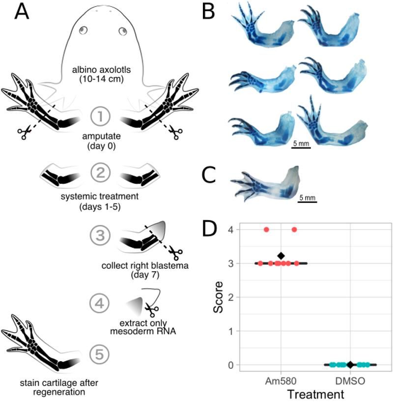
                       3.4. RNA-Sequencing of Blastema Mesenchyme Tissue Following Am580 Treatment
                       3.4.1. Systemic Am580 Treatment
                             To better understand the role of RA in respecifying positional information, we em-
                       ployed the Am580 agonist that selectively binds and activates RARα. The primary goal
                       of using a selective agonist instead of RA was to minimize activation of off-target genes
                       that are not involved in specifying positional identity. Twenty large juvenile axolotls
                       (10–13 cmlong) were selected for systemic Am580 or DMSO treatment and blastema mes-
                       enchyme tissue collection. After amputating both forelimbs and resting for 24 h, 10 axolotls
                       were placed in 25 nM Am580 in HS, and the remaining 10 axolotls were placed in an equal
                       volume of DMSO in HS (Figure 5A). After five days of treatment, the axolotls were returned
                       to HS for 24 h. The right forelimb blastemas were then collected from all 20 axolotls, the
                       epidermis was removed from the blastemas and the mesenchyme stored in RNALater.
                       The left forelimbs were left intact to regenerate to serve as observational controls for the
                       efficacy of respecification since contralateral limbs behave identically. At the completion of
                       regeneration, the left forelimb regenerates were collected, stained with Victoria blue, and
                       scored based on proximodistal duplication. Scores for 9 Am580-treated (1 axolotl died prior
                       to limb collection) and 10 control limbs show an increase in duplication of Am580-treated
                       axolotls compared to the DMSO control (Figure 5D).

                       3.4.2. RNA-Sequencing Metrics
                            The six limbs from the Am580 treatment group that showed the greatest level of
                       duplication (Figure 5B) as well as six limbs from the DMSO control group, all scoring 0 with
                       no duplication, were selected for RNA extraction and RNA-sequencing. Approximately
                       642 million paired-end, 150 bp reads were obtained from 12 libraries (mean of 53.5 M
                       read pairs per library), with each library constructed from total RNA collected from the
                       mesenchyme tissue of an individual blastema. All 12 libraries passed quality control
                       analysis with fastQC [12], showed improvement following quality and adapter trimming
                       with Trimmomatic, and were retained for downstream analyses.

                       3.4.3. Transcriptome Assembly and Differential Expression Analysis
                            Using the axolotl genome (version AmexG_v3.0.0) as a reference, a transcriptome was
                       assembled from reads in both the Am580 and DMSO treatment groups. Trimmed, paired-
                       end reads were aligned to the genome using HISAT2 and the transcripts assembled using
                       StringTie. A transcriptome was created using gffRead and gffCompare. The final assembly
                       is composed of 193,947 total transcripts, including 11,962 novel transcripts identified
                       by StringTie.
Cells 2021, 10, 2174                                                                                                                        9 of 17

                                        From 36,917 genes identified by featureCounts, 3637 were determined by edgeR to
                                  be differentially expressed in the Am580-treated animals compared to the DMSO-control
                                  groups at an adjusted p-value of 0.05. Only 2580 were matched to a significantly similar
                                  homolog by BLASTx, indicating that many of the identified genes are either novel to the
                                  axolotl or simply not yet annotated in the SwissProt database. Of these 2580 differentially
                                  expressed genes, 1177 are upregulated and the remaining 1403 are downregulated. A
 Cells 2021, 10, x FOR PEER REVIEWrestricted list of 505 genes was created by selecting all genes with at least a twofold change
                                                                                                                            9 of 17
                                  in expression, and the top thirty differentially expressed genes are presented in Table 1.

                                Figure      (A) Experimental
                                 Figure5.5. (A)   Experimentaldesign
                                                                   designforforboth
                                                                                bothDMSO
                                                                                       DMSO      control
                                                                                              control   (n (n = 10)
                                                                                                           = 10) andand25 nM25 nM
                                                                                                                               RARαRARα   (n =treat-
                                                                                                                                     (n = 10)   10)
                                 ments. BothBoth
                                treatments.     forelimbs    are amputated
                                                      forelimbs               mid-zeugopod
                                                                  are amputated     mid-zeugopod  at day
                                                                                                       at 0day
                                                                                                            and0 treated   systemically
                                                                                                                  and treated           with either
                                                                                                                                systemically  with
                                 DMSO
                                either   or Am580
                                       DMSO            on days
                                                or Am580        1–5. The
                                                            on days   1–5.right blastema
                                                                           The right        is collected
                                                                                       blastema            on day
                                                                                                  is collected     7, the
                                                                                                               on day    7,epidermis removed,
                                                                                                                           the epidermis        and
                                                                                                                                         removed,
                                 RNA   extracted    from   the mesoderm.     The  left  blastema    serves  as an  observational
                                and RNA extracted from the mesoderm. The left blastema serves as an observational control. (B) Leftcontrol. (B) Left
                                 forelimbregenerates
                                forelimb   regeneratescollected
                                                           collectedfrom
                                                                      fromthe
                                                                            thesix
                                                                                sixaxolotls
                                                                                     axolotlsthat
                                                                                                thatwere
                                                                                                      wereselected
                                                                                                            selectedforforRNA-sequencing;
                                                                                                                           RNA-sequencing;(C) (C)AA
                                 representative left forelimb regenerate collected from a DMSO-treated axolotl. (D) Limb duplication
                                representative left forelimb regenerate collected from a DMSO-treated axolotl. (D) Limb duplication
                                 scores for all left limbs in the Am580-treatment and DMSO-control groups. The Am580-treated
                                scores for all left limbs in the Am580-treatment and DMSO-control groups. The Am580-treated limbs
                                 limbs scored 3.2, an increase in PD duplication compared to DMSO controls (mean score of 0).
                                scored 3.2, an increase in PD duplication compared to DMSO controls (mean score of 0).
                                 3.4.2. RNA-Sequencing Metrics
                                      The six limbs from the Am580 treatment group that showed the greatest level of du-
                                 plication (Figure 5B) as well as six limbs from the DMSO control group, all scoring 0 with
                                 no duplication, were selected for RNA extraction and RNA-sequencing. Approximately
                                 642 million paired-end, 150 bp reads were obtained from 12 libraries (mean of 53.5 M read
                                 pairs per library), with each library constructed from total RNA collected from the mes-
                                 enchyme tissue of an individual blastema. All 12 libraries passed quality control analysis
                                 with fastQC [12], showed improvement following quality and adapter trimming with
                                 Trimmomatic, and were retained for downstream analyses.
Cells 2021, 10, 2174                                                                                                        10 of 17

      Table 1. Top 30 differentially expressed transcripts. Transcript name is given as “AMEXTC” if found in the reference
      genome or “MSTRG” if identified as novel by StringTie. UniProt identification and gene names are given for transcripts
      with a significant BLASTx result.

                       Transcript Name                          UniProt ID            Gene Name                  log2FC
         AMEXTC_0340000164913_cytochrome                      CP26A_CHICK                CP26A                  15.382878
       AMEXTC_0340000004000_LOC108712603                     MMP21_CYNPY                MMP21                  8.68081722
            AMEXTC_0340000018651_retinoic                    RARB_HUMAN                  RARB                  8.34534428
                        MSTRG.18535                            RS25_SHEEP                RPS25                 6.96185493
         AMEXTC_0340000220729_hypothetical                    APMAP_RAT                 APMAP                  6.31133061
                        MSTRG.12045                           ATTY_MOUSE                  TAT                  6.13262763
                        MSTRG.12046                           ATTY_MOUSE                  TAT                  5.72243555
                        MSTRG.2544                                   -                      -                  5.61165445
             AMEXTC_0340000053364_LOXe                       LX15B_HUMAN               ALOX15B                 5.26551299
              AMEXTC_0340000039558_TAT                        ATTY_MOUSE                  TAT                  5.26351744
                        mRNA01059                                    -                      -                  5.21247926
       AMEXTC_0340000032012_LOC108645441                             -                      -                  5.15846088
           AMEXTC_0340000247464_ripply3.L                    DSCR6_XENLA                RIPPLY3                5.05107637
                        MSTRG.17898                          NDNF_HUMAN                  NDNF                  5.04336304
       AMEXTC_0340000179221_LOC102944588                    GIMA4_HUMAN                 GIMAP4                 5.02378211
           AMEXTC_0340000012549_fibroblast                    FGF8_CHICK                 FGF8                  −4.8925275
                        MSTRG.4054                                   -                      -                  −4.9088476
                        MSTRG.23812                                  -                      -                  −4.9178374
                        MSTRG.16115                                  -                      -                  −4.966291
                        MSTRG.23482                           RTJK_DROME                  POL                  −5.0176166
                        MSTRG.4440                             TPH_XENLA                  TPH                  −5.2716375
       AMEXTC_0340000028898_LOC108413561                      RTJK_DROME                  POL                  −5.2869995
          AMEXTC_0340000081353_OCSTAMP                      OCSTP_HUMAN                OCSTAMP                 −5.3164526
                        MSTRG.1117                                   -                      -                  −5.5152443
                        MSTRG.16116                           LIN1_NYCCO                  LIN1                 −5.6941371
                        MSTRG.13846                                  -                      -                  −5.702535
       AMEXTC_0340000024275_LOC101872464                      B3A2_PONAB                SLC4A2                 −5.8790351
             AMEXTC_0340000007120_HTR6                       5HT6R_PANTR                 HTR6                  −6.3188017
                        MSTRG.5087                                   -                      -                  −6.5656262
                        MSTRG.25599                          LORF2_HUMAN                  LIN1                 −6.8496992

                                   3.5. Differentially Expressed Genes
                                   3.5.1. Integrin Signaling
                                        In a pathway analysis conducted using all significant differentially expressed genes
                                   from our Am580-treated blastemas, the top canonical pathway identified by IPA is integrin
                                   signaling. A total of 63 genes associated with integrin signaling are found among our
                                   differentially expressed genes, including 10 transmembrane receptors (Table 2). All but
                                   two integrin signaling receptor genes, itgav and itgb3, are upregulated following RARα
                                   activation. Integrins are heterodimeric glycoprotein complexes formed from an alpha and
                                   beta subunit, and they function as receptors on the cell surface to mediate cell migration,
                                   adhesion, and the construction of extracellular matrix (ECM). One of the most common
Cells 2021, 10, 2174                                                                                             11 of 17

                       integrin ligands is fibronectin, and we identified Lrfn5, the gene for a fibronectin type
                       III protein, as downregulated 1.28-fold following Am580 treatment. The upregulation of
                       nine integrin subunit genes and Flrt2 (upregulated 0.51-fold), a fibronectin receptor gene,
                       and the downregulation of the ligand Lrfn5 suggest that integrin signaling may play an
                       important role in the establishment of new, proximalized positional information following
                       RA treatment.

                       Table 2. Integrin signaling genes. Identified as a top canonical pathway by IPA.

                                Gene Symbol                    Entrez Gene Name                     log2 Ratio
                                     CAV1                           caveolin 1                        0.642
                                    ITGA2                    integrin subunit alpha 2                 1.139
                                    ITGA4                    integrin subunit alpha 4                 1.139
                                    ITGA5                    integrin subunit alpha 5                 0.492
                                    ITGA6                    integrin subunit alpha 6                 0.608
                                   ITGA2B                   integrin subunit alpha 2b                 1.001
                                    ITGAV                    integrin subunit alpha V                 −1.224
                                    ITGAX                    integrin subunit alpha X                 1.723
                                    ITGB2                     integrin subunit beta 2                 −0.904
                                    ITGB3                     integrin subunit beta 3                 −1.307

                       3.5.2. Extracellular Matrix Genes
                            Positional information in the axolotl limb is known to be located in the connective
                       tissue mesenchyme, but recent evidence suggests that it may also be present in the extra-
                       cellular matrix (ECM). A gain-of-function assay was conducted in vivo using the ALM
                       technique and decellularized axolotl ECM [30]. It was found that grafting ECM with dis-
                       parate positional information to an innervated wound could maintain blastema outgrowth
                       and form rudimentary limb elements. Decellularized mouse ECM from corresponding
                       limb locations was also able to induce blastema growth in the axolotl, indicating similar
                       mechanisms of positional identity interpretation. To find avenues for further exploration
                       of the roles that the ECM may play in PD positional identity, we identified several collagen
                       and matrix metalloproteinase (MMP) genes that are differentially expressed following
                       Am580 treatment. Three identified proteinases, Mmp9, Mmp14, and Mmp28, are downregu-
                       lated 2.27, 0.64, and 0.83, respectively, and Mmp21 is upregulated 8.68-fold. As previously
                       discussed, Mmp9 is upregulated by Prod1 treatment in urodeles, so its downregulation here
                       may prove an interesting avenue for study. The ECM itself is made of mostly collagens, and
                       we identified seven differentially expressed collagen components. Five components, Col5a1,
                       Col5a3, Col6a1, Col6a2, and Col6a6, are upregulated 3.63-, 2.09-, 1.07-, 0.85-, and 1.98-fold,
                       respectively. The remaining two components, Col4a5 and Col4a6, are downregulated 1.11-
                       and 1.38-fold, respectively.

                       3.5.3. Cell Membrane and Secretory Protein Coding Genes
                            To identify candidate genes with direct roles in establishing, maintaining, and inter-
                       preting positional information, we searched for upregulated genes with at least a twofold
                       change that encode secretory proteins or membrane-bound proteins with a domain ex-
                       posed to extracellular space. Positional information is thought to be maintained on the cell
                       surface of mesodermal, fibroblast-like connective tissue cells, such as Prod1 in the newt,
                       and discovering a suitable membrane protein in the axolotl would be a substantial finding.
                       While this assay did not reveal a single obvious gene candidate, 15 upregulated membrane
                       proteins were found with known functions in cell signaling and another 7 with structural
                       roles involving cell adhesion (Table 3). Proteins secreted into the extracellular space could
                       be involved in cell–cell signaling that relay positional information to nearby cells, so a
                       thorough search for genes that encode potentially secreted proteins was performed. We
                       identified a total of 51 differentially expressed, secretory protein-coding genes, including
                       29 genes upregulated at least twofold (Table 4).
Cells 2021, 10, 2174                                                                                                       12 of 17

      Table 3. Genes encoding membrane-bound proteins. The transcript name is given as “AMEXTC” if the transcript is found
      in the reference genome or “MSTRG” if identified as novel by StringTie. The UniProt ID and gene name are given for the
      most similar homolog as determined by BLASTx search. The log2 fold change was determined by edgeR, with positive
      values indicating upregulated expression following Am580 treatment.

                        Transcript                             UniProt ID               Gene Name                log2 FC
         AMEXTC_0340000038644_LOC108803933                   P2RY1_MOUSE                   P2RY1                   4.48
              AMEXTC_0340000062820_GEM                       GEM_HUMAN                     GEM                     4.32
                       MSTRG.21040                           HRH2_MOUSE                    HRH2                    3.32
                       MSTRG.22927                              TLR4_PIG                   TLR4                    3.09
         AMEXTC_0340000034284_LOC102448090                  M4A4A_HUMAN                  MS4A4A                    3.05
              AMEXTC_0340000013374_htr2b                    5HT2B_HUMAN                   HTR2B                    2.93
              AMEXTC_0340000062083_FGD2                      FGD2_MOUSE                    FGD2                    2.83
                       MSTRG.8675                            FGD2_HUMAN                    FGD2                    2.71
                       MSTRG.3167                             FSHR_CAIMO                   FSHR                    2.47
             AMEXTC_0340000014713_EPHA5                     EPHA5_HUMAN                   EPHA5                     2.4
            AMEXTC_0340000042498_ADRA2A                       ADA2A_PIG                  ADRA2A                     2.3
         AMEXTC_0340000030234_LOC109141183                  UNC5A_HUMAN                   UNC5A                    2.24
         AMEXTC_0340000057843_LOC108718813                   MRC1_HUMAN                    MRC1                     2.2
              AMEXTC_0340000156303_BLNK                       BLNK_CHICK                   BLNK                    2.12
               AMEXTC_0340000062611_RGR                       RGR_BOVIN                     RGR                    2.06
            AMEXTC_0340000150384_cadherin                    CAD17_MOUSE                  CDH17                    3.28
              AMEXTC_0340000048099_CDH4                      CADH4_CHICK                   CDH4                    3.11
         AMEXTC_0340000004184_LOC102348750                  CEAM5_MOUSE                 CEACAM5                    2.66
             AMEXTC_0340000028915_ICAM5                      ICAM5_MOUSE                  ICAM5                    2.44
               AMEXTC_0340000049914_Sell                    LYAM2_HUMAN                    SELE                    2.39
              AMEXTC_0340000220626_PLLP                      PLLP_HUMAN                    PLLP                     2.3
         AMEXTC_0340000220764_LOC101952950                   MYO10_BOVIN                  MYO10                    2.06

                                 3.5.4. Unknown Genes
                                       Many differentially expressed genes could not be linked to a known protein sequence
                                 either from the genome annotation file or an additional BLASTx analysis. Twenty-three
                                 unannotated transcripts with at least a fourfold change following Am580 treatment were
                                 selected for BLASTn analysis to determine if these transcripts are derived from genes
                                 novel to the axolotl or if they are conserved in other species. It is possible that key genes
                                 involved in limb regeneration and positional information may be novel to the axolotl or
                                 the genus Ambystoma, and these genes will be overlooked if only annotated transcripts are
                                 analyzed. A standard BLASTn analysis returned no significant result for 14 transcripts,
                                 hits limited to species of the genus Ambystoma for 4 transcripts, and hits to only known
                                 axolotl sequences for the remaining 5 transcripts. While some BLASTn results included
                                 homologous matches to sequences in other organisms, the regions matching to the query
                                 were very short. All hits with a match smaller than 10 percent of the query length were
                                 ignored and not included in this analysis. These 23 genes cannot be identified through
                                 homology with known sequences and their function in limb regeneration and positional
                                 identity remain a complete mystery. Future work should consider investigating these genes
                                 as they undergo considerable differential expression during Am580 treatment.
Cells 2021, 10, 2174                                                                                                       13 of 17

      Table 4. Genes encoding secretory proteins. Transcript name is given as “AMEXTC” if found in the reference genome or
      “MSTRG” if identified as novel by StringTie. Gene names are left blank for UniProt entries without a listed gene name.

                        Transcript                             UniProt ID                Gene Name                log2FC
         AMEXTC_0340000004000_LOC108712603                   MMP21_CYNPY                   MMP21                    8.68
                       MSTRG.17898                          NDNF_HUMAN                     NDNF                     5.04
                       MSTRG.6144                            FCNV4_CERRY                      -                     4.36
              AMEXTC_0340000030622_PROZ                       PROZ_BOVIN                    PROZ                    4.22
              AMEXTC_0340000250114_c4a.L                       CO4_BOVIN                     C4                     3.87
         AMEXTC_0340000209130_LOC105403673                   CO5A1_MOUSE                  COL5A1                    3.63
            AMEXTC_0340000025257_HAPLN4                      HPLN4_MOUSE                  HAPLN4                    3.55
                       MSTRG.15067                            VCO3_NAJKA                      -                     3.42
         AMEXTC_0340000036400_LOC100552635                   FCNV1_VARKO               FCNV1_VARKO                  3.41
             AMEXTC_0340000044633_PKDCC                     PKDCC_HUMAN                    PKDCC                    3.29
          AMEXTC_0340000062173_Angiogenin                    ANGI_MOUSE                     ANG                     3.22
                       MSTRG.6402                              MSMB_PIG                    MSMB                     3.17
               AMEXTC_0340000056401_ostn                     OSTN_HUMAN                     OSTN                    3.09
                       MSTRG.829                              IL12B_FELCA                   IL12B                   2.85
          AMEXTC_0340000250115_complement                       CO4_RAT                      C4                      2.6
              AMEXTC_0340000233392_CETP                       CETP_CHICK                    CETP                     2.6
           AMEXTC_0340000044847_Coiled-coil                  WNT6_MOUSE                     WNT6                    2.48
            AMEXTC_0340000048200_TSPEAR                     TSEAR_HUMAN                    TSPEAR                   2.47
                       MSTRG.13661                             IL18_CHICK                    IL18                   2.36
                       MSTRG.10022                             IL8_CHICK                     IL8                    2.32
            AMEXTC_0340000000382_fibroblast                   FGF2_BOVIN                    FGF2                    2.14
                       MSTRG.1555                            GDF6A_DANRE                   GDF6A                    2.14
            AMEXTC_0340000053380_COL27A1                    CO5A3_HUMAN                   COL5A3                    2.09
         AMEXTC_0340000232220_LOC107293967                  CHIT1_HUMAN                    CHIT1                    2.07
         AMEXTC_0340000170356_LOC102365657                     SFTPD_RAT                   SFTPD                    2.05
               AMEXTC_0340000064262_ndnf                     NDNF_XENTR                    NDNF                     2.04
                       MSTRG.5658                             SDF1_XENLA                    SDF1                    2.03
           AMEXTC_0340000010931_ADAMTS3                      ATS3_HUMAN                  ADAMTS3                    2.03
                       MSTRG.22754                          HMCN1_HUMAN                    HMCN1                    2.01

                                 4. Discussion
                                      We have shown here that the effect of RA on respecifying the PD axis of the regenerat-
                                 ing axolotl limb and thus duplicating the elements that are regenerated is transduced by
                                 only one of the three RARs, namely RARα. We arrived at this conclusion by decreasing
                                 the concentration of each of the RAR agonists until complete selectivity was obtained, at
                                 a concentration of 25 nM. At this concentration, the RARα agonist fully duplicated the
                                 regenerating limb such that after amputating through the middle of the lower limb, the
                                 regenerate contained all the elements of the limb, beginning from the proximal stylopod
                                 instead of just replacing the elements that were amputated. In contrast, the other two RAR
                                 agonists are completely inactive at this concentration and had no effect on the regenerating
                                 limb. At a 10-fold higher concentration, however, all three agonists were active at inducing
                                 duplications and this is due to the typical loss of selectivity of pharmacological agents
                                 which occurs at saturating conditions [11]. This explains why a previous use of one of the
Cells 2021, 10, 2174                                                                                           14 of 17

                       RAR agonists, a RARγ agonist, concluded that this was the receptor involved, but without
                       comparing any of the other agonists nor performing a concentration experiment [9].
                             We have previously shown by microarrays in the neural stem cells of the mouse
                       brain that these RAR agonists induce a different, although not unique, set of downstream
                       gene targets [31]. For example, after treatment with a RARα agonist, a total of 3155 genes
                       were responsive to the treatment, of which 1439 were specific to RARα, the remainder
                       overlapping with RARβ and RARγ agonist treatment. So, while not generating a unique
                       dataset, the use of this RARα agonist certainly reduces the gene targets compared to the
                       use of the pan-agonist RA as has been used in previous experiments of this type [6,9].
                       Therefore, we proceeded to identify the gene targets of RARα during limb duplication
                       and, in addition, as a further refinement, we used RNA isolated only from the blastemal
                       mesenchyme rather than the whole blastema, since the mesenchyme is the tissue which is
                       responsible for the effects of RA and not the epidermis [32].
                             We identified 2580 differentially expressed genes, less than half of which were upreg-
                       ulated and more than half were downregulated. The top upregulated gene was Cyp26A,
                       closely followed by Rarβ, both of which are in the RA regulation pathway. Interestingly, in
                       a microarray experiment [9] where blastemas were treated with RA, rather than the RARα
                       agonist, a more extensive range of genes in the RA pathway were induced. For example,
                       all three Rars and Crabp2 were induced in this work, but we only saw one receptor, Rarβ,
                       and neither of the Crabp genes, suggesting that we have indeed identified a sub-set of RA
                       responsive genes.
                             In terms of known positional genes, Meis1 and Meis2 are upregulated following PD
                       limb duplication initiated by RA treatment, and their overexpression proximalizes limb
                       tissue [33]. In our Am580-treated samples, we identified Meis1 and Meis2 as significantly
                       upregulated by 0.72- and 2.73-fold, respectively. We also identified Pbx1, a transcription
                       factor that dimerizes with Meis proteins, as upregulated 1.83-fold. Fgf8 plays a role in
                       keeping limb bud cells in a distal state as it represses Meis expression, represses distal Hox
                       gene expression and is itself repressed by RA. The top downregulated gene in our RARα
                       analysis was Fgf8 and also downregulated was the distal Hox gene a-13. The upregulated
                       Hox genes we identified were Hoxb-6, known to be upregulated by a pan-RAR agonist in
                       the chick limb bud [34] and Hoxd-3. The latter Hox gene has been shown to increase the
                       adhesiveness of human hematopoietic cell lines and induce the formation of cell aggregates
                       by upregulating integrin a2/b3 [35]. Remarkably, integrins were the top canonical pathway
                       by IPA seen here and, furthermore, the formation of cell aggregates is precisely the behavior
                       that distal blastemal cells undergo during the proximalization process induced by RA [5].
                       These data indicate the proximalization of distal blastema tissue both in terms of its cellular
                       behavior and positional control on the RARα pathway and imply a uniformity of patterning
                       mechanisms across developing and regenerating vertebrate limbs.
                             Identifying genes related to the cell surface was the center of our analysis, not only
                       because of this known change in adhesive behavior of blastemal cells, perhaps induced
                       by Hoxd-3, but also because this is considered to be the mechanism by which positional
                       information is assessed during regeneration (see Introduction). In support of this concept,
                       as mentioned above, the top canonical pathway identified by IPA from our dataset was
                       integrin signaling and six integrins genes were upregulated. Among the extracellular
                       matrix-related genes, Mmp21 was the second highest upregulated gene and the collagen
                       5 and 6 genes were upregulated along with the downregulation of collagen 4. The cell
                       membrane genes upregulated include EphA5, Unc5a, a netrin receptor, two cadherins Cdh17
                       and Cdh4 and genes for secreted proteins include neuron-derived neurotropic factor, a
                       hyaluronan link protein, angiogenin, Wnt4, Wnt6, Fgf2 and Gdf6a.
                             In relation to other genes considered to be responsible for the assessment of PI, we
                       surprisingly did not find any differential expression of Prod1, the GPI-linked cell surface
                       molecule identified in the newt, Notophthalmus [6]. This may be because in the axolotl, the
                       homolog to newt Prod1 has multiple stop codons generating two protein products with
                       truncated C-terminals, both of which lack a GPI-anchor and are secreted instead of being
Cells 2021, 10, 2174                                                                                                     15 of 17

                       localized to the cell surface [8]. Perhaps axolotl Prod1 is not involved in positionally related
                       events; rather, it may be involved in proliferation as a binding partner to nAG [36].
                             A more recent experiment in axolotls using a single cell sequencing dataset of connec-
                       tive tissue cells has identified a gene called Rarres1 (retinoic acid receptor responder 1) or
                       Tig1 [37]. This gene is expressed in a shallow gradient in the mature limb, is upregulated by
                       RA and is localized to the cell surface, having a transmembrane domain and a hyaluronic
                       acid binding motif characteristic of its human homologue. Transfection of Tig1 into the
                       blastema induces the translocation of distal cells to proximal regions and an antibody to
                       TIG1 prevents the engulfment of distal blastemas by proximal blastemas, precisely the
                       behavior required of a specifier of positional information. We see the upregulation by
                       1.84-fold of Tig1 in our dataset, suggesting that this gene is a downstream target of RARα
                       which acts to regulate PD identity. Since this was a relatively low level of upregulation
                       (and below our 2-fold cut-off), it suggests that searching for genes on the basis of their high
                       levels of upregulation may not the most successful strategy.
                             Much work will be required to test the function of the individual genes we have
                       identified above in controlling positional information, their pathways from the genome to
                       the cell surface and ECM and how these molecules interact, but our results described here
                       provide continuing supporting for the concept that in regeneration, the cell surface is the
                       seat of positional information recognition.

                       5. Conclusions
                             Our data suggest that the RARα transcription factor is the dominant receptor which
                       transduces the endogenous RA signal to control PD identity in the regenerating limb. The
                       complex transcriptional network, which is instigated in respecifying PI, involves many
                       components: (1) the upregulation of retinoic acid metabolism processes such as the Cyp26s
                       to autoregulate the RA signal; (2) the downregulation of cell cycling; (3) the downregulation
                       of distal positional genes such as Hoxa-13 and of distal regulatory secreted molecules such
                       as Fgf-8; (4) the upregulation of proximal identity genes such as Meis1 and 2; (5) the
                       upregulation of ECM regulators such as Mmp21 and ECM molecules such as collagen 5
                       and 6; (6) the upregulation of cell surface molecules such as integrins, cadherins, ephrins;
                       (7) the release of secreted molecules such as Wnts, neurotrophic factors and angiogenic
                       factors. How these pathways interact will be a major task for continuing research on the
                       nature and control of positional information in the regenerating limb.

                       Author Contributions: Conceptualization, M.M.; Data curation, T.P.; Formal analysis, T.P.; Funding
                       acquisition, M.M.; Investigation, T.P.; Methodology, T.P.; Project administration, M.M.; Visualization,
                       T.P.; Writing—original draft, T.P.; Writing—review and editing, M.M. Both authors have read and
                       agreed to the published version of the manuscript.
                       Funding: This research was funded by NSF, grant number IOS 1558017.
                       Institutional Review Board Statement: The study was conducted according to the guidelines of the
                       Declaration of Helsinki, and approved by the Institutional Review Board of University of Florida
                       (protocol 201810351).
                       Informed Consent Statement: Not applicable.
                       Data Availability Statement: The data is now publicly available and has been assigned accession
                       GSE182296 at https://www.ncbi.nlm.nih.gov/geo/query/acc.cgi?acc=GSE182296 (accessed on 19
                       August 2021).
                       Conflicts of Interest: The authors declare no conflict of interest. The funders had no role in the design
                       of the study; in the collection, analyses, or interpretation of data; in the writing of the manuscript, or
                       in the decision to publish the results.
You can also read