TWO DOSES OF SARS-COV-2 VACCINATION INDUCE MORE ROBUST IMMUNE RESPONSES TO EMERGING SARS-COV-2 VARIANTS OF CONCERN THAN DOES NATURAL INFECTION.
←
→
Page content transcription
If your browser does not render page correctly, please read the page content below

Two doses of SARS-CoV-2 vaccination induce more
robust immune responses to emerging SARS-CoV-2
variants of concern than does natural infection.
Donal T. Skelly
 Peter Medawar Building for Pathogen Research, Nuffield Department of Clinical Neurosciences,
University of Oxford, UK https://orcid.org/0000-0002-2426-3097
Adam C. Harding
 Sir William Dunn School of Pathology, University of Oxford, Oxford, UK https://orcid.org/0000-0003-
1479-959X
Javier Gilbert-Jaramillo
 Sir William Dunn School of Pathology, University of Oxford, Oxford, UK https://orcid.org/0000-0003-
1268-2304
Michael L. Knight
 Sir William Dunn School of Pathology, University of Oxford, Oxford, UK https://orcid.org/0000-0002-
8780-1630
Stephanie Longet
 Public Health England, Porton Down, UK https://orcid.org/0000-0001-5026-431X
Anthony Brown
 Peter Medawar Building for Pathogen Research, Nuffield Department of Medicine, University of Oxford,
UK
Sandra Adele
 Peter Medawar Building for Pathogen Research, Nuffield Department of Medicine, University of Oxford,
UK https://orcid.org/0000-0003-4458-1751
Emily Adland
 Peter Medawar Building for Pathogen Research, Department of Paediatrics, University of Oxford, Oxford,
UK
Helen Brown
 Peter Medawar Building for Pathogen Research, Nuffield Department of Medicine, University of Oxford,
UK
Medawar Laboratory Team
Tom Tipton
 Public Health England, Porton Down, UK https://orcid.org/0000-0002-0573-528X
Lizzie Stafford

                                               Page 1/28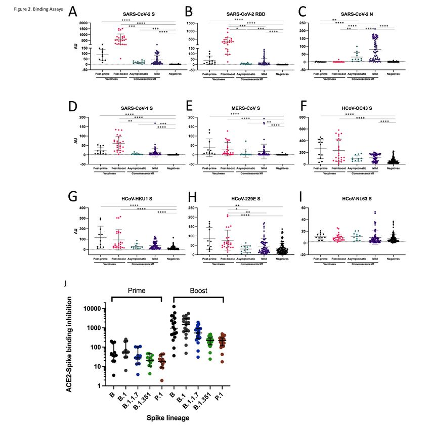
Nuffield Department of Medicine, University of Oxford, Oxford, UK https://orcid.org/0000-0002-1610-
5136
Síle A. Johnson
 Medical Sciences Division, University of Oxford, Oxford, UK https://orcid.org/0000-0002-4100-8522
Ali Amini
 Peter Medawar Building for Pathogen Research, Nuffield Department of Medicine, University of Oxford,
UK https://orcid.org/0000-0002-6837-8881
OPTIC Clinical Group
Tiong Kit Tan
 MRC Human Immunology Unit, MRC Weatherall Institute of Molecular Medicine, University of Oxford, UK
 https://orcid.org/0000-0001-8746-8308
Lisa Schimanski
 MRC Human Immunology Unit, MRC Weatherall Institute of Molecular Medicine, University of Oxford, UK
Kuan-Ying A. Huang
 Department of Infectious Diseases, Taoyuan General Hospital, Ministry of Health and Welfare, Taoyuan,
and Taipei Medical University, Taipei, Taiwan
Pramila Rijal
 MRC Human Immunology Unit, MRC Weatherall Institute of Molecular Medicine, University of Oxford, UK
 https://orcid.org/0000-0002-9214-9851
PITCH Study Group
CMORE/PHOSP-C Group
John Frater
 Peter Medawar Building for Pathogen Research, Nuffield Department of Medicine, University of Oxford,
UK
Philip Goulder
 Peter Medawar Building for Pathogen Research, Department of Paediatrics, University of Oxford, Oxford,
UK
Christopher P. Conlon
 Nuffield Department of Medicine, University of Oxford, Oxford, UK
Katie Jeffery
 Oxford University Hospitals NHS Foundation Trust, Oxford, UK https://orcid.org/0000-0002-6506-2689
Christina Dold
 Oxford Vaccine Group, Department of Paediatrics, University of Oxford, Oxford, UK
Andrew J. Pollard
 Oxford Vaccine Group, Department of Paediatrics, University of Oxford, Oxford, UK
 https://orcid.org/0000-0001-7361-719X
Alex Sigal
 Africa Health Research Institute, Durban 4001, South Africa https://orcid.org/0000-0001-8571-2004
Tulio de Oliveira

                                                Page 2/28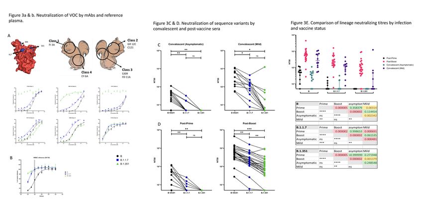
KwaZulu-Natal Research Innovation and Sequencing Platform, Durban, South Africa, 4001
https://orcid.org/0000-0002-3027-5254
Alain R. Townsend
MRC Human Immunology Unit, MRC Weatherall Institute of Molecular Medicine, University of Oxford, UK
https://orcid.org/0000-0002-3702-0107
Paul Klenerman
Peter Medawar Building for Pathogen Research, Nuffield Department of Medicine, University of Oxford,
UK https://orcid.org/0000-0003-4307-9161
Susanna J . Dunachie
Peter Medawar Building for Pathogen Research, Nuffield Department of Medicine, University of Oxford,
UK https://orcid.org/0000-0001-5665-6293
Eleanor Barnes
Peter Medawar Building for Pathogen Research, Nuffield Department of Medicine, University of Oxford,
UK https://orcid.org/0000-0002-0860-0831
Miles W. Carroll ( miles.carroll@phe.gov.uk )
Public Health England, Porton Down, UK
William S. James ( william.james@path.ox.ac.uk )
Sir William Dunn School of Pathology, University of Oxford, Oxford, UK https://orcid.org/0000-0002-
2506-1198
Research Article
Keywords: vaccine, immunology, virology, SARS-CoV-2, COVID-19, variants of concern, VOC, antibodies,
neutralizing antibodies, T cells, homotypic immunity, heterotypic immunity
Posted Date: March 29th, 2021
DOI: https://doi.org/10.21203/rs.3.rs-226857/v2
License: This work is licensed under a Creative Commons Attribution 4.0 International License.
Read Full License
Version of Record: A version of this preprint was published at Nature Communications on August 17th,
2021. See the published version at https://doi.org/10.1038/s41467-021-25167-5.
Page 3/28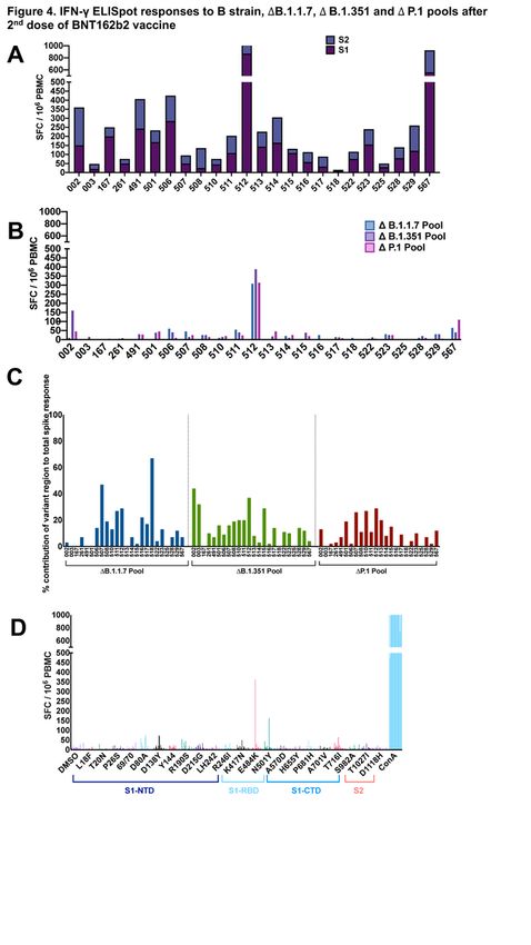
Abstract
Both natural infection with SARS-CoV-2 and immunization with vaccines induce protective immunity.
However, the extent to which such immune responses protect against emerging variants is of increasing
importance. Such variants of concern (VOC) include isolates of lineage B.1.1.7, first identified in the UK,
and B.1.351, first identified in South Africa. Our data confirm that VOC, particularly those with
substitutions at residues 484 and 417, escape neutralization by antibodies directed to the ACE2-binding
Class 1 and the adjacent Class 2 epitopes but are susceptible to neutralization by the generally less
potent antibodies directed to Class 3 and 4 epitopes on the flanks of the receptor-binding domain. To
address the potential threat posed by VOC, we sampled a SARS-CoV-2 uninfected UK cohort recently
vaccinated with BNT162b2 (Pfizer-BioNTech, two doses delivered 18-28 days apart), alongside a cohort
sampled in the early convalescent stages after natural infection in the first wave of the pandemic in
Spring 2020. We tested antibody and T cell responses against a reference isolate of the original
circulating lineage, B, and the impact of sequence variation in the B.1.1.7 and B.1.351 VOC. Neutralization
of the VOC compared to B isolate was reduced, and this was most evident for the B.1.351 isolate. This
reduction in antibody neutralization was less marked in post-boost vaccine-induced responses compared
to naturally induced immune responses and could be largely explained by the potency of the homotypic
antibody response. After a single vaccination, which induced only modestly neutralizing homotypic
antibody titres, neutralization against the VOC was completely abrogated in the majority of vaccinees.
Importantly, high magnitude T cell responses were generated after two vaccine doses, with the majority of
the T cell response directed against epitopes that are conserved between the prototype isolate B and the
VOC. These data indicate that VOC may evade protective neutralizing responses induced by prior
infection, and to a lesser extent by immunization, particularly after a single vaccine dose, but the impact
of the VOC on T cell responses appears less marked. The results emphasize the need to generate high
potency immune responses through vaccination in order to provide protection against these and other
emergent variants.

Introduction
The emergence of new lineages of severe acute respiratory syndrome coronavirus 2 (SARS-CoV-2) on
three continents towards the end of 2020, and their rapid expansion at the expense of the previously
dominant lineages, poses significant challenges to public health (WHO | SARS-CoV-2 Variants) 1. In order
to address these challenges effectively, there is an urgent need to understand the biological
consequences of the mutations found in these lineages, and the consequential impact on their
susceptibility to current control measures, including vaccines, drugs and non-pharmaceutical
interventions.

Three variants (B.1.1.7, B.1.351 and P1) have been termed variants of concern (VOC). All three variants
share the N501Y substitution in the receptor-binding domain (RBD) of spike glycoprotein (S), which
increases binding affinity of S with the virus’s cellular receptor, angiotensin-converting enzyme 2 (ACE2) 2
(see Figure 1). As of 1 March 2021, N501Y is present globally in 77% of currently sequenced samples
                                                  Page 4/28
(data from GISAID - Initiative). Lineage B.1.1.7, first identified in the UK in September 2020 (0F0F[1]), is
characterized by additional mutations in S, such as deletion of residues 69 & 70 and the P681H
substitution, for which plausible effects on the virus biology are proposed, as well as five other mutations
in S, a premature stop codon in ORF8, three substitutions and a deletion in ORF1 and two amino acid
substitutions in nucleoprotein (N), of as-yet unknown significance. Lineage B.1.351 3 was first identified
in November 2020 in South Africa and is characterized by two additional substitutions of likely
significance in RBD, namely, K417N and E484K. The former is predicted to disrupt a salt bridge with D30
of ACE2, a characteristic of SARS-CoV-2 in distinction to severe acute respiratory syndrome coronavirus
(SARS-CoV-1), but may not impact on binding, whereas the latter, which might disrupt the interaction of
RBD with K31 of human ACE2, may enhance ACE2 binding 24. On 1 March 2021, this lineage accounted
for 5% of all current sequences globally, and 100% of those identified in South Africa. The third variant of
concern, P.1 (formerly B.1.1.28.1) is characterized by K417T, in addition to E484K and N501Y, and
accounted for 80% of all viruses sequenced in Brazil on 1 March 2021. In early 2021, E484K had been
detected first in lineage B.1.1.7 in the United Kingdom (UK) 5 and subsequently in lineages A23.1, B.1 and
B.1.177, as well as in imported cases of B.1.51 and P.21F1F[2] . Our data confirm that VOC, particularly
those such as B.1.351 with substitutions at residues 484 and 417, escape neutralization by antibodies
directed to the ACE2-binding Class 1 and the adjacent Class 2 epitopes, but are susceptible to
neutralization by the generally less potent antibodies directed to Class 3 and 4 epitopes on the flanks of
the RBD
The immune correlates of protection against infection and disease caused by SARS-CoV-2 are
imperfectly understood (reviewed by 6,7). Classically, neutralization by antibody, measured by reduction in
plaque or infectious foci by authentic virus in vitro is considered a major component of protection, though
indirect effects of antibody, such as complement activation and opsonization may also play a role in
vivo. Recent studies have demonstrated that symptomatic re-infection within six months after the first
wave in the UK was very rare in the presence of anti-S or anti-N IgG antibodies 8,9. Virus-specific
lymphocytes may play an important direct role in protection, in addition to their indirect effect mediated
through help to antibody-producing cells. Robust T cell immune responses (with CD4+ T cells
dominating) to S, M, N and some ORF antigens are readily detected after infection, correlate with disease
severity and are durable for at least several months 10–12. Furthermore, CD8 depletion studies in non-
human primate (NHP) challenge studies suggest T cells also play a protective role especially when
antibody levels are low 13,14 15. Nevertheless, passive infusion of neutralizing antibody has been shown
to be sufficient to mediate effective protection against SARS-CoV-2 in these NHP studies14. Although
studies in NHPs of both adenovirus-26 and DNA-based vaccine candidates found that levels of
neutralizing antibodies but not of T cells were significantly correlated with viral clearance 13,16 recent
reports involving subunit vaccine candidates in NHP found not only neutralizing antibodies, but also N-
specific CD4+ responses were a statistically significant correlate of protection 1713,16, recent reports
involving subunit vaccine candidates in NHP found not only neutralizing antibodies, but also N-specific
CD4+ responses were a statistically significant correlate of protection 17.
Page 5/28Multiple vaccines have been reported to have efficacy against COVID-19 (coronavirus disease 2019) in
phase III clinical trials. Of these, three – Pfizer/BNT162b2, Moderna/mRNA-1273 and Sputnik V – that
were reported to have efficacies against symptomatic infection in the mid-90% range, had also induced
classical neutralizing antibody titres substantially higher than those found on average in convalescent
patients 18–20. In contrast, one – CoronaVac – that showed approximately 50% efficacy, had been
reported to induce neutralizing titres several-fold lower than those found in convalescent patients 21. The
two remaining vaccines, Sinopharm/BBIBP-CorV and AstraZeneca/AZD1222 (ChAdOx-1 nCoV-19), had
intermediate values of both clinical efficacy against symptomatic infection and relative potency in
generating neutralizing antibody responses 22,23. mRNA and adenovirus-vectored vaccines generate high
magnitude SARS-CoV-2 multispecific CD4+ and CD8+ T cells responses. Reports of vaccines assessed in
South Africa where B.1.351 dominates are currently emerging and include Ad26.COV2.S (single dose
Ad26 vectored vaccine) 24, Novavax (recombinant spike/adjuvant) 25 and AZD1222 26. Each of the
studies report reduced efficacy in South African populations. Vaccine correlates of protection, and the
relative contribution of T cell and humoral immunity, are yet to be precisely defined since detailed
immune analysis in people with vaccine breakthrough infections is lacking.
In pseudotype virus neutralization assays, it appears that convalescent sera from patients exposed to
prototype strain of SARS-CoV-2, in distinction to vaccine-elicited responses, may not be effective in
neutralizing lineage B.1.351 27,28. As the lineage-defining substitutions include changes in previously
identified antibody epitopes and regions of S associated with its processing and rearrangement during
cellular infection, this is a very plausible observation. In order to test whether convalescent sera and sera
from vaccine recipients were similarly affected in their ability to neutralize authentic virions, we have
undertaken classical neutralization assays against reference isolates of both B.1.1.7 and B.1.351
compared to the early pandemic B isolate. We find that, while cross-neutralization of B.1.1.7 is only
modestly reduced compared to that of the prototype B lineage, cross-neutralization of B.1.351 may be
markedly reduced in convalescent sera, and after a single vaccine dose. However, both the neutralization
of VOC and the generation of viral specific T cells, is significantly enhanced by a boost vaccination. In
addition, vaccination not only induces enhanced reactivity to S from endemic human coronaviruses, but
also results in significant cross-reactivity to both SARS-CoV-1 and Middle East respiratory syndrome-
related coronavirus (MERS-CoV).
Since viral mutations may also affect T cell recognition, we also evaluate the contribution of T cells that
target epitopes located at sites of amino acid substitution in the spike glycoproteins of VOC. We show
that the majority of T cell responses in recipients of two doses of the BNT162b2 vaccine are generated by
epitopes that are invariant between the prototype B lineage virus and VOC.
Whilst the T cell data is encouraging, the loss of neutralizing antibody recognition against VOC suggest
that reformulation of vaccines to address new variant lineages ought to be considered and indicates that
seasonal re-vaccination might be required for this virus.
Page 6/28Footnotes:
[1] Preliminary genomic characterisation of an emergent SARS-CoV-2 lineage in the UK defined by a novel
set of http://filogeneti.ca/covizu/ spike mutations - SARS-CoV-2 coronavirus / nCoV-2019 Genomic
Epidemiology - Virological
[2] Updated regularly at Nextstrain / groups / neherlab / ncov / S.E484
Methods
Volunteer samples
Volunteers were recruited at Oxford University Hospitals NHS Foundation Trust in ethically approved
studies. Healthcare Workers (HCWs) with asymptomatic SARS-CoV-2, defined as being SARS-CoV-2 PCR
positive on screening without symptoms (mean 28 days post-PCR testing, range 24 – 34 days) and mild
symptomatic COVID-19, defined as being SARS-CoV-2 PCR positive and having symptoms not requiring
O2 support/hospitalization (mean 28 days post-symptom onset, range 24 – 37 days) were recruited
under the OPTIC Study: Oxford Translational Gastrointestinal Unit GI Biobank Study 16/YH/0247 [[REC at
Yorkshire & The Humber – Sheffield]. HCWs not known to be previously infected with SARS-CoV-2, were
recruited after vaccination with the COVID-19 mRNA Vaccine BNT162b2 (Pfizer). 11 participants were
recruited post-prime (mean 29 days after a single dose, range 18-41). 25 participants were recruited post-
boost (mean 8 days after the second dose, range 7-17 days) and assessed again for T cell reactivity 28
days boost. An additional 13 unvaccinated, non-SARS-CoV-2 exposed HCW were recruited and assessed
for T cell reactivity. Four unvaccinated participants were recruited under the Observational Biobanking
study approvals SthObs (18/YH/0441) and assessed for neutralizing antibodies. Pre-pandemic negative
control sera, used for binding assays, were obtained from a prior vaccine study of the National Vaccine
Evaluation Consortium performed in 2017. Ethics approval from NHS Heath Research Authority – NRES
committee London City and East 2017. A summary table in supplementary materials shows the details
for each sample in terms of days since vaccination or infection and the assays that each sample were
run on. The study was conducted according to the principles of the Declaration of Helsinki (2008) and the
International Conference on Harmonization (ICH) Good Clinical Practice (GCP) guidelines. Written
informed consent was obtained for all patients enrolled in the study.
Virus isolates
Prototype isolate (PANGO lineage B) was Victoria/01/2020 29 , received at P3 from Public Health England
(PHE) Porton Down (after being supplied by the Doherty Centre Melbourne) in April 2020, passaged in
VeroE6/TMPRSS2 cells, used here at P5, and confirmed identical to GenBank MT007544.1, B hCoV-
19_Australia_VIC01_2020_ EPI_ ISL_ 406844_ 2020-01-25.
Page 7/28B.1.1.72F2F[3] (20I/501Y.V1.HMPP1) isolate, H204820430, 2/UK/VUI/1/2020, received in Oxford at P1
from PHE Porton Down in December 2020, passaged in VeroE6/TMPRSS2 cells (NIBSC reference
100978), used here at P4.
B.1.351 (20I/501.V2.HV001) isolate30 was received at P3 from the Centre for the AIDS Programme of
Research in South Africa (CAPRISA), Durban, in Oxford in January 2021, passaged in VeroE6/TMPRSS2
cells (NIBSC reference 100978), used here at P4.
For all isolates, identity was confirmed by deep sequencing at the Wellcome Trust Centre for Human
Genetics, University of Oxford.
Microneutralization Assay (MNA)
The study was performed in the containment level 3 facility of the University of Oxford operating under
license from the Health and Safety Authority, UK, on the basis of an agreed Code of Practice, Risk
Assessments (under the Advisory Committee on Dangerous Pathogens guidance) and Standard
Operating Procedures. The microneutralization assay determines the concentration of antibody that
produces a 50% reduction in infectious focus-forming units of authentic SARS-CoV-2 in Vero CCL81 cells.
Quadruplicate serial dilutions of serum or monoclonal antibody (20 μL) were preincubated with 100-200
FFU (20 μL) of SARS-CoV-2 for 30 minutes at room temperature. After pre-incubation, 100 μL of Vero
CCL81 cells (4.5 x 10⁴) were added and incubated at 37°C, 5% CO2. After 2 hours, 100 μL of a 1.5%
carboxymethyl cellulose-containing overlay was applied to prevent satellite focus formation. Eighteen
(B.1.351) or 23 hours (B, B.1.1.7) post-infection, the monolayers were fixed with 4% paraformaldehyde,
permeabilized with 2% Triton X-100 and stained for the nucleocapsid (N) antigen or spike (S) antigen
using monoclonal antibodies (mAbs) EY 2A and EY 6A, respectively 31. After development with a
peroxidase-conjugated antibody and TrueBlue peroxidase substrate, infectious foci were enumerated by
ELISpot reader. Data were analysed using four-parameter logistic regression (Hill equation) in GraphPad
Prism 8.3.
Expression and purification of monoclonal antibodies
Monoclonal antibodies FI 3A (Class 1), GR 12C (Class 2), FD 11A (Class 3) and EY 6A (Class 4) used in
this study were isolated from convalescent patients as previously described 32. In brief, plasmablasts
from hospitalised RT-PCR-confirmed SARS-CoV-2 infected patients (day 14 to day 22 post onset of
symptoms) were isolated. Freshly separated or thawed PBMCs were stained with fluorescent-labelled
antibodies to cell surface markers; Pacific blue anti-CD3 (clone UCHT1, Cat. No. 558117, 420 BD),
Fluorescein isothiocyanate anti-CD19 (clone HIB19, Cat. No. 555412, BD), 421 Phycoerythrin-Cy7 anti-
CD27 (clone M-T271, Cat. No. 560609, BD), 422 Allophycocyanin-H7 anti-CD20 (clone L27, Cat. No.
641396, BD), Phycoerythrin423 Cy5 anti-CD38 (clone HIT2, Cat. No. 555461, BD) and Phycoerythrin anti-
Page 8/28human IgG (clone G18-145, Cat. No. 555787, BD). The CD3neg CD19pos CD20neg CD27hi CD38hi IgGpos
plasmablasts were gated as single cells.
Sorted single cells were used to produce human IgG mAbs, as previously described 33. Briefly, the variable
region genes from each single cell were amplified in a reverse transcriptase polymerase chain reaction
(RT-PCR: QIAGEN, Germany) using a cocktail of sense primers specific for the leader region and antisense
primers to the Cγ constant region for heavy chains and Cκ and Cλ for light chains. The RT-PCR products
were amplified in separate PCR for the individual heavy and light chain gene families using nested
primers to incorporate unique restriction sites at the ends of the variable gene as previously described 33.
Monoclonal antibodies C121 (Class 2) and S309 (Class 3) were derived from the published sequences
34,35
by gene synthesis (GeneArt). These variable genes were then cloned into expression vectors for the
heavy and light chains. Plasmids were transfected into the Expi293F cell line for expression of
recombinant full-length human IgG mAbs in serum-free transfection medium. The mAbs were then
affinity purified using a MabSelectSure column (Cytiva, USA) according to the manufacturer’s protocol
and buffer exchanged into 1xPBS using a 10k MWCO Amicon Ultracentrifugal Unit.
NIBSC 20/130 reference serum was obtained from the National Institute for Biological Standards and
Control, UK. It is human plasma from a donor recovered from COVID-19.
Mesoscale Discovery (MSD) binding assays
IgG responses to SARS-CoV-2, SARS-CoV-1, MERS-CoV and seasonal coronaviruses were measured using
a multiplexed MSD immunoassay: The V-PLEX COVID-19 Coronavirus Panel 3 (IgG) Kit (Cat # K15399U)
from Meso Scale Diagnostics, Rockville, MD USA. A MULTI-SPOT® 96-well, 10 Spot Plate was coated with
three SARS CoV-2 antigens (S, RBD, N), SARS-CoV-1 and MERS-CoV spike trimers, as well as spike
proteins from seasonal human coronaviruses HCoV-OC43, HCoV-HKU1, HCoV-229E, HCoV-NL63 and
bovine serum albumin. Antigens were spotted at 200−400 μg/mL (MSD ® Coronavirus Plate 3). Multiplex
MSD Assays were performed as per the instructions of the manufacturer. To measure IgG antibodies, 96-
well plates were blocked with MSD Blocker A for 30 minutes. Following washing with washing buffer,
samples diluted 1:500 in diluent buffer, as well as the reference standard and internal controls, were
added to the wells. After 2-hour incubation and a washing step, detection antibody (MSD SULFO-TAG™
Anti-Human IgG Antibody, 1/200) was added. Following washing, MSD GOLD™ Read Buffer B was added
and plates were read using a MESO® SECTOR S 600 Reader. Statistical analysis was performed using
Kruskal-Wallis one-way ANOVA.
A multiplexed MSD immunoassay (MSD, Rockville, MD) was also used to measure the ability of human
sera to inhibit ACE2 binding to SRAS-CoV-2 spike (B, B.1, B.1.1.7, B.1.351 or P.1). A MULTI-SPOT® 96-well,
10 Spot Plate was coated with five SARS-CoV-2 spike antigens (B, B.1, B.1.1.7, B.1.351 or P.1). Multiplex
MSD Assays were performed as per manufacturer’s instructions. To measure ACE2 inhibition, 96-well
plates were blocked with MSD Blocker for 30 minutes. Plates were then washed in MSD washing buffer,
Page 9/28and samples were diluted 1:10 – 1:100 in diluent buffer. Importantly, an ACE2 calibration curve which
consists of a monoclonal antibody with equivalent activity against spike variants was used to interpolate
results as arbitrary units. Furthermore, internal controls and the WHO international standard were added
to each plate. After 1-hour incubation recombinant human ACE2-SULFO-TAG™ was added to all wells.
After a further 1-hour plates were washed and MSD GOLD™ Read Buffer B was added, plates were then
immediately read using a MESO® SECTOR S 600 Reader.
Peptides used in IFN-γ ELISpot assays
Peptides corresponded to SARS-CoV2 prototype lineage B isolate, VIC01., 15-18 amino-acids overlapping
by 10 amino-acids and spanning the entire spike region, were used in IFN-γ ELISpot assays. Spike
peptides were used in two pools (S1 and S2) (Mimotopes). CMV, EBV, influenza and tetanus antigens
(CEFT) were used in single pools as positive control antigens (2 µg/mL: Proimmune, Oxford, UK). Single
peptides (Mimotopes, Victoria Australia) that mapped to sites containing substitutions in lineages B.1.1.7
(n=17), B.1.351 (n=21) and P.1 (n=22) with reference to B were used in single peptides or pooled by
individual VOC. T cell responses to original B strain peptides covering the areas of known
sequence/amino acid mutations/deletions in the VOC (B.1.1.7, B.1.351 and P.1) relative to B are
assessed. Three peptides, each of which span a single mutational site/region, were used in these assays:
i) firstly in pools to cover all mutation regions within each VOC and then ii) mapped to single mutational
regions. T cell responses to the peptide pools that span the mutational regions are also assessed alone
and in relation to the total T cell response against the entire spike antigen.
IFN-γ T cell ELISpot assays
Peripheral blood mononuclear cells (PBMCs) were isolated by density gradient centrifugation using
LymphoprepTM (p=1.077 g/mL, Stem Cell Technologies), washed twice with RPMI (Roswell Park
Memorial Institute)-1640 (Sigma, St. Louis, MO, USA) containing 10% heat-inactivated FCS (fetal calf
serum) (Sigma), 1 mM Pen (100U/ml)/Strep (100 ug/ml) and 2 mM L-glutamine (Sigma) and
resuspended in R10 and counted using the Guava® ViaCountTM assay on the Muse Cell Analyzer
(Luminex Cooperation). PBMCs were frozen and stored in liquid nitrogen before use.
96-well Multiscreen-I plates (Millipore, UK) were coated for 3 hours with 10 μg/mL GZ-4 anti-human IFN-γ
(Mabtech, AB, Sweden) at room temperature. PBMC were added at 2x10⁵ cells in 50 μL per well and
stimulated with 50 μL of SARS-CoV-2 peptide pools (2 ug/mL per peptide) in duplicate. R10 with dimethyl
sulfoxide (DMSO) (final concentration 0.4%, Sigma) was used as negative control and CEFT ((2 µg/mL,
Proimmune)/ Concanavalin A (5 µg/mL final concentration, Sigma) were used as positive control
antigens. After 16-18 hours at 37⁰C PBMC were removed and secreted IFN-γ detected using anti-IFN-γ
biotinylated mAbs at 1 μg/mL (7-B6-1-biotin, Mabtech) for 2-3 hours, followed by streptavidin alkaline
phosphatase at 1 μg/mL for 1-2 hours (SP-3020, Vector Labs). The plates were developed using
Page 10/28BCIP/NBT (5-bromo-4-chloro-3-phosphatase/ nitro blue tetrazolium) substrate (Thermo Scientific/Pierce Biotechnology, Rockford, Il) according to the manufacturer's instructions. ELISpot plates were read using an AID ELISpot Reader (v.4.0). Results were reported as spot-forming units (SFU)/106 PBMC. Background (mean SFU in negative control wells) was subtracted from antigen stimulated wells to give the final result. Footnotes: [3] https://virological.org/t/preliminary-genomic-characterisation-of-an-emergent-sars-cov-2-lineage-in- the-uk-defined-by-a-novel-set-of-spike-mutations/563 Results Spike protein sequence differences in SARS-CoV-2 lineages. The primary structure of the spike glycoprotein (S), and the characteristic sequence variants of the current three lineages of concern are illustrated in Figure 1. In this study, we analysed the homotypic neutralization of the prototypic, PANGO lineage B isolate, VIC001 (hereafter referred to simply as “B”), by mAbs, sera from convalescent individuals following SARS-CoV-2 infection, and recipients of the BNT162b2 (Pfizer) vaccine, which are each induced by prototypic S antigen. We then assessed heterotypic neutralization of two new VOC (B.1.1.7 and B.1.351). In Figure 1, we indicate the residues of S at which the respective lineage – as well as a third lineage of concern, P.1 – differ from lineage B. Binding of antibodies to coronavirus proteins, and inhibition of ACE2-spike binding We probed the antibody-binding properties of sera from vaccinated, convalescent and pre-pandemic control sera using a customised MSD coronavirus antigen immunoassay (Figure 2). We observed that sera from individuals receiving two doses of the Pfizer vaccine showed a non-significant increase in binding to SARS-CoV-2 spike and RBD compared to those receiving single dose and a significant difference from sera of convalescent individuals one month after infection (Figures 2A, and 2B, respectively, p
suggesting that the vaccine can induce a broad response to widely shared epitopes, such as those
exemplified by EY 6A 32 and CR3022 36.

We also screened for antibody binding to the spike antigen of the four common circulating coronaviruses
(Figure 2 F-I). There is a significant increase in binding to the Betacoronavirus clade A isolates, HCoV-
HKU1 and HCoV-OC43, in vaccinated and COVID-19 convalescent sera (p30-fold, p < 0.001 by Mann Whitney comparison) in those sera
derived from individuals receiving the boost vaccination compared to prime. Furthermore, sera from
boosted individuals had a >3-fold and 10-fold lower mean inhibitory activity for B.1.351 and B.1.1.7
respectively compared to the heterosubtypic B lineage spike. Following vaccine boost, the mean inhibitory
activity of B differs significantly from B.1.351 and P.1 but not B.1.1.7 (Friedman test, p
of 1,280 quoted on the 20/130 data sheet, neutralization of B.1.1.7 was decreased to 125 (86 – 164), and
of B.1.351 to 14 (0.1 – 51).

Neutralization by sera from convalescent COVID-19
individuals
Sera from convalescent individuals neutralized prototype B virus with highly variable potency (NT50
range  1/100, Figure 3D), whereas 2/25 individuals showed more modest titres (10 <
NT50 < 100). These sera neutralized the B.1.1.7 isolate with a significantly lower potency (average NT50
= 320; p< 0.0001, Kolmogorov-Smirnov test); the same 23/25 had NT50 titres > 100 and 2/25 NT50 titres
10-100. The decline in neutralization potency against the B.1.351 isolate was further significantly reduced
(NT50 = 171; P= 0.000001), but 12/25 retained NT50 titres>100, 11/25 NT50 10-100 with only the 2/25
with modest homotypic neutralization potency having undetectable heterotypic neutralizing potency.

                                                  Page 13/28
The relationship of the neutralizing titre of each individual’s serum to B to the corresponding titre against each variant apparent in Figure 3D is significant. Spearman correlation coefficients (r) are: 0.76 (B to B.1.1.7, CL 0.52 – 0.98; P = 0.0000092); 0.74 (B to B.1.351, CL 0.48 – 0.88. P = 0.00002); and 0.79 (B.1.1.7 to B.1.351, CL 0.57 – 0.91, P = 0.000002). T cell responses to spike antigens in prototypic B strain and VOC Following two doses of BNT162b2, spike-specific T cells were detected in all individuals against spike antigens covering the prototypic B strain, assessed in IFN-γ ELISpot assays peaking 7 days after the second vaccine (mean magnitude 561, range 110-1717 SFC/106 PBMC) (Figure 4A and S4). Spike specific T cells could not be detected in unvaccinated SARS-CoV-2 unexposed HCW (S3). T cells Assessing the contribution of T cells that target epitopes located at the site of B.1.1.7, B.1.351 and P.1 spike mutation sites, we find that T cells target epitopes spanning mutation sites in 18/24 individuals (Figure 4B). In each individual, T cells targeted 0-19 (mean 6) epitopes located at mutation sites (Supplementary Table S2) with a total of 8, 9 and 10 epitopes targeted in lineage B.1.1.7, B.1.351 and P.1 respectively. The overall contribution of T cells targeting mutation regions to the total spike specific response is (mean and range) 13% (0-67%) for B.1.1.7, 14% (0-44%) for B.1. 351 and 10% (0-29%) for P1 (Figure 4C). Although the overall contribution of T cell responses to mutational regions/total spike responses was low, in general multiple individuals had T cells that targeted each of the mutational regions, spanning all spike domains (Figure 4D and Supplementary Table S2). T cell responses to total spike and mutation sites were further assessed in a small number of vaccinees after only a single vaccine; here low magnitude T cell responses were detected (Figure S5A), with T cells targeting mutational regions in 3/5 vaccinees (Figure S5B). Similar to post boost responses, the relative contribution of these to total spike was low (% mean contribution and range; 24% (2-34%) for B.1.1.7, 11% (0-20%) for B.1. 351 and 7% (0-23%) for P1) (Figure S5C). Prediction of heterotypic neutralization by immunoassay Authentic virus neutralization assays require specialist staff and facilities that are not widely available, and access to reference isolates of virus that are laborious to distribute. Accordingly, we asked whether high throughput ELISA-style immunoassays could provide a degree of predictive value for heterotypic neutralization following two vaccine doses. We performed Spearman non-parametric correlation analysis between the neutralization results, the spike-binding, and ACE2-spike binding-inhibition results obtained from the same sera, and the degree of T cell response to whole S protein determined by ELISPOT analysis from the same donors, as detailed in the foregoing sections. The results (summary heatmap in Figures 5A and 5B, in table form in Supplementary Table 3) show that there is a consistently highly significant correlation (P
spike binding-inhibition activity and authentic virus neutralization. For example, the Spearman r between
neutralization by serum of lineage B virus and the binding activity to lineage B RBD is 0.68 (95% CI 0.5 to
0.8, n = 56, P = 1 e-10), and the r between neutralization of lineage B.1.351 and binding to B RBD is 0.71
(0.5 to 0.8, n=56, P = 8 e-10). The correlation between neutralization and ACE2-spike binding-inhibition is,
if anything, slightly stronger, with r = 0.71 (0.5 to 0.9, n = 35, P = 4 e-6) for lineage B, and r = 0.79 (0.6 to
0.9, n = 35, P = 2 e-8) for lineage B.1.351. (NB in this assay, the spike sequences correspond to the virus
lineage in the neutralization assay.)

Interestingly, binding activity to SARS-CoV-2 S predicted binding to both SARS-CoV-1 S and MERS-CoV S
very well (r = 0.92 (0.86 to 0.95), n = 56, P = 2 e-23; and r = 0.55 (0.3 to 0.7), n=56, P = 9 e-6). Moderate
correlations (r of the order of 0.5) were seen with binding to the spike of endemic human
betacoronaviruses and to the spike of alphacoronavirus OC43, but not to that of alphacoronavirus HCoV-
HKU1. No significant correlations were observed between humoral and T cell responses (see
Supplementary table 3c).

Discussion
Our results show that both binding and neutralization by antibodies induced by the S protein of prototypic
lineage B is diminished to S from recent VOC; B.1.351 to a greater extent than B.1.1.7. This broad trend
masks both qualitative and quantitative differences in antibody responses by individuals, whose serum
may contain differing proportions of antibodies to neutralizing epitopes that we show here are
sometimes conserved between lineages.

Given the cost and difficulty of authentic virus neutralization assays, it is encouraging that in our hands,
both a high-throughput spike-binding assay and a spike-ACE2 binding-inhibition assay provide a
significant correlation with the neutralizing potency – both homotypic and heterotypic – of human sera.

It is also reassuring to find that the majority of T cell responses in recipients of two doses of the
BNT162b2 vaccine are generated by epitopes that are invariant between the prototype and two of the
current VOC (B.1.1.7 and B.1.351). This data is compatible with a recent preprint report that the epitope
sequences of the vast majority of SARS-CoV-2 T cell epitopes are not affected by the mutations found in
the B.1.1.7 or B.1.351 variants 40, with no significant differences observed in CD4 and CD8 responses to a
pool of S peptides corresponding to the ancestral sequence and those corresponding to the different
variants. T cell responses to SARS-CoV-2 are known to target a wide range of regions in spike 41.
Consistent with this, our data show that neutralization of sera and T cell activity are independent
16
  .Moreover, in over 90% of the recipients of two vaccine doses, heterotypic neutralizing titres (NT50)
remain comfortably above the level associated with immune protection in non-human primate challenge
studies 13,16. However, in a majority of individuals whose homotypic neutralization titres were more
modest – including over 50% of convalescent COVID-19 individuals and recipients of a single dose of
vaccine – heterotypic neutralization dropped to negligible levels. This loss of cross-neutralization was
particularly notable against B.1.351 with potential implications for vaccine effectiveness in populations
                                                     Page 15/28
where this VOC dominates and when only moderate levels of S antibodies are generated after
vaccination.
It should be noted that neutralization escape, observed in a well of a micro-titre plate, is not direct
evidence of vaccine failure. Non-neutralizing antigen-specific antibodies, T cells and innate lymphocytes
clearly have the potential to contribute to vaccine efficacy 42. The acceptance that prior infection with
influenza virus results in reduced disease against subsequent infection with heterosubtypic strains, in
both human and animal challenge studies, provides further evidence that cellular components and non-
neutralizing antibodies make an important contribution to protection 43–45. We also note that the recent
South African and UK vaccine clinical trials for Novavax reportedly showed 60 and 85.6% protective
efficacy against infection for the B.1.351 and B.1.1.7 VOC respectively, with no cases of vaccinated
individuals requiring hospitalization due to severe disease 46. Ongoing analysis of real-world vaccine roll
out will illuminate the extent of vaccine breakthrough with VOC, although there is already evidence that
two-dose regimen of AZD1222 does not protect against mild-to-moderate COVID-19 caused by B.1.351 47.
Nevertheless, our results re-emphasize the urgent need to deploy the most effective vaccine strategies as
widely and rapidly as possible in order to provide population protection against the emerging lineages of
concern of SARS-CoV-2. Our findings show clearly that the weaker responses generated for example by
natural infection or single doses of vaccine, do not provide adequate cross-neutralization. The results
support the recommendations by Pfizer, the FDA and EMA for a two-dose vaccine regimen.
Declarations
Acknowledgements.
Variant B.1.1.7 was isolated and rapidly shared by Kevin Bewley and colleagues within the National
Infection Service at Public Health England, Porton Down UK. The customised coronavirus ELISA plates
were a gift from Meso Scale Diagnostics, Rockville, MD USA. We thank OUH COVID research nurses and
ISARIC. We are grateful for the advice of Professor EC Holmes, University of New South Wales, for advice
on the lineage assignment of the isolates used in this study.
Competing Interests:
The authors declare no competing interests.
Funding Statements:
The views expressed in this article are those of the authors and not necessarily those of the National
Health Service (NHS), the National Institutes for Health Research (NIHR), or the Medical Research Council
(MRC).
This work was supported by the UK Department of Health and Social Care as part of the PITCH
(Protective Immunity from T cells to Covid-19 in Health workers) Consortium, the UK Coronavirus
Page 16/28Immunology Consortium (UK-CIC) and the Huo Family Foundation. Department of Health and Social Care
(DHSC)/UKRI/NIHR COVID-19 Rapid Response Grant (COV19-RECPLA). E.B. and P.K. are NIHR Senior
Investigators and P.K. is funded by WT109965MA and NIH (U19 I082360). S.D. is funded by an NIHR
Global Research Professorship. M.C., S.L. and T.T. are funded by a USA FDA grants
HHSF223201510104C & 75F40120C00085 Characterization of severe coronavirus infection in humans
and model systems for medical countermeasure development and evaluation. A.C.H. and W.J. are
supported by University of Oxford Rapid COVID Response Fund, for which the contribution of donors is
gratefully acknowledged. D.S. is supported by the NIHR Academic Clinical Fellow programme in Oxford.
J.G-J is supported by Ecuadorian National Government Scholarship, M.L.K. is supported by the BBSRC.
A.A. is a Wellcome Clinical Research Training Fellow (216417/Z/19/Z). P.K. and M.C. are in the National
Institute for Health Research Health Protection Research Unit (NIHR HPRU) in Emerging and Zoonotic
Infections (NIHR200907) at University of Liverpool in partnership with Public Health England (PHE), in
collaboration with Liverpool School of Tropical Medicine and the University of Oxford. The C-MORE
authors’ work was supported by NIHR Oxford Biomedical Research Centre, British Heart Foundation (BHF)
Oxford Centre of Research Excellence (RE/18/3/34214), United Kingdom Research Innovation. The C-
MORE Study is also funded by the Medical Research Council and Department of Health and Social Care/
National Institute for Health Research Grant (MR/V027859/1) ISRCTN number 10980107, as part of the
collaborative research programme entitled PHOSP-COVID Post-hospitalisation COVID-19 study: a national
consortium to understand and improve long-term health outcomes.
Data Availability:
Data relating to the findings of this study are available from the corresponding author upon request.
Source data are provided with this paper.
References
1. Rambaut, A. et al. Preliminary genomic characterisation of an emergent SARS-CoV-2 lineage in the
UK defined by a novel set of spike mutations. virological.org (2020).
2. Starr, T. N. et al. Deep Mutational Scanning of SARS-CoV-2 Receptor Binding Domain Reveals
Constraints on Folding and ACE2 Binding. Cell 182, 1295–1310.e20 (2020).
3. Tegally, H. et al. Emergence and rapid spread of a new severe acute respiratory syndrome-related
coronavirus 2 (SARS-CoV-2) lineage with multiple spike mutations in South Africa. medRxiv (2020).
4. Verkhivker, G. M., Agajanian, S., Oztas, D. & Gupta, G. Computational Analysis of Protein Stability and
Allosteric Interaction Networks in Distinct Conformational Forms of the SARS-CoV-2 Spike D614G
Mutant: Reconciling Functional Mechanisms through Allosteric Model of Spike Regulation. bioRxiv
2021.01.26.428331 (2021). doi:10.1101/2021.01.26.428331
5. Collier, A. et al. SARS-CoV-2 B.1.1.7 escape from mRNA vaccine-elicited neutralizing antibodies 1 2
Dami. (2021). doi:10.21203/RS.3.RS-156101/V1
Page 17/286. Dai, L. & Gao, G. F. Viral targets for vaccines against COVID-19. Nature Reviews Immunology 21, 73–
82 (2021).
7. Koch, T., Mellinghoff, S. C., Shamsrizi, P., Addo, M. M. & Dahlke, C. Correlates of Vaccine-Induced
Protection against SARS-CoV-2. Vaccines 9, 238 (2021).
8. Mahase, E. Covid-19: Past infection provides 83% protection for five months but may not stop
transmission, study finds. BMJ 372, n124 (2021).
9. Lumley, S. F. et al. Antibody Status and Incidence of SARS-CoV-2 Infection in Health Care Workers. N.
Engl. J. Med. (2020). doi:10.1056/nejmoa2034545
10. Ogbe, A. et al. T cell assays differentiate clinical and subclinical SARS-CoV-2 infections from cross-
reactive antiviral responses. medRxiv 2020.09.28.20202929 (2020).
doi:10.1101/2020.09.28.20202929
11. Dan, J. M. et al. Immunological memory to SARS-CoV-2 assessed for up to 8 months after infection.
Science (80-.). 371, eabf4063 (2021).
12. Peng, Y. et al. Broad and strong memory CD4 + and CD8 + T cells induced by SARS-CoV-2 in UK
convalescent individuals following COVID-19. Nat. Immunol. 21, 1336–1345 (2020).
13. Yu, J. et al. DNA vaccine protection against SARS-CoV-2 in rhesus macaques. Science (80-.). 369,
806–811 (2020).
14. McMahan, K. et al. Correlates of protection against SARS-CoV-2 in rhesus macaques. Nature 590,
630–634 (2020).
15. Gooch, K. et al. One or two dose regimen of the SARS-CoV-2 synthetic DNA vaccine INO-4800
protects against respiratory tract disease burden in nonhuman primate challenge model. (2021).
doi:10.21203/rs.3.rs-269242/v1
16. Mercado, N. B. et al. Single-shot Ad26 vaccine protects against SARS-CoV-2 in rhesus macaques.
Nature 586, 583–588 (2020).
17. Arunachalam, P. S. et al. Adjuvanting a subunit SARS-CoV-2 nanoparticle vaccine to induce protective
immunity in non-human primates. bioRxiv 2021.02.10.430696 (2021).
doi:10.1101/2021.02.10.430696
18. Logunov, D. Y. et al. Safety and efficacy of an rAd26 and rAd5 vector-based heterologous prime-boost
COVID-19 vaccine: an interim analysis of a randomised controlled phase 3 trial in Russia. Lancet 0,
(2021).
19. Baden, L. R. et al. Efficacy and Safety of the mRNA-1273 SARS-CoV-2 Vaccine. N. Engl. J. Med.
(2020). doi:10.1056/nejmoa2035389
20. Polack, F. P. et al. Safety and Efficacy of the BNT162b2 mRNA Covid-19 Vaccine. N. Engl. J. Med.
383, 2603–2615 (2020).
21. Zhang, Y. J. et al. Immunogenicity and safety of a SARS-CoV-2 inactivated vaccine in healthy adults
aged 18–59 years: Report of the randomized, double-blind, and placebo-controlled phase 2 clinical
trial. medRxiv 2020.07.31.20161216 (2020). doi:10.1101/2020.07.31.20161216
Page 18/2822. Barrett, J. R. et al. Phase 1/2 trial of SARS-CoV-2 vaccine ChAdOx1 nCoV-19 with a booster dose
induces multifunctional antibody responses. Nat. Med. (2020). doi:10.1038/s41591-020-01179-4
23. Folegatti, P. M. et al. Safety and immunogenicity of the ChAdOx1 nCoV-19 vaccine against SARS-
CoV-2: a preliminary report of a phase 1/2, single-blind, randomised controlled trial. Lancet 1–13
(2020). doi:10.1016/S0140-6736(20)31604-4
24. Janssen Biotech, I. Janssen Ad26.COV2.S Vaccine for the Prevention of COVID-19. (2021).
25. Novavax. Novavax COVID-19 Vaccine Demonstrates 89.3% Efficacy in UK Phase 3 Trial. (2021).
26. Madhi, S. A. et al. Safety and efficacy of the ChAdOx1 nCoV-19 (AZD1222) Covid-19 vaccine against
the B.1.351 variant in South Africa. Alex Sigal 13, 2 (2021).
27. Wibmer, C. K. et al. SARS-CoV-2 501Y.V2 escapes neutralization by South African COVID-19 donor
plasma. bioRxiv 2021.01.18.427166 (2021). doi:10.1101/2021.01.18.427166
28. Muik, A. et al. Neutralization of SARS-CoV-2 lineage B.1.1.7 pseudovirus by BNT162b2 vaccine-
elicited human sera. bioRxiv 2021.01.18.426984 (2021). doi:10.1101/2021.01.18.426984
29. Caly, L. et al. Isolation and rapid sharing of the 2019 novel coronavirus (SARS-CoV-2) from the first
patient diagnosed with COVID-19 in Australia. Med. J. Aust. (2020). doi:10.5694/mja2.50569
30. Cele, S. et al. Escape of SARS-CoV-2 501Y.V2 variants from neutralization by convalescent plasma.
medRxiv 2021.01.26.21250224 (2021). doi:10.1101/2021.01.26.21250224
31. Huang, K.-Y. A. et al. Breadth and function of antibody response to acute SARS-CoV-2 infection in
humans. bioRxiv 2020.08.28.267526 (2020). doi:10.1101/2020.08.28.267526
32. Huang, K.-Y. A. et al. Breadth and function of antibody response to acute SARS-CoV-2 infection in
humans. PLOS Pathog. 17, e1009352 (2021).
33. Huang, K. Y. A. et al. Structure–function analysis of neutralizing antibodies to H7N9 influenza from
naturally infected humans. Nat. Microbiol. 4, 306–315 (2019).
34. Robbiani, D. F. et al. Convergent antibody responses to SARS-CoV-2 in convalescent individuals.
Nature 584, 437–442 (2020).
35. Pinto, D. et al. Cross-neutralization of SARS-CoV-2 by a human monoclonal SARS-CoV antibody.
Nature 583, 290–295 (2020).
36. Ter Meulen, J. et al. Human monoclonal antibody combination against SARS coronavirus: synergy
and coverage of escape mutants. PLoS Med. 3, e237 (2006).
37. Huang, K.-Y. A. et al. Breadth and function of antibody response to acute SARS-CoV-2 infection in
humans. bioRxiv 2020.08.28.267526 (2020). doi:10.1101/2020.08.28.267526
38. Barnes, C. O. et al. Structures of Human Antibodies Bound to SARS-CoV-2 Spike Reveal Common
Epitopes and Recurrent Features of Antibodies. Cell 182, 828–842.e16 (2020).
39. Wang, Z. et al. mRNA vaccine-elicited antibodies to SARS-CoV-2 and circulating variants 2 3. bioRxiv
2021.01.15.426911 (2021). doi:10.1101/2021.01.15.426911
40. Alison Tarke, A. et al. Negligible impact of SARS-CoV-2 variants on CD4 + and CD8 + T cell reactivity
in COVID-19 exposed donors and vaccinees. bioRxiv 2021.02.27.433180 (2021).
Page 19/28doi:10.1101/2021.02.27.433180
41. Grifoni, A. et al. Targets of T Cell Responses to SARS-CoV-2 Coronavirus in Humans with COVID-19
Disease and Unexposed Individuals. Cell 181, 1489–1501.e15 (2020).
42. Provine, N. M. et al. MAIT cell activation augments adenovirus vector vaccine immunogenicity.
Science (80-.). 371, 521–526 (2021).
43. Gooch, K. E. et al. Heterosubtypic cross-protection correlates with cross-reactive interferon-gamma-
secreting lymphocytes in the ferret model of influenza. Sci. Rep. 9, 1–10 (2019).
44. Sridhar, S. et al. Cellular immune correlates of protection against symptomatic pandemic influenza.
(2013). doi:10.1038/nm.3350
45. Wilkinson, T. M. et al. Preexisting influenza-specific CD4 + T cells correlate with disease protection
against influenza challenge in humans. Nat. Med. 18, 274–280 (2012).
46. Mahase, E. Covid-19: Novavax vaccine efficacy is 86% against UK variant and 60% against South
African variant. The BMJ 372, (2021).
47. Madhi, S. A. et al. Efficacy of the ChAdOx1 nCoV-19 Covid-19 Vaccine against the B.1.351 Variant. N.
Engl. J. Med. NEJMoa2102214 (2021). doi:10.1056/NEJMoa2102214
Figures
Page 20/28Figure 1
Sequence variation in spike glycoprotein. The open reading frame encoding spike is illustrated, with the
position of key features of processing and function indicated to approximate scale (residue number
indicated above). During co-translational translocation to the endoplasmic reticulum (ER), the short
leader peptide (LP) is proteolytically removed. Following folding, trimer assembly and glycosylation in the
ER and Golgi, the trans-Golgi localized protease, furin, cleaves the boundary between the S1 and S2
Page 21/28polypeptides. Following binding of the receptor-binding domain (RBD, cyan) to ACE2 on host cells, cell-
surface TMPRSS2 proteolytically cleaves the S2' site, facilitating conformational changes to spike that
result in fusion of the virus envelope with the plasma membrane. Variant residue positions are indicated
below, and their approximate location on the S polypeptide is indicated. Residue identities are shown at
each of these positions for a prototype lineage B isolate, and at each position in the three lineages of
interest (B.1.1.7, B.1.351, and P.1) at which the respective lineage differs from prototype. Δ indicates
deletion of one or more residues. Note, there are lineage-defining substitutions outside RBD, in the N-
terminal domain (NTD) and C-terminal domain (CTD) of S1 (dark blue), and in S2 (tan). These may
include changes that directly or indirectly affect antibody-mediated neutralization by loss or altered
dynamics of epitope, respectively.
Page 22/28Figure 2
Binding assays. IgG antibodies specific to; A-C SARS-CoV-2 (S, RBD, N), D-E SARS-CoV-1 S, MERS-CoV S,
F-I HCoV-OC43 S, HCoV-HKU1 S, HCoV-229E S, HCoV-NL63 S, were measured using an MSD technology
platform customised array. Sera analysed was from vaccinees (post-prime and post-boost),
asymptomatic (mean 27 days post-PCR positive test, range 22-33 days) and mild COVID-19 convalescent
sera (mean 29 days post-symptom onset, range 18-40 days) and a cohort of prepandemic sera collected
between 2014 and 2018 negative for SARS-CoV-2 (negatives). Data are displayed as calculated
Page 23/28concentrations which use an MSD standard reference curve to interpret Arbitrary Units (AU). J. Inhibition analysis between ACE2 and recombinant spike from the designated homotypic and heterotypic lineages. Sera derived from individuals receiving prime or boost vaccination. Statistical difference between the groups was performed using a Kruskal-Wallis one-way ANOVA. Vaccinees post-prime n=11; vaccinees post-boost n=25; negatives n=103; asymptomatic COVID-19 convalescents n=11; mild COVID-19 convalescents n=62. The dashed lines in A-C show the cut-offs determined as the mean of negatives + 3SD. *p
by a trend line on the respective plot, and NT50 values used in Figure S1 and main results. NT50 against
B established in our assay indicated by the vertical dashed line with grey bars indicating the 95%
confidence interval. C. Neutralization by convalescent sera from asymptomatic patients (left) and those
with mild symptoms (right) against B, B.1.1.7 and B.1.351 isolates. D. Neutralization by sera from
recipients of a single dose (left) and both prime and boost doses (right) of BNT162b2 vaccine. E.
Homotypic and heterotypic neutralization potencies of the three sources of antibody against the three
isolates, shown by individual and sub-population mean and SD of NT50 values (upper panel). For each
isolate, pairwise comparisons of average NT50 estimates were made between groups of serum using the
Kolmogorov-Smirnov non-parametric test. P values for the r statistic are shown (lower panel), both
numerically and symbolically. P > 0.05 in green and “ns”. No results for 0.05 > P > 0.01. 0.01 > P > 0.001
in yellow and **. P < 0.001 in red and ****.

                                                 Page 25/28
Figure 4
ELISpot responses to prototype, B.1.1.7, B.1.351 and P.1. T cell responses were measured using IFN-γ
ELISpot assays in 24 healthy volunteers, 7-17 days after receiving the 2nd dose of BNT162b2. A. T cell
responses to 15-18-mer peptides in B strain overlapping by 10 amino-acids and spanning the entire spike
region. B. Summed T cell responses to peptides from B strain that mapped to sites with mutations in
B.1.1.7 (n=17 peptides), B.1.351 (n=21 peptides) and P.1 (n=22 peptides). C. Percentage contribution T
Page 26/28cells (using B peptides) that target mutational regions within B.1.1.7, B.1.351 and P.1, relative to the total
T cell spike response in each of the 24 volunteers. D. T cell responses to 22 individual peptides in B strain
that have corresponding mutations in B.1.1.7, B.1.351 and P.1 variants. Each bar represents one volunteer
with a positive response (defined as a response to the peptide minus the background that was greater
than twice the background). SFC/106 PBMC = spot forming cells per million peripheral blood
mononuclear cells, with background subtracted.
Figure 5
Cross-correlation of immune parameters following two vaccine doses. For each serum, pairwise
Spearman correlation analyses were undertaken between the value of binding of serum antibody to
coronavirus antigens, the ACE2-spike binding-inhibition potency (see Figure 2), and the homotypic and
heterotypic neutralizing titre of the same sera (see Figure 3). A. Heatmap of Spearman’s r parameter for
each comparison in which spike binding data was available (n = 56). Colour mapping is dual gradient
from Blue (r = 1.0) through White (r = 0.5) to Red (r = 0). Values outside this range are Black. B. Heatmap
Spearman’s r parameter for each comparison in which ACE2-spike binding-inhibition data were available
(n = 35). Colour mapping as in A.
Page 27/28Supplementary Files
This is a list of supplementary files associated with this preprint. Click to download.
SupplementaryFigure1.tiff
SupplementaryFigure2ab.tiff
SupplementaryFigure1.tiff
SupplementaryFigure2de.tiff
SupplementaryFigure3.tiff
SupplementaryFigure4.tiff
SupplementaryFigure5.tiff
SupplementaryFigure6.tiff
SupplementaryTable1.docx
SupplementaryTable1.docx
SupplementaryTable3.docx
Page 28/28You can also read