Distribution Characteristics of ST-Segment Elevation Myocardial Infarction and Non-ST-Segment Elevation Myocardial Infarction Culprit Lesion in ...
←
→
Page content transcription
If your browser does not render page correctly, please read the page content below
Hindawi
Computational and Mathematical Methods in Medicine
Volume 2022, Article ID 2420586, 7 pages
https://doi.org/10.1155/2022/2420586

Research Article
Distribution Characteristics of ST-Segment Elevation Myocardial
Infarction and Non-ST-Segment Elevation Myocardial Infarction
Culprit Lesion in Acute Myocardial Infarction Patients Based on
Coronary Angiography Diagnosis

          Guanglin Cao ,1 Zheng Zhao ,2,3 and Zesheng Xu                                1

          1
            Department of Cardiovascular Disease, Cangzhou Central Hospital of Tianjin Medical University, Tianjin 300000, China
          2
            Department of Cardiovascular Disease, First Central Clinical College of Tianjin Medical University, Tianjin 300000, China
          3
            Department of Cardiology, Tianjin First Central Hospital, Tianjin 300000, China

          Correspondence should be addressed to Zesheng Xu; 201607591126@stu.yznu.edu.cn

          Guanglin Cao and Zheng Zhao contributed equally to this work.

          Received 15 October 2021; Revised 10 December 2021; Accepted 7 January 2022; Published 2 February 2022

          Academic Editor: Osamah Ibrahim Khalaf

          Copyright © 2022 Guanglin Cao et al. This is an open access article distributed under the Creative Commons Attribution License,
          which permits unrestricted use, distribution, and reproduction in any medium, provided the original work is properly cited.

          This research was aimed at exploring the application value of coronary angiography (CAG) based on a convolutional neural
          network algorithm in analyzing the distribution characteristics of ST-segment elevation myocardial infarction (STEMI) and
          non-ST-segment elevation myocardial infarction (NSTEMI) culprit lesions in acute myocardial infarction (AMI) patients.
          Methods. Patients with AMI treated in hospital from June 2019 to December 2020 were selected as subjects. According to the
          results of an echocardiogram, the patients were divided into the STEMI group (44 cases) and the NSTEMI group (36 cases).
          All patients received CAG. All images were denoised and edge detected by a convolutional neural network algorithm. Then,
          the number of diseased vessels, the location of diseased vessels, and the degree of stenosis of diseased vessels in the two groups
          were compared and analyzed. Results. The number of patients with complete occlusion (3 cases vs. 12 cases) and collateral
          circulation (5 cases vs. 20 cases) in the NSTEMI group was significantly higher than that in the STEMI group, and the
          difference was statistically significant, P < 0:05. There was a statistically significant difference in the number of lesions between
          the distal LAD (1 case vs. 10 cases) and the distal LCX (4 cases vs. 11 cases), P < 0:05. There was a statistically significant
          difference in the number of patients with one lesion branch (1 vs. 18) and three lesion branches (25 vs. 12) between the two
          groups, P < 0:05. The image quality after the convolution neural network algorithm is significantly improved, and the lesion is
          more prominent. Conclusion. The convolutional neural network algorithm has good performance in DSA image processing of
          AMI patients. STEMI and NSTEMI as the starting point of AMI disease analysis to determine the treatment plan have high
          clinical application value. This work provided reference and basis for the application of the convolutional neural network
          algorithm and CAG in the analysis of the distribution characteristics of STEMI and NSTEMI culprit lesions in AMI patients.

1. Introduction                                                         disease causes a range of clinical manifestations. Common
                                                                        types include angina pectoris, acute coronary syndrome
Cardiovascular disease has always been the primary cause                (ACS), arrhythmia, and even sudden cardiac death [4–6].
threatening the life and health of people in developed coun-            Acute myocardial infarction (AMI) is one of the most seri-
tries, and it will also become the main cause of death in               ous cases in the development of coronary heart disease,
developing countries in the future. Therefore, its diagnosis            which poses a great threat to the life and health of patients
and treatment have gradually become the focus of research               [7]. At present, the main treatment principle for cardiovas-
and attention [1–3]. Coronary atherosclerotic heart (CAD)               cular disease is to repair the damaged myocardium and
2 Computational and Mathematical Methods in Medicine
quickly restore the blood supply to the heart. Therefore, 2. Materials and Methods
accurate diagnosis of AML at an early stage is very impor-
tant for diagnosis and treatment. Clinical studies showed 2.1. Research Objects. Eighty patients with non-ST-segment
that AMI can be divided into ST-segment elevation myocar- elevation AMI who were treated in hospital from June
dial infarction (STEMI) and non-ST-segment elevation 2019 to December 2020 were selected as the subjects. There
myocardial infarction (NSTEMI) according to ST-segment were 60 males and 20 females, aged 37-88 years old, with an
elevation [8]. ST-segment depression and no offset of average age of 60:33 ± 11:6 years old. Inclusion criteria: (1)
NSTEMI electrocardiogram (ECG) are objective, and the patients’ persistent ischemic chest pain time more than half
risk of death is different in the two types of patients [9]. At an hour, sublingual nitrate drugs, but chest pain does not
present, the most effective method for the diagnosis of car- relieve. (2) As time goes on, TnI or TnT is at least twice
diovascular diseases is coronary angiography (CAG), which the maximum. (3) ECG showed ST-segment elevation less
is regarded as the gold standard for the diagnosis of coro- than l mv, ST-segment depression greater than or equal to
nary artery diseases [10]. However, it takes a long time from 0.05 mv, or T wave inversion. (4) All patients underwent
the onset of AMI to the time of first medical contact and emergency interventional treatment within 72 h of onset.
then to the opening of blood vessels, so the implementation (5) All patients have complete medical records without miss-
of CAG requires corresponding equipment and professional ing important inspection items. Exclusion criteria: (1) old
operation [11, 12]. myocardial infarction, left main coronary artery disease,
In recent years, the rapid development of artificial intel- and ST-segment elevation AMI patients; (2) patients with
ligence and big data has gradually penetrated into the med- other serious heart diseases such as rheumatic heart disease
ical field. How to extract effective features from massive and primary cardiomyopathy; (3) patients cannot be per-
medical images to provide basis for clinical disease diagnosis formed CAG; (4) incomplete patient information; and (5)
and treatment has become the focus of current research. patients with AMI onset more than 72 h.
Traditional learning algorithms are limited in feature extrac- The selected patients were divided into the STEMI group
tion of medical images due to their weak feature expression (44 cases) and the NSTEMI group (36 cases) according to
ability caused by the shallow level of the training model. A whether ST-segment of body surface ECG was depressed.
depth learning algorithm is proposed under the background STEMI diagnostic criteria: chest pain combined with
of advanced CPU, continuous improvement of machine troponin concentration or creatine kinase isoenzyme con-
algorithm, and massive medical images, which makes up centration exceeded the normal value, and ECG showed
for the defects of a traditional learning algorithm [13]. A ST-segment arch upward elevation and had one or more
large number of research data show that a supervised of the following cases: persistent ischemic chest pain, an
deep-learning algorithm has a good application effect in echocardiogram showing abnormal segmental wall motion,
the field of image analysis. In particular, a deep convolu- and abnormal CAG. NSTEMI diagnostic criteria: chest
tional neural network algorithm is a method with the most pain with troponin concentration or creatine kinase isoen-
research value and potential in the field of image processing zyme concentration exceeding the normal value and had
and analysis [14]. At present, there are many researches on one or more of the following cases: persistent ischemic
the application of convolutional neural network in the field chest pain, ECG showing new ST-segment depression, an
of medical image, and the common applications are image echocardiogram showing abnormal segmental wall motion,
segmentation, image classification, image registration, and and abnormal CAG. This study had been approved by the
target detection [15]. Compared with other deep learning ethics committee of hospital, and all patients had signed
algorithms, the convolutional neural network algorithm the informed consent forms.
has a convolutional layer that can be directly manipulated
with two-dimensional data, so it has less robustness, and 2.2. Inspection Method. The digital electrocardiograph was
its error recognition rate is less than 0.1%. However, due to used to record the patient’s 18-lead electrocardiogram in
the complexity and low resolution of medical imaging, the the supine position and calm breathing. 18-lead ECG were
convolutional neural network model needs to be further performed within 10 minutes of admission, once every 4-8
optimized. To address the above problems, a method was hours on the first day and once every 12-24 hours on the sec-
proposed in this research for image classification based on ond day, for a total of 3 days. Interpretation analysis was
the existing convolutional neural network model, which conducted by two professional physicians, and the results
had little difference in accuracy but can effectively reduce of CAG were not informed to the analyst before ECG anal-
the parameter scale of the network. A complex convolutional ysis. If the two judgment results are the same or close, they
neural network that shortens the training time of the net- will be included, and if the difference is large, they will not
work was put forward, and various parameter optimization be included. The ECG with the most significant changes in
works were performed on it. ST-segment was selected, and the TP-segment of each lead
In this study, STEMI and NSTEMI patients were selected was observed first, and the equipotential line was set. The
as the research objects, and their CAG and echocardiogram starting point of the QRS wave group was set as the ST-
were processed by convolutional neural network technology, segment measurement base point (point J), the point 80 ms
and the imaging characteristics of patients were analyzed on after point J was used as the standard point of ST-segment
this basis, in order to provide reference and basis for early measurement, and the range of ST-segment offset was mea-
clinical diagnosis of AMI patients. sured (measured by small grids of elevation/depression, ifComputational and Mathematical Methods in Medicine 3
less than half grid, it was abandoned, and if more than half The noise is uncorrelated, and the mean is zero.
grid but less than one grid, it was counted as one grid),
and six ST-segments were measured continuously. The f ðx, yÞ = E½gðx, yÞ: ð3Þ
mean value of the six measurements was taken as the ST-
segment change value. E½gðx, yÞ is the expected value of gðx, yÞ, and M noisy
CAG process and IRA determination: CAG was inter- images are averaged.
preted by two associate chief physicians engaged in cardiol-
ogy interventional direction, and the results were interpreted 1 M
by superior chief physicians in case of disagreement. The ðx, yÞ =
f ðx, yÞ = E½gðx, yÞ ~ g 〠 g ðx, yÞ, ð4Þ
RSITJUN method was used, digital subtraction angiography M i=1 i
(DSA) was used to search for the coronary artery opening
and merge path by traversing the brachial trunk to the aortic δ2 gðx,yÞ = δ2 nðx,yÞ , ð5Þ
arch via the radial (or femoral) path. Conventional multipo-
sition projection (right coronary artery projection 3 posi- and n
where δ2 gðx,yÞ and δ2 nðx,yÞ are the variances of g
tions—left anterior oblique position, right anterior oblique at point ðx, yÞ.
position, left anterior oblique plus head position, left coro- Edge detection is as follows:
nary artery projection 6 positions—left anterior oblique
position, left anterior oblique plus head position, left ante- dθðxÞ
rior oblique plus foot position, right anterior oblique posi- ψ 1 ðx Þ = ,
dx
tion, right anterior oblique plus head position, right ð6Þ
anterior oblique plus foot position) was taken for selective d 2 θ ðx Þ
ψ ðx Þ =
2
:
left and right CAG to determine the location of coronary dx2
artery lesions, which were recorded as left anterior descend-
ing (LAD) branch, circumflex branch, and right coronary 2.4. Statistical Analysis. All data were analyzed by SPSS 19.0
artery. The lesions involving one branch were recorded as statistical software. The measurement data were expressed
single-branch lesions, involving two branches as double- by mean ± standard deviation. The test method was an inde-
branch lesions and involving three or more branches as pendent sample t-test, and the comparison between groups
multiple-branch lesions. If there were multiple-branch depended on the chi-square test. The descriptive samples
lesions, the most severe stenosis and thrombosis arteries were tested by the independent sample nonparametric
were identified as infarct-related arteries (IRA criteria were rank-sum Mann-Whitney U test. When P < 0:05, there was
thrombosis, residual stenosis, or spastic occlusion in the significant difference.
lumen). If the infarct-related artery spontaneously recanali-
zation, its most narrow location is occlusion. In the process 3. Results
of angiography, the occlusion of the infarct-related artery
can be judged by the following indirect signs: local filling 3.1. General Data Comparison. The general data of the two
of stenosis, defect signs, local contrast retention of stenosis, groups were compared as shown in Table 1. Table 1 shows
etc. Finally, the quantitative coronary analysis (QCA) system that a total of 80 patients were included in this study. The
was used to accurately measure IRA, calculate the degree of average age of the STEMI group was 63:11 ± 11:06. The
vascular stenosis, and record TIMI blood flow classification average age of the NSTEMI group was 59:88 ± 11:76. There
and TIMI myocardial perfusion classification. was no significant difference in age between the two groups,
P > 0:05. There was no significant difference in the risk fac-
2.3. Image Processing Algorithm. Denoising processing: the tors such as hypertension, hypercholesterolemia, diabetes,
array of image f ðx, yÞ is M × N, the processed image is smoking history, and coronary heart disease history between
g ðx, yÞ, and its gray level is determined by the average the two groups, P > 0:05. It shows that the two groups of
gray level of several pixels in the field of ðx, yÞ. The proc- patients are comparable.
essed image representation is as follows:
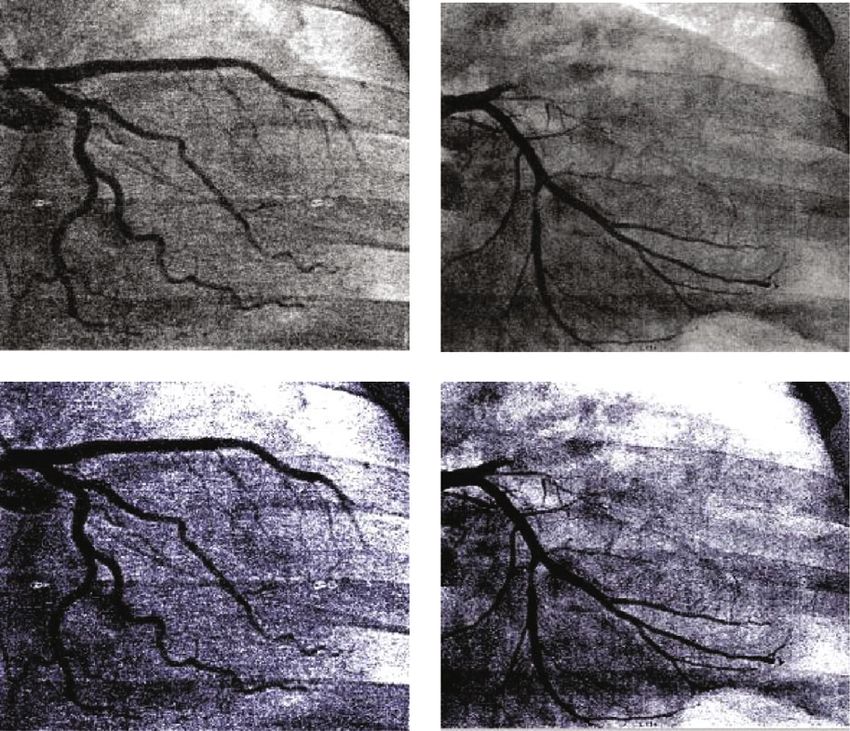
3.2. Display of Influence Data of Typical Cases. The images of
classic cases before and after DSA angiography in the two
1
gðx, yÞ = 〠 f ði, jÞ: ð1Þ groups are shown in Figure 1. CAG can clearly reflect the
M ði, jÞ∈s location and degree of coronary artery stenosis in patients.
After the neural network algorithm, the image quality is
x, y = 0, 1, 2 ⋯ , N − 1, S is the domain set centered on significantly improved, the lesion is more prominent, and
ðx, yÞ points, and M is the total number of coordinate the details of the lesion are also well displayed.
points in S. 3.3. Comparison of Coronary Stenosis Degree between the
For multiple images, if the original image is f ðx, yÞ Two Groups. The comparison of the degree of coronary
and the noise is n ðx, yÞ, then the noise image g ðx, yÞ is stenosis between the two groups is shown in Figure 2.
as follows: According to the analysis of Figure 2, the number of patients
with4 Computational and Mathematical Methods in Medicine
Table 1: Comparison of general data of two groups of patients.
STEMI NSTEMI
Item P
n = 44 n = 36
Age (years) 63:11 ± 11:06 59:88 ± 11:76 0.07
Hypertension (%) 53.88 55.9 0.32
Diabetes (%) 49.8 39.9 0.13
Smoking (%) 27.81 32.1 0.18
Heart rate (beat/min) 77:73 ± 10:22 68:03 ± 10:16 0.13
Systolic pressure (mmHg) 137:02 ± 18:31 136:14 ± 17:94 0.09
Diastolic blood pressure (mmHg) 85:07 ± 14:44 86:28 ± 9:66 0.14
White blood cells (×109) 8:53 ± 2:17 8:65 ± 2:09 0.13
Low density lipoprotein (mmol/L) 2:43 ± 0:81 2:64 ± 0:53 0.15
Triglyceride (mmol/L) 1:39 ± 0:46 1:48 ± 0:68 0.12
Fibrinogen (g/L) 2:83 ± 0:54 3:09 ± 0:71 0.13
Blood glucose (mmol/L) 9:11 ± 3:93 9:06 ± 4:43 0.23
CK-MB (U/L) 85:83 ± 20:15 84:09 ± 18:36 0.33
cTnT (ng/L) 1:36 ± 0:81 0:83 ± 0:68 0.069
NT-proBNP 1925:81 ± 2284:3 1918:84 ± 1147:2 0.38
EF (%) 55:66 ± 8:33 57:68 ± 9:33 0.33
Left ventricular end diastolic diameter (mm) 45:7 ± 3:77 49:35 ± 4:47 0.51
STEMI group NSTEMI group number of patients with complete occlusion (3 cases vs. 12
cases) and collateral circulation (5 cases vs. 20 cases) in the
NSTEMI group was significantly higher than that in the
STEMI group (P < 0:05). There was no significant difference
Before treatment
in the number of patients with stenosis degree between
75%~99% and 50%~74% between the two groups (P > 0:05).
3.4. Comparison of Lesion Sites between the Two Groups. The
comparison results of lesion sites between the two groups are
shown in Figure 3. Figure 3 shows 9 cases of proximal LAD,
5 cases of middle LAD, and 1 case of distal LAD in the
After processing
STEMI group. There were 5 proximal lesions, 9 middle
lesions, and 5 distal lesions in RCA. There were 6 cases of
proximal lesions and 4 cases of distal lesions in LCX. In
LAD of the NSTEMI group, there were 2 cases of proximal
lesions, 5 cases of middle lesions, and 10 cases of distal
lesions. There were 2 cases of proximal lesions, 1 case of
middle lesions, and 4 cases of distal lesions in RCA. There
Figure 1: DSA image display of typical cases.
was 1 case of proximal lesions and 11 cases of distal lesions
in LCX. There was significant difference between the two
groups in the lesion location between the distal end of
11. The number of patients with narrowing degree between LAD (1 case vs. 10 cases) and the distal end of LCX (4 cases
75% and 99% is 22, the number of patients with complete vs. 11 cases), P < 0:05.
occlusion is 3, and the number of patients with collateral
circulation is 5. The number of patients withComputational and Mathematical Methods in Medicine 5
Collateral circulation ⁎
Totally occluded ⁎
75%~99% ⁎
50%~74% ⁎6 Computational and Mathematical Methods in Medicine
short-term prognosis, high long-term mortality, and poor < 0:05. There was statistically significant difference in the
prognosis. The blood vessels of patients with NSTEMI are number of coronary artery lesions between the two groups
mostly incomplete occlusion lesions. The formation mecha- with 1 (1 vs. 18) and 3 (25 vs. 12), P < 0:05. This indicated
nism of collateral circulation is not clear. There are also that STEMI and NSTEMI as the entry point for diagnosis
many disputes in the treatment strategy and timing. There- and analysis of disease had high clinical application value.
fore, the understanding of AMI needs to be further The convolutional neural network algorithm shows better
improved [23]. For the diagnosis of AMI, CAG is still the performance in DSA image processing of AMI patients.
gold standard for its diagnosis [24]. In recent years, CAG This study provides a reference and basis for clinical diag-
has been widely used in clinical practice, but there are also nosis of AMI. However, due to the limited samples, this
many shortcomings and limitations of this technology [25, study is not comprehensive and in-depth enough. In
26]. In recent years, ultrasonic technology has been rapidly future study and work, it will be further expanded to con-
developed. Compared with CAG, it has the advantages of tinue to study this issue.
low cost, simple operation, and easy repetition. However,
an echocardiogram also has some limitations, such as its Data Availability
diagnostic accuracy for cardiovascular diseases with complex
conditions of multivessel disease is not high [27]. The data used to support the findings of this study are avail-
In recent years, the Internet and information technology able from the corresponding author upon request.
have flourished. The corresponding various image process-
ing technologies have also been rapidly developed, and med-
ical image processing technology is one of the hot and key of
Conflicts of Interest
research [28]. The deep convolution neural network algo- The authors declare no conflicts of interest.
rithm is one of the most valuable and potential methods in
the field of image processing and analysis. In the field of
medical imaging, it is mainly used for the segmentation of Authors’ Contributions
lesions or the establishment of prognosis models of some
Guanglin Cao and Zheng Zhao contributed equally to this
tumors and cerebral hemorrhage, but it is rarely used in
work.
the field of cardiac diseases [29, 30]. In this study, the imag-
ing data of different types of AMI patients such as DSA were
studied on the basis of image processing by a convolution References
neural network algorithm. It is found that the quality of
[1] E. Mahmud, H. L. Dauerman, W. FGP et al., “Management of
DSA image processed by a convolution neural network algo-
acute myocardial infarction during the COVID-19 pandemic:
rithm is more prominent. The most STEMI patients are cor- a consensus statement from the Society for Cardiovascular
onary artery stenosis of 50%-74% and 75%-99%. Patients in Angiography and Interventions (SCAI), the American College
the NSTEMI group had the most coronary artery stenosis of Cardiology (ACC), and the American College of Emergency
with complete occlusion and collateral circulation. The Physicians (ACEP),” Catheterization and Cardiovascular
lesions of the STEMI group were mostly concentrated in Interventions, vol. 96, no. 2, pp. 336–345, 2020.
the proximal LAD and middle RCA. The lesions of the [2] C. Vahdatpour, D. Collins, and S. Goldberg, “Cardiogenic
NSTEMI group were mostly concentrated in the distal shock,” Journal of the American Heart Association, vol. 8,
LAD and distal LCX. The number of diseased coronary no. 8, article e011991, 2019.
arteries in the STEMI group was two and three. The number [3] C. V. Pollack, A. Amin, T. Wang et al., “Contemporary
of diseased coronary arteries in the NSTEMI group was NSTEMI management: the role of the hospitalist,” Hosp Pract
mostly 1. In summary, the digital silhouette technology (1995), vol. 48, no. 1, pp. 1–11, 2020.
based on the convolutional neural network algorithm had a [4] G. Scalone, G. Niccoli, and F. Crea, “Editor’s Choice-Patho-
good effect and application value on the distribution charac- physiology, diagnosis and management of MINOCA: an
teristics of STEMI and NSTEMI culprit lesions in AMI update,” European Heart Journal Acute Cardiovascular Care,
patients. This was consistent with the conclusions of previ- vol. 8, no. 1, pp. 54–62, 2019.
ous related studies. [5] N. Kim, J. H. Lee, S. Y. Jang et al., “Korea Acute Myocardial
Infarction Registry-National Institute of Health Investigators.
Intravascular modality-guided versus angiography-guided
5. Conclusion percutaneous coronary intervention in acute myocardial
infarction,” Catheterization and Cardiovascular Interventions,
In this study, the imaging data of different types of AMI vol. 95, no. 4, pp. 696–703, 2020.
patients such as DSA were studied on the basis of a convolu- [6] K. Ameloot, P. Jakkula, J. Hästbacka et al., “Optimum blood
tion neural network algorithm. The number of patients with pressure in patients with shock after acute myocardial infarc-
complete occlusion (3 cases vs. 12 cases) and collateral circu- tion and cardiac arrest,” Journal of the American College of
lation (5 cases vs. 20 cases) in the NSTEMI group was signif- Cardiology, vol. 76, no. 7, pp. 812–824, 2020.
icantly higher than that in the STEMI group, with statistical [7] D. Tedeschi, A. Rizzi, S. Biscaglia, and C. Tumscitz, “Acute
significance (P < 0:05). There was statistical difference in the myocardial infarction and large coronary thrombosis in a
number of lesions in distal LAD (1 case vs. 10 cases) and patient with COVID-19,” Catheterization and Cardiovascular
distal LCX (4 cases vs. 11 cases) between the two groups, P Interventions, vol. 97, no. 2, pp. 272–277, 2021.Computational and Mathematical Methods in Medicine 7
[8] J. A. Dodson, A. Hajduk, J. Curtis et al., “Acute kidney injury risk patients with acute chest pain,” BMJ Evidence-Based Med-
among older patients undergoing coronary angiography for icine, vol. 24, no. 5, article e5, 2019.
acute myocardial infarction: the SILVER-AMI study,” The [23] M. K. Mahowald, F. Alqahtani, and M. Alkhouli, “Comparison
American Journal of Medicine, vol. 132, no. 12, pp. e817– of outcomes of coronary revascularization for acute myocar-
e826, 2019. dial infarction in men versus women,” The American Journal
[9] M. K. Erol, M. Kayıkçıoğlu, M. Kılıçkap et al., “Treatment of Cardiology, vol. 132, pp. 1–7, 2020.
delays and in-hospital outcomes in acute myocardial infarc- [24] A. Tamenishi, Y. Fujimoto, and C. Teramoto, “Giant right cor-
tion during the COVID-19 pandemic: a nationwide study,” onary artery aneurysm causing acute myocardial infarction;
Anatolian Journal of Cardiology, vol. 24, no. 5, pp. 334–342, report of a case,” Kyobu Geka, vol. 72, no. 8, pp. 616–618, 2019.
2020.
[25] M. Chyrchel, T. Gallina, O. Szafrański, Ł. Rzeszutko,
[10] T. S. Gilhofer and J. Saw, “Spontaneous coronary artery dissec- A. Surdacki, and S. Bartuś, “Comparison of the characteristics
tion: update 2019,” Current Opinion in Cardiology, vol. 34, of coronary interventions performed during day and night
no. 6, pp. 594–602, 2019. shifts in patients with acute myocardial infarction,” Interna-
[11] M. B. Rasmussen, C. Stengaard, J. T. Sørensen et al., “Compar- tional Journal of Environmental Research and Public Health,
ison of acute versus subacute coronary angiography in patients vol. 17, no. 15, p. 5378, 2020.
with NON-ST-elevation myocardial infarction (from the [26] Y. Zou and B. Zhang, “Clinical characteristics and prognosis of
NONSTEMI trial),” The American Journal of Cardiology, myocardial infarction with non-obstructive coronary arteries,”
vol. 124, no. 6, pp. 825–832, 2019. Zhonghua Nei Ke Za Zhi, vol. 59, no. 7, pp. 546–549, 2020.
[12] B. El-Sabawi, I. S. Shadrin, G. S. Sandhu, J. A. Crestanello, and [27] D. Mukherjee, “Myocardial infarction with nonobstructive
A. S. Jaffe, “Acute myocardial infarction due to fixed coronary coronary arteries: a call for individualized treatment,” Journal
artery stenosis from myocardial bridging,” Cardiovascular of the American Heart Association, vol. 8, no. 14, article
Revascularization Medicine, vol. 21, no. 11S, pp. 91–93, 2020. e013361, 2019.
[13] L. Wang, J. Li, Y. Gao et al., “Association between coronary [28] P. Tornvall, A. Göransson, J. Ekman, and H. Järnbert-Petters-
dominance and acute inferior myocardial infarction: a son, “Myocardial infarction in systemic lupus erythematosus:
matched, case-control study,” BMC Cardiovascular Disorders, incidence and coronary angiography findings,” Angiology,
vol. 19, no. 1, p. 35, 2019. vol. 72, no. 5, pp. 459–464, 2021.
[14] Q. Sheng, H. Zhao, S. Wu, and R. Liu, “Underlying factors [29] A. Jánosi, T. Ferenci, Z. Kőszegi et al., “Obstruktív koszorúér-
relating to acute myocardial infarction for coronary artery betegség nélkül kialakuló heveny szívizominfarktus (MINOCA)
ectasia patients,” Medicine (Baltimore), vol. 99, no. 36, article – gyakoriság és prognózis [Myocardial infarction without
e21983, 2020. obstructive coronary artery disease (MINOCA) - prevalence
[15] C. Yerasi, B. C. Case, B. J. Forrestal et al., “Treatment of ST- and prognosis],” Orvosi Hetilap, vol. 160, no. 45, pp. 1791–
segment elevation myocardial infarction during COVID-19 1797, 2019.
pandemic,” Cardiovascular Revascularization Medicine, [30] M. J. A. Williams, P. R. Barr, M. Lee, K. K. Poppe, and A. J.
vol. 21, no. 8, pp. 1024–1029, 2020. Kerr, “Outcome after myocardial infarction without obstruc-
[16] J. R. McConaghy, M. Sharma, and H. Patel, “Acute chest pain tive coronary artery disease,” Heart, vol. 105, no. 7, pp. 524–
in adults: outpatient evaluation,” American Family Physician, 530, 2019.
vol. 102, no. 12, pp. 721–727, 2020.
[17] S. Haruta and K. Arai, “Acute myocardial infarction caused by
coronary spasm and dissection treated with medical therapy,”
International Heart Journal, vol. 61, no. 1, pp. 169–173, 2020.
[18] K. Wakabayashi, T. Nishikura, T. Shinke, and K. Tanno,
“Acute myocardial infarction caused by persistent coronary
spasm associated with high-grade macrophage accumulation,”
BML Case Reports, vol. 13, no. 3, article e234502, 2020.
[19] J. Legutko, Ł. Niewiara, S. Bartuś et al., “Decline in the number
of coronary angiography and percutaneous coronary interven-
tion procedures in patients with acute myocardial infarction in
Poland during the coronavirus disease 2019 pandemic,” Kar-
diologia Polska, vol. 78, no. 6, pp. 574–576, 2020.
[20] A. A. Harhash, J. J. Huang, S. Reddy et al., “aVR ST segment
elevation: acute STEMI or not? Incidence of an acute coronary
occlusion,” The American Journal of Medicine, vol. 132, no. 5,
pp. 622–630, 2019.
[21] N. Shah, V. Agarwal, P. C. Olson, A. Naniwadekar,
A. Agarwal, and N. C. Patel, “Trends and predictors of cor-
onary revascularization in patients with coronary artery
anomalies and acute myocardial infarction: a nationwide
analysis of 8131 patients,” Coronary Artery Disease,
vol. 31, no. 4, pp. 327–335, 2020.
[22] P. D. Adamson and M. P. Than, “CT coronary angiography
does not reduce mortality or myocardial infarction in low-You can also read