Effectiveness Of Different Corticosterone Administration Methods To Alter Corticosterone Levels In Serum And Depressive-Like Behavior In Female Rats
←
→
Page content transcription
If your browser does not render page correctly, please read the page content below
Wayne State University

Wayne State University Theses

1-1-2015

Effectiveness Of Different Corticosterone
Administration Methods To Alter Corticosterone
Levels In Serum And Depressive-Like Behavior In
Female Rats
Jennifer Marie Kott
Wayne State University,

Follow this and additional works at: http://digitalcommons.wayne.edu/oa_theses
     Part of the Psychology Commons

Recommended Citation
Kott, Jennifer Marie, "Effectiveness Of Different Corticosterone Administration Methods To Alter Corticosterone Levels In Serum
And Depressive-Like Behavior In Female Rats" (2015). Wayne State University Theses. Paper 418.

This Open Access Thesis is brought to you for free and open access by DigitalCommons@WayneState. It has been accepted for inclusion in Wayne
State University Theses by an authorized administrator of DigitalCommons@WayneState.
       EFFECTIVENESS OF DIFFERENT CORTICOSTERONE ADMINISTRATION
         METHODS TO ALTER CORTICOSTERONE LEVELS IN SERUM AND
                DEPRESSIVE-LIKE BEHAVIOR IN FEMALE RATS

                                         by

                              JENNIFER M. KOTT

                                      THESIS

                         Submitted to the Graduate School

                            of Wayne State University,

                                 Detroit, Michigan

                      in partial fulfillment of the requirements

                                  for the degree of

                               MASTER OF ARTS

                                        2015

                                        MAJOR: PSYCHOLOGY

                                        Approved By:

                                        ____________________________________
                                        Advisor                         Date

                                          i
                                     DEDICATION

       This thesis is dedicated to my parents, Paul and Diane Kott for their love, support,

and encouragement throughout my life and academic career, and to all of my little rats

that were sacrificed for this study and the advancement of scientific research.

                                             ii
                             ACKNOWLEDGEMENTS

       This work would not have been made possible without the guidance of my

advisor, Dr. Susanne Brummelte, and my thesis committee members, Dr. Scott Bowen

and Dr. Michelle Tomaszycki. I would also like to thank Dr. Scott Bowen and Dr.

George Borszcz who provided access to their labs and the necessary equipment, without

which portions of this study would have been impossible to conduct. I would also like to

thank members of Dr. Brummelte’s lab who provided invaluable assistance in data

collection and analysis: Sean Mooney-Leber, Fay Shoubah, Jill Young, Zach Bruckley,

Manell Aboutaleb, and Susan Woods.

                                          iii
                                  TABLE OF CONTENTS

Dedication                                               ii

Acknowledgements                                       iii

List of Tables                                         vii

List of Figures                                        viii

1. “Introduction”                                        1

       1.1. Administration by Subcutaneous Pellet        5

       1.2. Administration by Drinking Water             5

       1.3. Administration by Subcutaneous Injection     6

2. “Method”                                              7

       2.1. Subjects                                     7

       2.2. Hormone Preparation and Administration       7

                  2.2.1. Pellet Implantation             8

                  2.2.2. Subcutaneous Injection          9

                  2.2.3. Drinking Water                 9

       2.3. Vaginal Lavage                             10

       2.4. Blood Collection and CORT Assay            10

       2.5. Behavioral Testing                         11

                  2.5.1. Forced Swim Test              11

                  2.5.2. Open Field Test               12

       2.6. Immunohistochemistry                       12

       2.7. Cell Counting                              13

       2.8. Data Analysis                              14

                                               iv
3. “Results”                                                                      14

       3.1. Body Weight                                                           14

               3.1.1. Water Intake                                                15

       3.2. Serum CORT Levels                                                     15

       3.3. Forced Swim Test Behavior                                             17

       3.4. Open Field Test                                                       18

       3.5. Neurogenesis in the Dentate Gyrus                                     18

4. “Discussion”                                                                   18

       4.1. Route of CORT Administration Affects Body Weight                      19

       4.2. Different Administration Methods Lead to Varying CORT Serum Levels    20

       4.3. Only Injections of CORT Resulted in Increased Immobility in the FST   23

       4.4. Chronic CORT Administration Did Not Alter Neurogenesis Levels         25

       4.5. Considerations for Choosing a CORT Administration Method              26

References                                                                        29

Abstract                                                                          35

Autobiographical Statement                                                        36

                                          v
                        LIST OF TABLES

Table 1: OFT …………………………………………………………………………… 44

Table 2: Hippocampal Neurogenesis (DCX) …………………………………………... 45

                              vi
LIST OF FIGURES
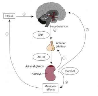
Figure 1: HPA axis …………………………………………………………………….. 38
Figure 2: Experimental timeline ……………………………………………………….. 39
Figure 3: Body weight …………………………………………………………………. 40
Figure 4: CORT levels in serum ……………………………………………………….. 41
Figure 5: CORT after FST ……………………………………………………………... 42
Figure 6: FST immobility ……………………………………………………………… 43
vii 1
1. INTRODUCTION
Stress is defined as a state that threatens or gives the perception of a threat to a
person’s physiological equilibrium (Weinstock, 2005). Stress is experienced when an
individual encounters a perceived threat and undergoes the biological response that
prepares the body for a reaction. A common term to describe this reaction is the “fight-or-
flight” response. In this response, stress from the environment activates the hypothalamus
to release corticotropin-releasing hormone (CRH). CRH then signals the pituitary gland
to release adrenocorticotropic hormone (ACTH), which stimulates the adrenal gland to
release stress hormones, most notably cortisol, into the bloodstream (see Figure 1). This
network of structures that prepares the body to react to the perception of a threat is known
as the hypothalamus-pituitary-adrenal (HPA) axis. It is through this network that stress,
especially when experienced chronically, is able to exert its detrimental effects on the
body. If there is an over-activation of this system, inflammatory markers may become
elevated, due to what is known as stress-induced immunocompromise. Elevated levels of
inflammatory markers are associated with pathophysiologic states such as an increased
risk for contracting infectious diseases and poor ability to heal (Stix, 2007).
The involvement of these inflammatory markers in the experience of stress makes
women especially vulnerable to the effects of stress, especially during pregnancy. This is
particularly important, as there is high comorbidity between stress and depression in
women, particularly during their reproductive years. Pregnant women undergoing high
levels of stress or depression are increasingly vulnerable to effects such as preterm birth
and lower birth weight of the infant. These results stem directly from the HPA axis
activation in response to stress. Increased inflammatory response stimulates lymphocytes 2
to release cytokines, prostaglandins, and interleukins. Cytokines and prostaglandins are
involved in controlling the contractions of uterine muscle, which may affect preterm
labor and delivery, and cortisol released in response to stress is also closely linked to the
process of childbirth. Most notably, cortisol is the most important modulator of the
timing of normal parturition (Bulletins--Obstetrics, 2003). Cortisol is also involved in
oxytocin release from the pituitary gland, which is a necessary process for the occurrence
of the uterine contractions required for childbirth. Stress can complicate this experience
by causing a premature release of cortisol, which could lead to preterm labor and
delivery. This possibility depends on the stressor itself, the duration of pregnancy at the
time of the stress, and other risk factors such as anxiety, depression, and other
psychosocial factors (Cardwell, 2013). Due to the unique role of these hormones in
women and the prevalence of stress and depression in women during pregnancy, it is
necessary to study the effects of stress particularly in women and not just generalize
findings from studies on the effects of stress in males.
Due to the subjective nature of stress in humans and the large variety of confounds
incorporated in human research, a rodent model to study the effects of stress on
pregnancy and the subsequent development of the offspring is essential. In the process of
developing this model to study development, it is first necessary to determine how best to
administer stress to female rodents to induce a depressive-like phenotype. This goal is
hindered by variation in the results in current research, both based on administration of
stress and differential results between stress administered to male rodents and stress
administered to females, making it difficult to draw conclusions. 3
There are many commonly used methods for inducing stress in rodents. Most
frequently, these involve paradigms such as restraint stress, chronic mild stress, or cold
stress. Stress by these methods are subject to several confounds, including variation in
techniques between labs and individual differences in rodents between stressful
measures. For example, one animal may be particularly sensitive to the effects of cold
stress but less so to restraint stress. There are also several different protocols for chronic
mild stress paradigms and each method likely produces variability in the results.
Furthermore, rats are readily habituate to predictable stressors which means these types
of stressors eventually do not elicit elevated levels of CORT over time (De Boer et al.,
1989; Herman, 2013). In order to avoid these pitfalls, we decided to employ an
alternative method of stress by administering exogenous corticosterone (CORT).
Corticosterone is the analog to cortisol, and both are the end-products of HPA activation
in humans and rats, respectively. Previous research has given CORT through 3 different
administration methods: a) time-release pellet implantation, b) in the drinking water, and
c) subcutaneous injection.
These 3 administration methods are frequently used and well-established in the
literature to induce a depressive-like phenotype in rodents. Interestingly, previous
research has shown that these administration methods produce differential effects in
females as compared to male rats (Dalla et al., 2010). While females appear to be more
sensitive to the stressful effects of the forced-swim test than are males, females display
less helplessness behavior in response to foot shock than do males (Dalla et al., 2008).
The sex differences in stress response may result from the unique physiology of females
and may be related to the female’s role as a caregiver in many mammalian species. 4
Indeed it has been hypothesized that females may have developed a different behavioral
response to stress than males that serves to be more adaptive during their vulnerable
states during pregnancy and nursing. Further, females have naturally higher levels of
circulating CORT as compared to males, which may contribute to the significant sex
difference in stress sensitivity between males and females. Due to these differences, it is
necessary to find a method to administer exogenous CORT that is effective to develop a
depressive-like phenotype in female rats.
A depressive-like phenotype in rodents has been characterized by several features:
elevated CORT levels in blood serum, decreased body weight, an increase in immobility
in the forced swim test, and a decrease in hippocampal neurogenesis. It has been
hypothesized that HPA axis dysregulation is incorporated in that pathology of depression,
and as such, is able to modulate the behavioral effects of stress. This theory is supported
in the human literature. One study in particular demonstrated that patients suffering from
depression whose HPA axis regulation improved after one week of pharmacological
treatment had lower depression scores and shorter admission times compared to patients
that continued to display HPA axis dysregulation (Schule et al., 2009). Therefore, it is
likely that the HPA axis serves to regulate the body’s response to stress and is
incorporated in its behavioral and physiological manifestations. As it may be impossible
to achieve a true model of depression in rodents, the goal of this research was to mimic as
many characteristics of the depressive-like phenotype of rodents as possible. To do this,
this study used 3 administration routes (a) time-release pellet implantation, b) in the
drinking water, and c) subcutaneous injection) to determine the efficacy of each in the
development of a depressive-like phenotype in female rodents. 5
1.1. Administration by Subcutaneous Pellet
Time-release pellets have been used in the literature to administer CORT in
rodents (Claflin et al., 2014; Claflin et al., 2005; Kraszpulski, et al., 2012; Murray et al.,
2008). Briefly, the rat is placed under anesthesia and the pellet is surgically implanted in
a pocket under the skin in the lateral neck area. The pellets are designed to release a
specific dose of CORT over 21 days before dissolving completely. In addition to the
surgical implantation and necessity of anesthesia, another factor that must be taken into
account with the time-release pellets is that the dose cannot be adjusted after
implantation, i.e., for weight loss or grain. However, there are no further invasive
procedures after the surgery and CORT is delivered at a constant dose throughout the
testing period. Research using these pellets has demonstrated their efficacy in increasing
CORT levels in serum, increasing immobility in the forced swim test, and decreasing
hippocampal neurogenesis in male rodents (Murray et al., 2008).
1.2. Administration by Drinking Water
While CORT has very limited water solubility, a related compound,
corticosterone hemisuccinate, is readily water soluble and can be easily incorporated into
a drinking water solution for administration to rodents, as demonstrated in previous
research (Casolini et al., 207; Catalani et al., 2002; Catalani et al., 2000; Catalani et al.,
1993). This method using CORT hemisuccinate has been frequently used to study the
effects of prenatal stress on development due to the minimally invasive nature of its
administration (Casolini et al., 207; Catalani et al., 2002; Catalani et al., 2000; Catalani et
al., 1993). Administration of CORT via drinking water has been shown to elevate levels
of CORT in serum in female rats (Catalani et al., 1993; Catalani et al., 2000; Catalani et 6
al., 2003) and increase immobility in the forced swim test in male rats (Donner et al.,
2012). A consideration that needs to be made for administration via drinking water is that
the solution will be consumed ad libitum, and as such, the dose or time of consumption
cannot be directly controlled by the researcher.
1.3. Administration by Subcutaneous Injection
Administering CORT through a subcutaneous injection has the advantage of
being under a great deal of control by the investigator. The experimenter is able to ensure
the exact dose is given and is able to adjust the dose in the event of weight loss or weight
gain throughout the course of the experiment. This method has been shown effective in
increasing immobility in the forced swim test in male rats (Hill et al., 2003) and alter
levels of hippocampal neurogenesis (Brummelte & Galea, 2010; Mirescu & Gould, 2006;
Wong & Herbert, 2006). A limitation of administration by injection, however, is that the
full dose of CORT is administered at the time of the injection and is not steadily
increased throughout the day.
This study aimed to evaluate the effectiveness of these 3 administration methods
to generate a depressive-like phenotype in female rats by chronic CORT administration
(21 days) at a dose of 40 mg/kg as used in the literature. As the majority of the literature
on this topic is in male rats, it is critical to elucidate the most effective method for CORT
administration in female rats. The goal of this experiment is to provide data for the future
investigation of the effects of stress before pregnancy on development, so in addition to
the efficacy of these methods, the levels of their invasiveness were also considered for
future use. The measures taken were: CORT levels in serum, body weight, immobility in
the forced swim test, and reduced levels of hippocampal neurogenesis using doublecortin 7
(DCX), which taken together are indicative of a depressive-like phenotype. To our
knowledge, this is the first study to compare across CORT administration methods in
female rodents on their ability to produce a depressive-like phenotype.
2. METHOD
2.1. Subjects
Thirty-six female rats (Sprague-Dawley, age 77-84 days) were purchased from
Charles River Laboratory (Portage, MI, USA) and were housed in a 12:12 hour light:dark
cycle (lights on at 06:00) with food (Rodent Lab Diet 5001; PMI, Nutrition International,
Inc., Brentwood, MO) and water available ad libitum. Animals were group-housed upon
arrival and single-housed after a 3-day acclimation period. CORT was administered via 3
different routes, each with a corresponding control group for which the female rats were
randomly assigned to 1 of 6 total groups: CORT pellet (n=6), control pellet (n=6), CORT
injection (n=6), vehicle injection (n=6), CORT in drinking water (n=6), and control
drinking water (n=6). All animals were weighed every 3 days.
2.2. Hormone Preparation and Administration
CORT or vehicle was administered for 23 days with a wean-off period from days
21 to 23 (see Fig. 2). The wean-off period for the injection and drinking water groups was
to mimic the potential decrease in CORT release in the implanted pellets. The dosages for
each administration group were calculated to reflect ~40 mg/kg/day (i.e., ~10 mg/day for
a 250 g female rat). Thus, the injection group received 40 mg/kg/day via injection. The
pellets contained 200 mg, which was designed to be released over 21 days (200 mg/21 d
= 9.52 mg/day; highest dose available). The dose for the drinking water (200 µg/mL) was
based on previous studies using CORT in the drinking water with the same concentration 8
which has previously been reported to result in 13.5 ± 1.5 mg daily CORT intake
(Catalani et al., 2002; Catalani et al., 2000; Catalani et al., 1993). An average daily water
consumption of 30-50 mL in rats would reflect ~10 mg/day of CORT exposure (200
µg/mL * 50 mL/day = 10mg/day; Ray & Behari, 1990; Stricker et al., 1997).
2.2.1. Pellet Implantation
Both CORT (#G-111, 200 mg, 21-day release) and placebo pellets (#C-111) were
purchased from Innovative Research of America (Sarasota, FL, USA) and implanted as
instructed by the manufacturer. Briefly, animals were anesthetized with isoflurane and
the flow rate was maintained at a rate of 2.5-3% throughout the procedure. While the use
of anesthesia may have been a potential limitation in these groups, it was required for the
implantation surgery. Once deeply anesthetized, eye-lubricant was applied and animals
received a subcutaneous injection of the non-steroidal anti-inflammatory analgesic,
Carprofen (5 mg/kg). The surgical site was shaved and disinfected and a small incision
(~2.5-3 cm) was made in the left shoulder area using aseptic techniques. The pellet was
placed in a subcutaneous pocket in the lateral neck area in accordance with the
manufacturer’s instructions. The incision site was sealed using sterile wound clips.
Animals were weighed before being returned to a clean cage. Post-surgical monitoring
was conducted every 5 minutes for 45-60 minutes until the animal was mobile and alert.
Monitoring continued at less frequent intervals after the animal was returned to the
standard housing cage and once daily for 3 weeks after surgery. Triple antibiotic ointment
was applied to the incision site daily for 3 days following surgery and as needed after the
third day. Wound clips were removed 7 to 10 days after the surgical procedure under 9
brief isoflurane anesthesia. As the pellets should have been fully dissolved after the 23-
day period, there was no attempt to surgically remove them at the end of this period.
2.2.2. Subcutaneous Injection
CORT (C2505, Sigma-Aldrich, St. Louis, MO, USA) was administered daily
subcutaneously at a dose of 40 mg/kg. CORT was prepared fresh every 2 to 3 days by
suspending 40 mg/ml CORT in sesame oil containing 10% ethanol. The control group
received an injection of sesame oil vehicle alone. Ethanol was deemed unnecessary to
incorporate into the control group, as it acts only to assist in dissolving the CORT into
solution and would only evaporate if incorporated into the sesame oil vehicle. Injections
were performed between 08:00 and 10:00 for 23 days. The dose of corticosterone was
decreased over the last injection days (days 21-d23) so that animals received 30 mg/kg on
day 21, 20 mg/kg on day 22, and 10 mg/kg on day 23 to mimic the wean-off period that
would be experienced with the pellet implantation.
2.2.3. Drinking Water
CORT hemisuccinate (#Q1562-000, Steraloids, Newport, RI, USA) was dissolved
at a dose of 200 µg/mL in drinking water. The use of CORT hemisuccinate was necessary
as CORT is difficult to dissolve in water. The added salt group in CORT hemisuccinate
makes it readily soluble in water and its efficacy has been demonstrated previously
(Catalani et al., 2002; Catalani et al., 2000; Catalani et al., 1993). The CORT-water was
administered in standard drinking bottles that were wrapped in aluminum foil due to the
potential light sensitivity of the solution. The control group received regular tap water,
also in bottles wrapped in aluminum foil to control for any effects of the novelty of
aluminum foil impacting drinking behavior. All bottles were weighed daily and refilled 10
with fresh CORT or control water every 4 days or as needed. After 20 days, the dose of
the CORT water was reduced to 150 µg/mL on day 21, 100 µg/mL on day 22, and 50
µg/mL on day 23 to mimic the wean-off period that would be expected with the pellet
administration. On day 24, all injections were stopped and all animals were returned to
normal tap water. We confirmed that all pellets were fully dissolved on the day of
sacrifice.
2.3. Vaginal Lavage
All animals were lavaged on the days of testing to determine the estrus cycle
phases. This data was used as a covariate in the analysis of the behavioral data to ensure
that the phase of the estrus cycle did not affect behavior in the tasks. Vaginal lavage was
performed by administering 3 to 4 drops of water into the vagina with an eyedropper and
immediately reabsorbing the fluid with the same eyedropper. The sample was then placed
on a glass microscope slide and stained with Cresyl Violet before the sample was dry.
Once completely dry, each sample was examined under a microscope and the estrous
cycle phase (metestrus, diestrus, proestrus, estrus) was determined based on the type of
cells present as previously described (Marcondes et al., 2002; McLean et al., 2012). This
data was later used to control for effects of phase of the estrous cycle on behavior in the
data analysis.
2.4. Blood Collection and CORT Assay
Blood samples were collected early during the CORT administration on day 3
(d3), near the end of the administration period (d18), and a full day after being weaned
off of the corticosterone (d25). Further, a blood sample was collected right after the first
forced swim test session on day 20 to measure the CORT levels evoked by the test as an 11
acute stressor. Blood was collected by poking the saphenous vein with a sterile 25-gauge
needle and allowing the sample to fall into a blood collection tube. The collection area
was shaved the day before collection to ensure rapid blood collection that lasted less than
3 minutes from the time the experimenter first touched the animal’s cage. CORT levels
quickly rise in response to stress, so the 3-minute time limit ensures consistent collection
points across animals with the stressful experience being the experimenter touching the
cage. All basal samples were collected in the morning between 08:00 and 10:00 to
ensure consistency in the CORT levels by being taken at the trough of the diurnal CORT
rhythm and kept on ice. Blood samples were allowed to clot overnight and spun down at
8,000G for 10 minutes the next day. Serum was extracted and stored at -20C until further
processing. Serum CORT was analyzed using a standard corticosterone EIA kit (Arbor
Assays; Cat. # K014-H5, Ann Arbor, MI, USA) following the manufacturer’s instruction.
2.5. Behavioral Testing
2.5.1. Forced Swim Test
Depressive-like behavior was assessed in the Porsolt Forced Swim Test (FST) on
days 20, 21, and 29 as previously described (Porsolt et al., 1978). Briefly, animals were
placed individually into a Plexiglas container (44cm deep, 30cm in diameter, Noldus
Information Technology, Leesburg, VA, USA) filled with tap water at 25 ± 1C. The
animal was placed in the tank for 15 minutes during the first session (d20) for habituation
to the task, and 10 minutes for sessions 2 (d21) and 3 (d29). Session 2 was conducted at
the end of the CORT administration period and session 3 was conducted after the
washout from the CORT administration. All sessions were videotaped and later manually
scored by an investigator blind to the animals’ conditions for swimming, struggling, or 12
immobile behavior. The investigator would press a key on the keyboard at the start of
each behavior and the Noldus computer software would total the duration of time spent in
each behavior. Behaviors were defined as (1) swimming – movement of the forelimbs or
hind limbs in a paddling fashion; (2) struggling – quick movements of the forelimbs with
the front paws breaking the surface of the water; or (3) immobility – floating with no
additional movement than what is required to stay afloat.
2.5.2. Open Field Test
The Open Field Test (OFT) was conducted on d19 to assess activity level of the
animals. This was conducted in a circular arena with a diameter of 180cm and 50cm high
walls (Jain et al., 2011). Movement was tracked during a 5-minute session using Noldus
Ethovision® XT Version 7.0 software (Leesburg, VA, USA). The arena was cleaned with
70% ethanol between animals. Measures taken were central entries, defined as entries
into the inner circle (30cm from the wall) and overall distance travelled.
2.6. Immunohistochemistry
To probe for effects of the different delivery methods on hippocampal
neurogenesis, we collected brains from all animals on d30. To do this, animals were
deeply anesthetized with sodium pentobarbital and perfused transcardially. Following
extraction, brains were post-fixed in 4% paraformaldehyde for 24 hours and then
transferred to 30% sucrose in phosphate buffered saline (PBS until sectioning. Coronal
sections (40µm) were obtained using a freezing microtome (Microm HM 450, Thermo
Scientific, Ann Arbor, MI, USA) and stored in antifreeze until staining. Every 10th slice
was processed for Doublecortin (DCX), an endogenous marker of new neurons. Slices
were rinsed in 0.1M PBS overnight to remove traces of antifreeze. If not stated otherwise, 13
tissue was rinsed 3 times for 8 minutes each in 0.1M PBS between each step. Tissue was
incubated in 0.3% hydrogen peroxide (H2O2) for 30 minutes, before being incubated in
goat anti-DCX (0.1 M PBS, 0.5% Triton-X, 1:1000 goat anti-DCX) for 24 hours on a
rotator at room temperature.
The following day, tissue was incubated in biotinylated rabbit anti-goat in 0.1M
PBS (1:200 Vector Laboratories, Burlingame, CA, USA) for 90 minutes on a rotator.
Afterward, tissue was incubated in ABC Complex (Vector Laboratories, Burlingame,
CA, USA) for an hour on a rotator at room temperature, before reacting with 3,3’-
daminobenzidine (DAB kit, Vector Laboratories, Burlingame, CA, USA) for 4 minutes
using the provided Nickel from the DAB kit to enhance the staining. Tissue was mounted
on glass microscope slides. Once dry, slides were dehydrated and then cover-slipped
using Permount.
2.7. Cell Counting
Counting was performed by an experimenter blind to the animal’s group
designation. DCX-positive cells were counted at 400x magnification. Due to the potential
functional differences between the dorsal and ventral hippocampus, we examined the
density of cells in both the dorsal and ventral regions separately For this, all slices on
which the ventral portion of the dentate gyrus (DG) at the bottom of the slice was
obviously present (-5.2 mm to -6.7 mm below Bregma according to the dorso–ventral
coordinates of Paxinos and Watson (2005)) were considered “ventral,” while all slices
rostral to this section were considered “dorsal,” (-1.8 mm to -5.2 mm below Bregma) as
previously described (Brummelte & Galea, 2010). The area of the dentate gyrus was
measured using image analysis software (ImageJ, NIH, Bethesda, MD, USA), and the 14
total volume for the dorsal and ventral portion of the DG was estimated using Cavalieri’s
principle (Gundersen et al., 1978), i.e. by multiplying the total area measured by the
distance between measured sections (400 µm). Densities were calculated by dividing the
total number of cells for each area (estimated by multiplying the total number counted by
10, as we counted every 10th slice) by the volume of that area (dorsal or ventral).
2.8. Data Analysis
Repeated measures analysis of variance (ANOVA) were used to analyze body
weight (d1, d6, d13, d20, and d23 of CORT administration as within-subject factors),
immobility in the FST (sessions 2 and 3 as within subject factors), volume of the dentate
gyrus, and density of DCX-positive cells (dorsal and ventral as within subject factors).
Treatment (control, CORT) and administration route (injection, pellet, drinking water)
were treated as the between-subjects factors. Further, factorial ANOVAs with treatment
(control, CORT) and administration route (injection, pellet, drinking water) as the
between-subjects factors were performed to analyze body weight on d30, serum CORT
levels taken on d3, d18, d25, and after the FST on d20, and open field test behaviors for
anxiety-like behavior. After initial analysis, estrous-cycle phase was added as a covariate
to the analysis to test whether it had an impact on the reported group differences. All
post-hoc tests utilized Fisher LSD. Data was analyzed using STATISTICA (Statsoft,
Tulsa, OK, USA).
3. RESULTS
3.1 Body Weight
A factorial repeated-measures ANOVA revealed no main effect of CORT
treatment or route (p > 0.11, for both). There was, however, a significant interaction of 15
CORT treatment and administration route (F(2,26) = 3.95, p < 0.03), as well as an
interaction of days and CORT treatment (p < 0.001), and days and administration route (p
< 0.04). An LSD post-hoc test further revealed that these interaction effects were due to
the CORT injection group, which had significantly lower body weight compared to their
control group (p < 0.004) and compared to the other 2 CORT treatment groups (p < 0.04).
Looking at the post-hoc results by day revealed that this lower body weight began on d6
(p < 0.04) of CORT administration and continued throughout the rest of the experiment
with the difference between the groups increasing towards d23 (p < 0.001) (see Fig. 3A).
Body weight on the day of perfusions (d30) was analyzed with a factorial
ANOVA, as the wash-out period between d24 and d30 would disqualify this measure
from being incorporated in the repeated-measures ANOVA. This analysis specifically
assessed whether or not the difference in body weight remained constant between groups
after the wash-out period. The factorial ANOVA for body weight on d30 revealed a
significant CORT by administration route interaction effect (F(2,23) = 4.84, p < 0.02). A
LSD post-hoc test revealed that this effect was driven by the CORT injection group,
which still had lower body weight compared to all other groups (p < 0.04), except the
water control group (p < 0.06) (see Fig. 3B).
3.1.1. Water Intake
There was no difference in average daily water intake between the 2 groups
receiving either normal tap water or CORT in their drinking water (p < 0.38, No CORT:
39.06mL ± 1.5mL; CORT: 40.97mL ± 1.4mL). This amount of daily water consumption
means that animals received only ~8mg of CORT daily (40ml* 200µg/ml = 8mg).
3.2 Serum CORT Levels 16
Serum CORT levels were analyzed separately for each collection time point (d3,
d18, and d25) with factorial ANOVAs, as day 25 was after the wash-out period, and due
to the fact that CORT levels can vary substantially between days due to environmental
circumstances. The use of factorial ANOVAs aided in reducing any confounds created by
intermittent behavioral testing on CORT serum levels. On d3 there was a significant main
effect of treatment (F(1,28) = 150.2; p < 0.001) and route of administration (F(2,28) =
85.1, p < 0.001), and an interaction effect of CORT and route (F(2,28) = 80.8, p < 0.001).
The post-hoc test revealed that the CORT pellet and injection groups had significantly
elevated levels compared to their own and all other vehicle groups (p’s < 0.001).
Interestingly, the CORT-water group had similar levels to all the control groups (p <
0.37).
On d18 there was a significant effect of route (F(2,22) = 11.76, p < 0.001) and
treatment (F(1, 22) = 9.23, p < 0.006), and an interaction effect of route and CORT
treatment (F(2,22) = 14.53, p < 0.001). The post-hoc test showed that only the injection
group had significantly elevated CORT levels compared to all control groups (p’s <
0.001).
On d25, one full day after the last reduced dose CORT administration, there was a
significant interaction effect of route and CORT treatment (F(2,23) = 4.22, p < 0.027),
but no significant main effect. The post-hoc test revealed that the injection group had still
slightly higher CORT levels compared to their control group (p < 0.01) (See Fig. 4).
CORT levels were analyzed in response to the stress of the first swim test session
(d20). The factorial ANOVA revealed a significant main effect of treatment (F(1,26) =
19.5, p < 0.001) and route of administration (F(2,26) = 4.1, p < 0.028), but no interaction 17
effect. Post-hoc tests showed that CORT-treated animals had significantly lower stress
CORT levels than their controls (pellet: (p < 0.04), water: (p < 0.01), injection: (p <
0.007)). We repeated the analysis and included the estrous cycle phase for the first FST
session to control for potential variation in stress CORT levels due to the estrous phase,
however, the estrous cycle phase was a non-significant covariate (p < 0.87) and did not
impact the significant effects (See Fig. 5).
3.3 Forced Swim Test Behavior
Animals were tested in the FST at the end of the CORT administration period
(d20/21) and again 6 days after weaning off the CORT (d29). A repeated measures
ANOVA with time spent immobile in FST 2 and FST 3 as the within subject factor
revealed a significant CORT by route of administration interaction effect (F(2,26) = 9.5,
p < 0.001) and a significant effect of the test session (F(1,26) = 11.3, p < 0.002), with
animals being generally more immobile in the third compared to the second FST session.
The post-hoc test further showed that CORT injection animals spent significantly more
time immobile compared to their vehicle control (p < 0.001). Further, the CORT in
drinking water group spent significantly less time immobile compared to their control
group (p < 0.02). However, this may be an alpha error as the water control group had
significantly higher immobility scores compared to the other 2 control groups (p < 0.04
and 0.006, respectively; See Fig. 6).
To account for variability in FST performance due to estrous-cycle phase, we
repeated the analysis separately for each session while including the estrous-phase as a
co-variant. The estrous cycle was a non-significant contributor to FST 3 performance (p < 18
0.98). However, estrous cycle was a significant co-variable in the FST 2 immobility
scores analysis (p < 0.002), but with no effect on the main outcomes.
3.4. Open Field Test
There was no significant difference for treatment or route of administration for the
distance travelled in the open field between the groups (all p’s > 0.19), nor was there a
difference in the time spent in the center (all p’s > 0.07; See Table 1).
3.5. Neurogenesis in the Dentate Gyrus
A repeated-measures ANOVA (controlling for the estrous cycle) revealed no
significant main effects of treatment (p < 0.90), route (p < 0.69), or their interaction (p <
0.21), and no effect by area (dorsal versus ventral; all p’s > 0.21) for the density of DCX-
positive cells (Table 2). Also, the estrous cycle did not significantly impact these results
(p < 0.68) as almost 80% of the animals were either in estrus or pro-estrus at the time of
sacrifice.
4. DISCUSSION
The present study is the first to compare methods of administering exogenous
CORT in female rodents on their efficacy in producing a depressive-like phenotype. It
was found that CORT via injection alone was able to produce an increased level of
CORT in serum, decreased body weight, and increased immobility in the forced swim
test. None of the methods investigated impacted hippocampal neurogenesis. Results from
the present study indicate that body weight, CORT serum levels, and FST behavior were
very different depending on the method used for CORT administration. Female rats that
were administered CORT through injections weighed less, were more immobile in the
FST, and had the highest CORT serum levels, which is consistent with previous 19
publications (Brummelte & Galea, 2010; Gregus et al., 2005). Conversely, animals that
received CORT in their drinking water or through subcutaneously inserted 21-day release
pellets showed no significant weight loss or increased immobility in the FST. Only the
pellet group had transiently increased serum levels of CORT that returned back to normal
on day 18.
4.1 Route of CORT Administration Affects Body Weight
Starting on the 6th day of CORT administration, injection animals weighed less
than their controls and this difference remained significant until the last day of the
experiment, even after the animals were weaned off the CORT. This effect is consistent
with previous reports on reduced body weight after CORT injections (Brummelte et al.,
2006; Gregus et al., 2005). However, we did not observe significant reductions in body
weight in the CORT pellet or drinking water group. Previous studies using CORT in the
drinking water found that male animals receiving CORT via drinking water at 40 and 100
µg/mL displayed significantly less weight gain as compared to control groups while
animals receiving 400 µg/mL showed significant weight loss compared to control
animals (Donner et al., 2012; Pekary et al., 2008). Similarly, several previous studies
using CORT pellets have reported some reductions in body weight (Claflin et al., 2005).
This apparent discrepancy with our results may be due to the fact that we used only
females in our study. Females are known to have naturally higher circulating CORT
levels and adult females do not show the continued significant weight increases observed
in males. Thus, CORT delivered by drinking water to the female rats in our study may
not have been as effective in altering body weight as it is males. For the pellet group,
there was a trend for reduced body weight on day 6 which became significant if the pellet 20
group was analyzed separately without the injection group included in the analysis. It is
not surprising that this effect disappears after day 6, as it seems that all CORT may have
been released from the pellet too early.
The larger effect of CORT on body weight when given via injection correlates
with the higher serum levels observed in this group. The ability of stress to exert
physiological effects through the HPA axis could result in this observed reduced body
weight and decreased weight gain. Interestingly, s.c. injections of 10mg/kg led to
significantly elevated CORT serum levels and reduced body weight in males in a
previous investigation, but non-significant changes in females in the present
investigation, again suggesting that females are less sensitive to lower levels of CORT
(Brummelte & Galea, 2010). This highlights the importance of taking sex differences into
account as many studies have investigated the effects of chronic CORT exposure in only
males (Bush et al., 2003; Gregus et al., 2005).
4.2 Different Administration Methods Lead to Varying CORT Serum Levels
Rats in the injection group had elevated CORT levels on days 3 and 18 of the
experiment, while animals in the drinking water group did not reveal any alterations in
basal CORT levels during the administration period. Animals that received CORT pellets
had higher CORT serum levels 3 days after the implantation, but levels were back to
baseline on day 18. All doses were originally calculated to reflect ~10mg of CORT per
day; however, there were certain limitations on ensuring that all groups received exactly
the same amount of CORT. The pellet animals received ~9.5mg per day if the pellet
released CORT at a constant rate, while the drinking water group ended up taking in
~8mg a day based on ~40mL of water intake per day. Further, with the 21-day release 21
pellets and CORT in the drinking water, it is impossible to adjust the dose of CORT
given for weight changes in the animals during the experiment. All but the CORT
injection group continued to gain weight throughout the experiment, so that by the last
day of CORT administration animals in these 2 groups may have received less CORT per
body weight than they received in the beginning. These slightly lower doses in the pellet
and water group and the unintentional but unavoidable tapering off of the dose due to
increasing body weight, may partly explain why these animals had lower serum
concentrations and seemed better adjusted to the CORT treatment.
Our study was designed to compare the different administration methods of
CORT exposure as well as compare our results to previously used doses in other studies
investigating the developmental effect of CORT exposure (Catalani et al., 2002; Catalani
et al., 2000). These papers reported serum levels of 4.3 ± 0.5 to 9.5 ±1.8µg/100mL
(equivalent to 43-95ng/mL) after 200µgl/mL in the drinking water in postpartum female
rats (Catalani et al., 1993). These levels are within the normal range of basal CORT
serum levels for adult females, and thus in line with our findings in the present study of
similar CORT levels in control and CORT water females. This effect was also displayed
in male rats at doses varying from 25µg/L to 400µg/L, where CORT levels in serum were
only elevated at the 400µg/L, but not at 25µg/L (Pekary et al., 2008). Other studies using
400µg/mL of CORT in drinking water for 21 days in males reported significant increases
in morning CORT levels, but not beyond the natural evening peak in CORT (Magarinos
et al., 1998). These studies collectively are in agreement with our finding in the present
study that CORT administered via drinking water did not significantly increase CORT
levels in serum. It should be noted that the drinking water group in the present study 22
received CORT hemisuccinate, instead of CORT, which does not go into a water-based
solution as easily. The use of CORT hemisuccinate versus CORT may have also
contributed to the lower serum levels observed in the present group. Also, the timing of
blood collection (early morning, in this study) compared to the peak drinking time of the
animals (beginning of dark cycle) may explain why increases in CORT serum levels are
not consistently found across studies using water drinking administration.
The pellet group displays elevated CORT serum levels on day 3, but these levels
were less than half the CORT serum levels of the injection group. The fact that the CORT
serum levels were back to normal by day 18 indicates that the pellets may have released
its contents earlier than the 21-day period. This idea is in line with previous studies that
have found similar effects with these pellets releasing most of their CORT within a few
days rather than over the period of 21 days (Claflin et al., 2014; Claflin et al., 2005;
Kraszpulski, et al., 2012; LaPlante & Tomaszyki, 2012). However, it is possible that
these results may be specific to the brand of pellet that was used and other pellets may be
more reliable in their delivery.
Interestingly, all 3 CORT groups revealed lower stress-induced CORT levels after
the FST on day 20 as compared to the control groups. This indicates that even the groups
that did not show measurable increases in baseline CORT levels experienced alterations
in HPA axis functioning in response to the exogenous CORT administrations. The fact
that CORT levels were lower may suggest that the animals down-regulated their
endogenous CORT production as a consequence of the excess amount of exogenous
CORT circulating in their systems. All groups were still able to mount a stress-induced
response, though the magnitude was lower in the injection group, probably due to the 23
already high baseline levels. These results agree with previous research showing that high
levels of CORT are able to alter HPA axis function (Dallman et al., 1994). For instance,
exposing rats to a series of acute stressors for 14 days resulted in reduced HPA response
following acute immobilization stress on day 15 (Roth et al., 2012).
One limitation of our study is that we did not measure levels of Corticosteroid-
binding globulin (CBG). While only free CORT is biologically active and can induce
effects in the body, serum CORT levels reflect the total amount of CORT (i.e., free and
CBG-bound CORT). We cannot exclude the possibility that the CORT-treated animals
up-regulated their CBG levels to bind most of the excessive CORT. However, the fact
that we did observe behavioral (FST) and body weight changes in at least the injection
group, suggests that the elevated total levels of CORT did have an impact on the animals’
brain and behavior. Another potential limitation is that only one dose of CORT was used
in this study. However, this dose has been frequently and successfully used in the
literature, so we don’t think this is a major limitation in this study. Another limitation in
this study is the inconsistent delivery of CORT in the CORT pellet group. It seems that
the pellets released too much CORT too soon and had stopped working before the end of
the experiment, as evidenced by the CORT levels in serum which were only significant at
the first time point. It is also noteworthy that the rats receiving the CORT placebo pellets
developed fluid-filled seromas around the site of the pellet which may have impacted
their body weight. A valuable future direction may include repeating the study with the
inclusion of males or a different brand of pellets to test the theory that some of these
methods are effective in males at similar doses, but less so in females. 24
4.3. Only Injections of CORT Resulted in Increased Immobility in the Forced Swim
Test
High levels of CORT have repeatedly been shown to induce depressive-like
behavior in the FST in males and females (Galea et al., 2008; Gregus et al., 2005; Hill et
al., 2003). However, this was only evident if using sufficiently high doses. For instance,
10mg/kg of CORT s.c. injections into postpartum dams did not increase immobility
levels in female rats (Brummelte et al., 2012) whereas 20mg/kg per day for 20 days
increased immobility in males, but not female rats (Hill et al., 2003).
Similar results to this study have been previously reported for chronic CORT
administered via pellet implantation. Male mice implanted with CORT pellets showed an
increase in immobility in the FST 7 days after implantation, but this effect was abolished
by days 14 and 21 (Murray et al., 2008). Studies administering CORT via drinking water
demonstrated an increase in immobility in the FST at doses of 100 and 400µg/mL after
18 days of CORT treatment. However, this study utilized exclusively male rats and these
results may reflect sex specific differences in CORT-sensitivity and behavior in the FST
(Donner et al., 2012). One study using 80ug/ml CORT in drinking water of female mice
failed to demonstrate differences in immobility in the FST based on CORT
administration, further emphasizing the importance of assessing females in the FST
(Zoratto et al., 2011). Thus, it is conceivable that females need higher levels of
circulating CORT before their FST performance is affected, which may explain why we
only saw an effect in our injection group that had significantly higher serum
concentrations than the other groups. 25
4.4. Chronic CORT Administration Did Not Alter Neurogenesis Levels
A growing body of research indicates that heightened levels of stress or CORT in
rodents can change levels of neurogenesis in the brain (Brummelte & Galea, 2010;
Mirescu & Gould, 2006; Wong & Herbert, 2006). For example, previous work has shown
that 40mg/kg of CORT led to slightly fewer Ki67-positive and DCX-positive cells in the
ventral dentate gyrus of females rats, while the same dose led to fewer DCX-positive
cells in the ventral and dorsal hippocampus of male rats (Brummelte & Galea, 2010).
Surprisingly, none of the CORT-treated groups in the current study showed any
alterations in the number of DCX-positive cells in the dentate gyrus. However, it should
be mentioned that the previous study measures were taken immediately after the last day
of injections while in the current study, females were weaned off the CORT for 3 days
and sacrificed a week after the last reduced dose injection. The wean-off period in the
present study was deemed necessary as the subcutaneous pellet would have a slow decay
in CORT release and this effect needed to be mimicked in the other groups for
consistency. Thus, neurogenesis levels may have recovered and returned to normal by the
point we took measurements. In a previous study a lower dose of CORT via s.c. injection
(10mg/kg) did not result in altered cell proliferation or neurogenesis levels (Brummelte &
Galea, 2010) consistent with our current finding of no difference in the water or pellet
group. However, there may be other changes to the hippocampal structure as others found
that 21 days of 400µg/mL of CORT in the drinking water led to reduced apical dendrites
in the CA3 region and reduced number of DCX-positive cells in the piriform cortex layer
II (Magarinos et al., 1998; Nacher et al., 2004). 26
Other studies focusing on the effects of CORT administration via pellet implantation on
hippocampal neurogenesis have demonstrated that a 100mg CORT pellet was able to
significantly reduce hippocampal neurogenesis after 21 days of implantation, which
remained significant after a 7-day washout on day 28 (Murray et al., 2008). This study
did measure CORT levels in serum at day 21, and saw a significant effect of CORT pellet
implantation on CORT in serum, and also measured CORT levels at day 28 after a seven-
day washout period where this effect was abolished. These results may differ from those
of the present study due to the fact that in our study, CORT serum levels were not
significantly elevated 18 days after pellet implantation. Thus, our animals had plenty of
time to regain normal levels of neurogenesis by day 30 when they were sacrificed. The
study (Murray et al., 2008) demonstrating significant differences in levels of
neurogenesis may have utilized pellets that did not release their content entirely before
the end of the 21-day period, as is theorized ours may have.
Significant sex differences exist in the effects of CORT administration or stress
on neurogenesis levels. For example, adult males show diminished cell survival in
response to chronic stress, whereas the same stressors in females demonstrate enhanced
cell survival, further demonstrating the sex-specific effects of stress on hippocampal
neurogenesis in adulthood (Kuipers et al., 2006). These studies suggest that we cannot
generalize findings from males to females and need to pay particular attention to the
unique physiology of females that may result in different or opposite effects of stress and
corticosterone.
4.5 Considerations for Choosing a CORT Administration Method 27
Overall, CORT pellets and water administration were less controllable methods in
regards to managing the CORT dose, but even if the final dose in these 2 groups was
slightly lower than anticipated (due to less water intake or weight gain), it does not
account for the drastic difference in CORT serum levels between these groups and the
CORT injection group. More likely, the different pharmacokinetics involved with each
administration method may play a role. The absorption and distribution of CORT from an
oral route will be much slower and probably less effective compared to subcutaneous
administrations, which may explain why the water group seems to be the least affected by
the CORT treatment. Further, the timing and the natural diurnal rhythm of CORT are
essential considerations. While the pellets provide a supposedly constant supply of
exogenous CORT, the injections deliver one acute burst in increased CORT levels per
day, and the drinking water dose results in varying amounts depending on the water
consumption during a given day. Considering that the artificially increased amount of
CORT in the system will lead to some form of HPA axis reprogramming or adjustment, it
is conceivable that the HPA axis reacts to each delivery method differently.
Taken together, our least preferred administration method were the subcutaneous
pellets, as they were apparently not reliable in their release pattern. This could however
have been specific to the brand of pellet we used, as others have reported no such issues
(Claflin et al., 2014, Claflin et al., 2005). We also had some trouble with wound healing
in 3 animals of the CORT pellet group, and all of the placebo pellet rats developed a
fluid-filled pocket around the implant several days after the implantation, which led to the
exclusion of some of these animals from the study. None of the animals had infected
incision sites or any indication that this was caused by the surgical procedure according 28
the veterinarian consult, who further confirmed that the complications were unlikely due
to surgical complications, as only the placebo animals developed seromas, while the
CORT pellet group had trouble with wound healing. The extra fluid may also explain the
temporary weight gain seen in the placebo pellet animals between days 6 and 9. Besides
that, the placebo pellet animals looked very similar to the other 2 control groups in most
measures, suggesting no significant impact of these complications on our measures.
To our knowledge, this is the first study to compare CORT levels by
administration route in female rodents. The results confirm previous studies that only
high CORT levels in serum reduce FST behavior and body weight, but neurogenesis
levels seem to recover after a washout period. This study enables us to better understand
how CORT levels are impacted by administration route, and we plan to use this to further
investigate the effects of CORT administration during pregnancy on the development of
the offspring. This study also indicates the necessity of verifying CORT levels in the
system after administration regardless of administration route. 29
REFERENCES
Brummelte, S., & Galea, L. A. (2010). Chronic high corticosterone reduces neurogenesis
in the dentate gyrus of adult male and female rats. Neuroscience, 168(3), 680-690.
Brummelte, S., Lieblich, S. E., & Galea, L. A. (2012). Gestational and postpartum
corticosterone exposure to the dam affects behavioral and endocrine outcome of
the offspring in a sexually-dimorphic manner. Neuropharmacol, 62(1), 406-418.
Brummelte, S., Pawluski, J. L., & Galea, L. A. (2006). High post-partum levels of
corticosterone given to dams influence postnatal hippocampal cell proliferation
and behavior of offspring: A model of post-partum stress and possible depression.
Horm Behav, 50(3), 370-382.
Bulletins--Obstetrics, A. C. o. P. (2003). Acog practice bulletin. Management of preterm
labor. Number 43, may 2003. Int J Gynaecol Obstet, 82(1), 127-135.
Cardwell, M. S. (2013). Stress: Pregnancy considerations. Obstet Gynecol Surv, 68(2),
119-129.
Casolini, P., Domenici, M. R., Cinque, C., Alema, G. S., Chiodi, V., Galluzzo, M.,
.Musumeci, M., Mairesse, J., Zuena, A. R., Matteucci, P., Marano, G., Maccari,
S., Nicoletti, F., & Catalani, A. (2007). Maternal exposure to low levels of
corticosterone during lactation protects the adult offspring against ischemic brain
damage. J Neurosci, 27(26), 7041-7046.
Catalani, A., Casolini, P., Cigliana, G., Scaccianoce, S., Consoli, C., Cinque, C., Zuena,
A. R., & Angelucci, L. (2002). Maternal corticosterone influences behavior, stress
response and corticosteroid receptors in the female rat. Pharmacol Biochem
Behav, 73(1), 105-114.You can also read