Evaluation and implementation of a mannequin-based surgical simulator for margin-involving eyelid laceration repair - a pilot study - BMC Medical ...
←
→
Page content transcription
If your browser does not render page correctly, please read the page content below
Zhao et al. BMC Medical Education     (2021) 21:170
https://doi.org/10.1186/s12909-021-02600-3

 RESEARCH ARTICLE                                                                                                                                  Open Access

Evaluation and implementation of a
mannequin-based surgical simulator for
margin-involving eyelid laceration repair –
a pilot study
Jiawei Zhao1, Meleha Ahmad1, Emily W. Gower2, Roxana Fu3, Fasika A. Woreta1 and Shannath L. Merbs4*

  Abstract
  Background: Repair of margin-involving eyelid lacerations is a challenge for beginning ophthalmology residents,
  yet no commercially-available simulation models exist for learning this skill. The objective of the study was to
  modify a mannequin-based surgical simulator originally developed for trachomatous trichiasis surgery training to
  teach margin-involving eyelid laceration repair and to evaluate its success within a residency wet-lab environment.
  Methods: We modified a previously developed mannequin-based training system for trachomatous trichiasis
  surgery into a simulator for margin-involving eyelid laceration repair. Six ophthalmology residents from a tertiary
  care academic institution performed at least one simulated margin-involving eyelid laceration repair using the
  surgical simulator between September 2019 and March 2020. Each session was video recorded. Two oculoplastic
  surgeons reviewed the videos in a blinded fashion to assess surgical proficiency using a standardized grading
  system. Participants were surveyed on their comfort level with eyelid laceration repair pre- and post-completion of
  simulation. They were also queried on their perceived usefulness of the surgical simulator compared to past
  methods and experiences.
  Results: Six residents completed 11 simulation surgeries. For three residents who completed more than one
  session, a slight increase in their skills assessment score and a decrease in operative time over two to three
  simulation sessions were found. Self-reported comfort level with margin-involving eyelid laceration repairs was
  significantly higher post-simulation compared to pre-simulation (p = 0.02). Residents ranked the usefulness of our
  surgical simulator higher than past methods such as fruit peels, surgical skill boards, gloves, and pig feet (p = 0.03)
  but lower than operating room experience (p = 0.02). Residents perceived the surgical simulator to be as useful as
  cadaver head and emergency department/consult experience.
  (Continued on next page)

* Correspondence: smerbs@som.umaryland.edu
4
 Department of Ophthalmology and Visual Sciences, University of Maryland
School of Medicine, 419 W. Redwood St., Suite 420, Baltimore, MD 21201,
USA
Full list of author information is available at the end of the article

                                       © The Author(s). 2021 Open Access This article is licensed under a Creative Commons Attribution 4.0 International License,
                                       which permits use, sharing, adaptation, distribution and reproduction in any medium or format, as long as you give
                                       appropriate credit to the original author(s) and the source, provide a link to the Creative Commons licence, and indicate if
                                       changes were made. The images or other third party material in this article are included in the article's Creative Commons
                                       licence, unless indicated otherwise in a credit line to the material. If material is not included in the article's Creative Commons
                                       licence and your intended use is not permitted by statutory regulation or exceeds the permitted use, you will need to obtain
                                       permission directly from the copyright holder. To view a copy of this licence, visit http://creativecommons.org/licenses/by/4.0/.
                                       The Creative Commons Public Domain Dedication waiver (http://creativecommons.org/publicdomain/zero/1.0/) applies to the
                                       data made available in this article, unless otherwise stated in a credit line to the data.
Zhao et al. BMC Medical Education (2021) 21:170 Page 2 of 8
(Continued from previous page)
Conclusions: We developed a surgical simulator for teaching eyelid laceration repair and showed its utility in
developing trainees’ surgical skills. Our surgical simulator was rated to be as useful as a cadaver head but is more
readily available and cost effective.
Keywords: Surgical simulator, Resident education, Surgical training, Eyelid laceration repair, Oculoplastic surgery,
Ophthalmology
Background Methods
Surgical training across multiple specialties, including All study procedures were approved by the Institutional
ophthalmology, has traditionally been based on an ap- Review Board of the Johns Hopkins University School of
prenticeship model, resulting in significant variability in Medicine and adhered to the requirements of the Health
a trainee’s experience and exposure to different proce- Insurance Portability and Accountability Act.
dures. There has also been a decrease in resident auton-
omy as rules for ensuring patient safety have increased Mannequin-based simulator
[1]. A growing number of simulation-based models have A mannequin-based training system called the Human
become available in ophthalmology allowing for the Eyelid Analog Device for Surgical Training and skills
training and assessment of procedural skills without as- Reinforcement in Trichiasis (HEAD START, Ho’s Art
sociated patient risk [2, 3]. LLC, Yadkinville, NC) was previously developed for
Eyelid lacerations associated with ocular and perio- trachomatous trichiasis surgery training [10]. The man-
cular trauma often occur outside of regular clinic nequin head is made out of silicone (Fig. 1a) and has a
hours, when residents take primary call. Therefore, it removable orbit upon which a disposable eyelid cartridge
is important for an ophthalmology resident to be pro- is mounted (Fig. 1b-c). The four layers of the eyelid cart-
ficient at eyelid laceration repairs early during their ridge mimic the primary layers of the eyelid: skin,
training. Eyelid laceration repair has been prioritized muscle (gray line), tarsus and conjunctiva (Fig. 1d). The
as one of the top 10 procedures that should be prac- eyelid can be incised and sutures can be placed, allowing
ticed in a simulation-based manner to achieve profi- a trainee to perform each step of the surgical procedure
ciency before working with actual patients [4]. as they would in patient with the same type of sutures.
Lacerations involving the eyelid margin are particu- Modifications to the standard HEAD START eyelid
larly challenging to repair, and they require a com- cartridge were required to replicate a margin-involving
plex, layered closure with several different suture eyelid laceration. Instead of a tapered eyelid margin that
materials. Familiarity with the steps of the technique simulates cicatricial entropion in the standard trachoma-
and an appreciation for the feel of the tissues in- tous trichiasis cartridge, a square-edged design replicat-
volved can help the ophthalmology resident achieve a ing the architecture of a normal upper eyelid margin
well-reconstructed eyelid that is both functional and was created. In the new cartridge design, the “gray line,”
aesthetic. an important anatomic landmark for laceration repair, is
Very few simulation models exist for teaching oculo- visible. The original model had a tarsal layer that was
plastics procedures. Cadaver-based models are advanta- too friable to hold the partial-thickness tarsal sutures
geous because of their fidelity and realism, but they have that are used to repair a full-thickness eyelid laceration.
limited applications due to low availability and high cost Various materials were tried in eyelid cartridge proto-
[5, 6]. A surgical skill board has been employed to teach types to attain the greatest similarity to the feel of a hu-
simple wound closure in a 1-day surgical course run by man tarsus and to provide the tensile strength required
Moorfields Eye Hospital [7]. Animal models, such as pig to retain partial-thickness sutures. The final design in-
eyelid [8] and split pig-head models [9], have been used corporated fleece fabric as the tarsal layer. The
to teach eyelid laceration repair. However, there can be remaining layers were made out of different colors and
inconsistency in the quality of the animal tissue and im- thicknesses of silicone to represent the skin and con-
portant anatomic differences when compared to the hu- junctiva, and a fabric layer for the orbicularis (Fig. 1d).
man eyelid.
The aim of this study was to develop and evaluate a
cost-effective and reproducible surgical simulator to Perceived need for better surgical simulation model
teach margin-involving eyelid laceration repair. In In September 2019, ophthalmology residents of all post-
addition, we investigated the implementation of the graduate year (PGY) levels at the Johns Hopkins Hos-
simulator within a wet lab training environment. pital were given a questionnaire to assess their level of
experience and comfort with repairing simple andZhao et al. BMC Medical Education (2021) 21:170 Page 3 of 8
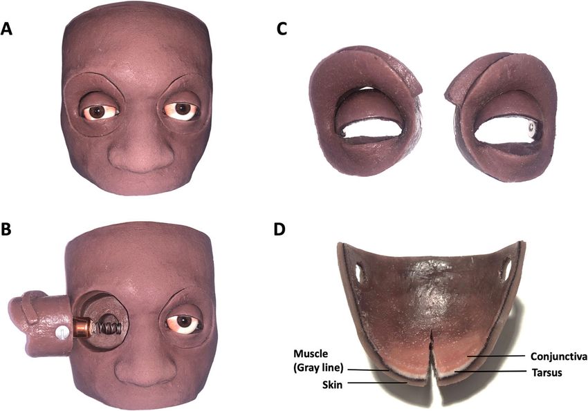
Fig. 1 a The mannequin-based surgical simulator for margin-involving eyelid laceration repair. b The orbit is removable. c The eyelid cartridge
attaches to the orbit. d A vertical incision made in the eyelid demonstrates the four layers of the simulator eyelid that mimic the primary layers of
the human eyelid: skin, orbicularis muscle, tarsus and conjunctiva
margin-involving eyelid lacerations (Supplementary file Identical, full-thickness vertical incisions of the eyelids
1A). Informed consent was obtained from all partici- were made before they were provided to the participants.
pants. Comfort with repair was rated on a 5-point Likert Two attending oculoplastic surgeons (S.M. and R.F.)
scale with 5 being most comfortable, and 1 being least reviewed the laceration repair session videos in a masked
comfortable. Demographic information, including age, fashion and random order. They assessed the surgical
gender, PGY level and completion status of the oculo- proficiency of each participant using a standardized
plastics rotation were recorded. Finally, participants were grading system (Supplementary file 2A). Operating time
queried on prior methods used to practice eyelid lacer- was calculated as the period from loading the first suture
ation repair and their satisfaction with existing methods. to the trimming the ends of the final suture.
Satisfaction with various practice methods was rated on
a 5-point Likert scale with 5 being very satisfied and 1
being very dissatisfied. Perceived comfort level post simulation and feedback on
simulator
Simulator implementation At the end of the pilot study (March 2020), a question-
Before the first simulation session, participants were naire was given to the participants that had completed
given a 1-h didactic session on eyelid anatomy and a one or more simulation sessions asking them to rate
demonstration of margin-involving eyelid laceration re- their comfort level with repair of simple and margin-
pair using the surgical simulation model. Documentation involving eyelid lacerations post-training with the surgi-
outlining the details of each procedural step and use of cal simulator and the usefulness of different models and
the simulator was made readily available to participants experiences for learning margin-involving eyelid lacer-
in the wet lab. Participants were instructed to perform ation repair (Supplementary file 3A). These models and
up to 5 simulated margin-involving eyelid laceration re- experiences included cadaver head, our surgical simula-
pairs using the model, 2 weeks apart. Each session was tor, past methods (fruit peels, surgical skill boards,
video recorded. Participants were provided with all the gloves, pig feet), as well as operating room and emer-
necessary surgical instruments, sutures, phone for video gency department/consults experiences. In addition, par-
recording, adjustable phone holder, and simulator eye- ticipants were queried on effectiveness and value of our
lids. Participants were instructed to wear gloves, to re- surgical simulator, and whether they would recommend
move any possible identifiers throughout the duration of inclusion of the simulator into a formal oculoplastics
the recording, and to mute the sound of the recording. curriculum.Zhao et al. BMC Medical Education (2021) 21:170 Page 4 of 8
Analysis interns and/or residents. Median Likert scale satis-
The Wilcoxon rank-sum test was used to compare faction with past methods was 3.
Likert scores of comfort level with eyelid laceration re-
pair pre-simulation and post-simulation. It was also used
to compare usefulness of different methods for prac- Simulator implementation
ticing margin-involving eyelid lacerations. Inter-rater re- Out of the 10 residents who completed the pre-
liability of the surgical skills assessment score of each training survey, 6 performed at least one simulation
simulated session was determined using the intraclass session. All 5 PGY2 residents participated (1 per-
correlation coefficient (intraclass correlation coefficient, formed 3 simulations, 1 performed 2 simulations,
two-way mixed-effects models, consistency). The Spear- and 3 performed 1 simulation). One PGY4 resident
man’s rank correlation coefficient was employed to performed 3 simulations. The time interval between
measure correlation between the average surgical skills simulation sessions varied between residents (aver-
assessment score and operating time. All statistical ana- age = 35 days, SD = 32 days). A total of 11 videos
lysis was performed using SPSS version 25 (IBM Corp., were assessed by the two attending oculoplastic sur-
Armonk, N.Y.). A p ≤ 0.05 was considered statistically geons. The inter-rater reliability among the raters
significant. was high (Intraclass Correlation Coefficient = 0.862,
P = 0.002).
Results Across all 6 participants, the average surgical skills as-
Perceived need for better surgical simulation model sessment score for the first session was 24 (SD = 7) out
A total of 10 residents completed the pre-training of maximum score of 42. We found no correlation be-
survey. Their demographic information and level of tween the first session operating time and the surgical
experience with performing simple, margin-involving, skills assessment score (averaged between 2 attendings)
and canalicular-involving eyelid laceration repairs are (Spearman’s rank correlation coefficient = − 0.232, p =
summarized in Table 1. As expected, before the 0.66) (Fig. 2). We plotted each resident’s average surgical
simulation practice, residents reported slightly more skills assessment score versus session number for the 3
comfort with repair of a simple eyelid laceration ver- residents (Resident 1, a PGY2; Resident 2, a PGY4, and
sus a margin-involving laceration (median Likert Resident 3, a PGY2) who completed more than one ses-
scale 3 vs. 2). Prior to the study, 60% of residents sion (Fig. 3a). A slight increase in their skills assessment
had practiced on cadavers, 40% on fruit peels, 40% score across simulation sessions was found while opera-
on gloves, 40% on pig feet and 20% on a surgical tive time declined across, most notably in Resident 1
skills board during their training as medical students, (Fig. 3b).
Table 1 Demographic data and level of experience with performing eyelid laceration repairs of the 10 residents that completed the
pre-training survey
Demographic Variable N (%)
Gender Male 5 (50%)
Female 5 (50%)
Training level PGY2 5 (50%)
PGY3 4 (40%)
PGY4 1 (10%)
Completion of oculoplastics rotation Yes 6 (60%)
No 3 (30%)
In progress 1 (10%)
Number of eyelid laceration repair(s) performed Average (SD)
Simple PGY 2 0.4 (0.5)
PGY3 + PGY4 8 (4)
Margin-involving PGY 2 0.2 (0.4)
PGY3 + PGY4 2 (1)
Canalicular-involving PGY 2 0.2 (0.4)
PGY3 + PGY4 3 (2)
PGY postgraduate yearZhao et al. BMC Medical Education (2021) 21:170 Page 5 of 8 Fig. 2 Operative time during first simulation session and average surgical skills assessment score among residents who completed at least one session Perceived comfort level post-training and feedback on oculoplastics curriculum. The remainder of the partici- simulator pants who responded with “Maybe” commented that The 6 participants who performed at least 1 simulation they would have had a favorable response with curricu- session completed the post-training questionnaire. Com- lum integration if time was specifically allocated for fort level with simple eyelid laceration repairs was sig- these practice sessions during the oculoplastic surgery nificantly higher post-simulation compared to pre- rotation. More variation in responses was seen regarding simulation (median Likert value 4 vs. 1, p = 0.02) and for the likelihood of the participants to use this simulator margin-involving eyelid laceration repair (median Likert for future eyelid laceration practice. value 3 vs 1; p = 0.02). Residents reported that they pre- ferred using our surgical simulator in learning margin- Discussion involving laceration repair more than past methods such A rising demand for increased patient safety has resulted as fruit peels, surgical skill boards, gloves, and pig feet in the rapid growth of simulation-based surgery models (p = 0.03), and they felt that the simulator was as useful in ophthalmology training [2, 3]. Prior studies have dem- as cadaver heads and emergency department/consult ex- onstrated improvement in operating room performance perience (Table 2). with the use of surgical simulation [11–13]. This study All 6 participants were somewhat satisfied or ex- successfully converted a mannequin-based surgical tremely satisfied with using the surgical simulator for su- simulator for trachoma surgery into a simulator for turing practice and for learning margin-involving eyelid margin-involving eyelid laceration repair and evaluated laceration repairs (Table 3). Fifty percent of participants its usefulness to teach laceration repair. The simulator recommended including the simulator into the formal provided a platform for practicing all the necessary steps Fig. 3 a Average session-specific surgical skills assessment score versus simulation session. b Operating time versus simulation session
Zhao et al. BMC Medical Education               (2021) 21:170                                                                                              Page 6 of 8

Table 2 Comparison of usefulness of the mannequin-based                                moving on to real-life experiences, such as the oper-
surgical simulator to other methods and experiences in learning                        ating room and emergency room.
margin-involving eyelid laceration repair                                                Animal models have been utilized to teach eyelid la-
                                          Median Likert Scalea          P valueb       ceration repair. Pfaff [8] described the use of pig eyelids.
Surgical simulator                        2.5                           –              This model involves half of a rubber ball to mimic the
Operating room                            5                             0.02           convexity of the globe, and steel screws arranged to
Emergency department/consult              4                             0.07
                                                                                       simulate canthal tendons and the arcus marginalis. Simi-
                                                                                       larly, Kersey [9] reported the use of a split pig-head
Cadaver head                              2.5                           0.74
                                                                                       model, which involves a vertical midline sagittal section
Past methods                              1                             0.03           of a pig head. On gross and histologic examination, hu-
a
 Usefulness of each method was ranked on a 5-point Likert scale (1 = least             man and pig eyelids were found to be similar. Neverthe-
useful, 5 = most useful)
b
 Likert scale of our surgical simulator was compared to operating room,                less, there are important differences, including thicker
emergency room/consults, cadaver head and past methods (fruit peels,                   skin and additional loose connective tissue in the pig
surgical skill boards, gloves, pig feet) using Wilcoxon rank-sum test
                                                                                       eyelid, and the technical performance of the trainees
                                                                                       using their models was not assessed.
of the procedure and permitted evaluation of trainee’s                                   Although the usefulness of the mannequin-based sur-
technical performance.                                                                 gical simulator and cadaver head were rated to be
  Overall, the feedback regarding the mannequin-                                       equivalent in our study, the costs are very different. The
based surgical simulator was positive. All participants                                reusable mannequin head has a one-time cost of $425,
were satisfied with using it for suturing and for prac-                                and the eyelid cartridge, the only part of the surgical
ticing margin-involving eyelid laceration repairs. As a                                simulator that needs to be replaced between simulation
model to teach repair of margin-involving eyelid lac-                                  practices, costs $12.50. This is in contrast to a cadaver,
erations, the surgical simulator was perceived to be                                   which can cost from $200 to upward of $5000 in pro-
equivalent to the emergency department/consult ex-                                     cessing fees per pair of eyelids [14, 15]. Our surgical
perience and cadaver head, but inferior to operating                                   simulator is a safer and a more easily manageable alter-
room experiences. It was perceived as superior to past                                 native to cadaver tissue, and is readily available and can
methods used, which included fruit peels, gloves, sur-                                 be used in more settings.
gical skill boards, and pig feet. Half of the partici-                                   Our surgical simulator was successfully integrated into
pants indicated that practice sessions with the                                        a wet lab environment following a didactic teaching ses-
surgical simulator should be part of the formal oculo-                                 sion. Participants’ surgical skills were assessed with a
plastics curriculum while the rest were ambivalent.                                    standardized grading system, which showed high inter-
Residents commented in the Post-Training Survey                                        rater agreement. Time to complete a procedure is fre-
(Supplementary file 3A) that the surgical simulator                                    quently used as an indicator of surgical experience [16,
was most useful for residents at the beginning of                                      17]. We did find that two of the three residents with
their training. In this way, it can help residents gain                                more than one simulation session showed reduction in
experience and confidence with use of surgical instru-                                 operative time between sessions. However, when analyz-
ments and different suturing techniques before                                         ing the first simulation session in all 6 residents, we did

Table 3 Post-simulation questionnaire responses (n = 6)
         Satisfaction with effectiveness of Satisfaction with          Realism of laceration                            Likelihood of using Inclusion of
         simulator for margin-involving     effectiveness of simulator repair with simulator                            simulator for future simulator into
         laceration repaira                 for suturing practicea     compared to real-lifea                           practicesa           formal
                                                                                                                                             curriculumb
Likert   N (%)                                      N (%)                             N (%)                             N (%)                     N (%)
Scale
1        0 (0)                                      0 (0)                             0 (0)                             0 (0)                     0 (0)
2        0 (0)                                      0 (0)                             1 (16.7)                          1 (16.7)                  3 (50)
3        0 (0)                                      0 (0)                             2 (33.3)                          3 (50)                    3 (50)
4        3 (50)                                     3 (50)                            3 (50)                            1 (16.7)
5        3 (50)                                     3 (50)                            0 (0)                             1 (16.7)
a
 Satisfaction with effectiveness, realism and likelihood of future use of simulator were rated on a 5-point Likert scale (1 = extremely unsatisfied/very unrealistic/
extremely unlikely, 5 = extremely satisfied/very realistic/extremely likely)
b
 There were 3 answer choices for the question regarding the inclusion of simulator into formal oculoplastics curriculum (1 = No, 2 = Maybe, 3 = Yes)
Zhao et al. BMC Medical Education (2021) 21:170 Page 7 of 8
not find a correlation between operative time and surgi- surgical simulator can be used to augment resident’s
cal skills assessment score in our cohort. Similar results training experience and allows for targeted supervision
were reported in a cleft palate surgery simulator, where and independent self-reflection before moving onto live
operative time did not correlate with level of experience surgery.
[18]. The researchers postulated that with increasing ex-
Abbreviations
perience came greater appreciation for difficult surgical HEAD START: Human Eyelid Analog Device for Surgical Training and skills
steps and the consequence of suboptimal repair [18]. Reinforcement in Trichiasis; PGY: Postgraduate year
Limitations of this study include the small sample size
and a single institution. None of the PGY3 residents per- Supplementary Information
formed a simulation session, given they had already The online version contains supplementary material available at https://doi.
org/10.1186/s12909-021-02600-3.
completed the oculoplastics rotation and were concen-
trating on learning cataract surgery. Additionally, they Additional file 1: Supplementary file 1A. Pre-Training Survey
had already performed laceration repairs in the emer- Additional file 2: Supplementary file 2A. Surgical skills assessment
gency department and operating room, so there was tool for evaluating proficiency in performing margin-involving eyelid
lower motivation to practice on a surgical simulator. lacerations
The suggested number of simulation sessions was five; Additional file 3: Supplementary file 3A. Post-training survey
however, none of the participating residents was able to
complete more than three sessions. This is likely due to Acknowledgements
We thank Jiangxia Wang, MS (Department of Biostatistics, Johns Hopkins
the busy clinical demands of the PGY2 year. A possible University School of Public Health, Baltimore, MD, USA) for her guidance in
solution is to build dedicated time for the simulation data analysis for this manuscript.
practices into the resident schedule at the beginning of
Authors’ contributions
residency. Many of the mechanical properties of the sur- JZ, SLM contributed to the conception, design of the study, acquisition,
gical simulator are remarkably similar to the human eye- analysis and interpretation of the data, and drafted and substantially revised
lid; however, the eyelid obviously does not bleed and the the manuscript. MA, EWG contributed to the design of the study, acquisition
and interpretation of the data, and revised the manuscript. RF, FAW
tissue planes do not separate the way they do in a hu- contributed to the acquisition and analysis of data, and revised the
man eyelid. At the time the residents completed the pre- manuscript. All authors have approved the submitted manuscript and have
training questionnaire about laceration repair experi- agreed both to be personally accountable for the authors’ own contributions
and to ensure that questions related to the accuracy or integrity of any part
ence, 3 of the 5 PGY-2 residents had not completed the of the work, even ones in which the author was not personally involved, are
oculoplastics rotation, but at the time of the first simula- appropriately investigated, resolved, and the resolution documented in the
tion session, only 1 of the 5 residents (Resident 1 in Fig. literature.
3) had not completed the oculoplastics rotation. Al- Funding
though anecdotal, it is interesting to note that Resident The Johns Hopkins Wilmer Research Award Grants, 2018–2020. Funding was
1, who had not started the oculoplastics rotation at the used to obtain the supplies for the study.
time of the first simulation session, had the longest ini-
Availability of data and materials
tial simulation session. He/she subsequently reduced All data generated or analyzed during this study are included in this
their session time by almost 50% by the third session. published article.
This highlights the particular usefulness of the model
Declarations
with inexperienced residents.
A multicenter study of this novel mannequin-based Ethics approval and consent to participate
training system is ongoing. The training system has been All study procedures were approved by the Institutional Review Board of the
Johns Hopkins University School of Medicine and adhered to the
integrated into the orientation curriculum at two differ- requirements of the Health Insurance Portability and Accountability Act. Risks
ent institutions, and nine PGY-2 residents have been in- and benefits of study enrollment were discussed with each participant, and
vited to participate. This is particularly timely given the informed consents were obtained.
importance of increased caution with person-to-person Consent for publication
contact during the SARS-CoV2 pandemic and resulting Not applicable.
limitations in residents’ direct patient care.
Competing interests
The authors declare that they have no competing interests.
Conclusion
In this study, we demonstrated the usefulness of a surgi- Author details
1
Wilmer Eye Institute, Johns Hopkins University School of Medicine,
cal simulator for teaching eyelid laceration repairs and Baltimore, MD, USA. 2Gillings School of Global Public Health and Department
showed its utility for developing trainees’ surgical skills. of Ophthalmology, University of North Carolina, Chapel Hill, NC, USA.
3
We successfully used this model to further evaluate Department of Ophthalmology, University of Pittsburgh Medical Center,
Pittsburgh, PA, USA. 4Department of Ophthalmology and Visual Sciences,
trainees’ technical performance with a standardized University of Maryland School of Medicine, 419 W. Redwood St., Suite 420,
grading system with high inter-rater reliability. This Baltimore, MD 21201, USA.Zhao et al. BMC Medical Education (2021) 21:170 Page 8 of 8
Received: 4 January 2021 Accepted: 4 March 2021 Publisher’s Note
Springer Nature remains neutral with regard to jurisdictional claims in
published maps and institutional affiliations.
References
1. Norrell K, Marasigan J, Bogener J. New paradigms in post-graduate surgical
education. Mo Med. 2017;114(4):278–82.
2. Lorch AC, Kloek CE. An evidence-based approach to surgical teaching in
ophthalmology. Surv Ophthalmol. 2017;62(3):371–7. https://doi.org/10.1016/
j.survophthal.2017.01.003.
3. Thomsen AS, Subhi Y, Kiilgaard JF, la Cour M, Konge L. Update on
simulation-based surgical training and assessment in ophthalmology: a
systematic review. Ophthalmology. 2015;122(6):1111–30 e1. https://doi.org/1
0.1016/j.ophtha.2015.02.028.
4. Thomsen ASS, la Cour M, Paltved C, Lindorff-Larsen KG, Nielsen BU, Konge L,
Nayahangan LJ. Consensus on procedures to include in a simulation-based
curriculum in ophthalmology: a national Delphi study. Acta Ophthalmol.
2018;96(5):519–27. https://doi.org/10.1111/aos.13700.
5. Gilbody J, Prasthofer AW, Ho K, Costa ML. The use and effectiveness of
cadaveric workshops in higher surgical training: a systematic review. Ann R
Coll Surg Engl. 2011;93(5):347–52. https://doi.org/10.1308/147870811X582
954.
6. Thomas MP. The role of simulation in the development of technical
competence during surgical training: a literature review. Int J Med Educ.
2013;4:48–58. https://doi.org/10.5116/ijme.513b.2df7.
7. Koukkoulli A, Chandra A, Sheth H, Okhravi N, Verma S, Sullivan P, Ezra DG.
Bridging the gap: theory-based design of a microsurgical skills course for
ophthalmology residents. J Surg Educ. 2015;72(4):585–91. https://doi.org/1
0.1016/j.jsurg.2014.12.015.
8. Pfaff AJ. Pig eyelid as a teaching model for eyelid margin repair. Ophthalmic
Plast Reconstr Surg. 2004;20(5):383–4. https://doi.org/10.1097/01.IOP.
0000134269.82082.A8.
9. Kersey TL. Split pig head as a teaching model for basic oculoplastic
procedures. Ophthalmic Plast Reconstr Surg. 2009;25(3):253. https://doi.org/1
0.1097/IOP.0b013e3181a394c1.
10. Gower EW, Kello AB, Kollmann KM. Training trichiasis surgeons: ensuring
quality. Community Eye Health. 2014;27(87):58.
11. Barsuk JH, McGaghie WC, Cohen ER, O'Leary KJ, Wayne DB. Simulation-
based mastery learning reduces complications during central venous
catheter insertion in a medical intensive care unit. Crit Care Med. 2009;
37(10):2697–701.
12. Crochet P, Aggarwal R, Dubb SS, Ziprin P, Rajaretnam N, Grantcharov T,
Ericsson KA, Darzi A. Deliberate practice on a virtual reality laparoscopic
simulator enhances the quality of surgical technical skills. Ann Surg. 2011;
253(6):1216–22. https://doi.org/10.1097/SLA.0b013e3182197016.
13. Palter VN, Grantcharov TP. Individualized deliberate practice on a virtual
reality simulator improves technical performance of surgical novices in the
operating room: a randomized controlled trial. Ann Surg. 2014;259(3):443–8.
https://doi.org/10.1097/SLA.0000000000000254.
14. Aboud ET, Aboud G, Aboud T. “Live cadavers” for practicing airway
management. Mil Med. 2015;180(3 Suppl):165–70. https://doi.org/10.7205/
MILMED-D-14-00396.
15. Tabas JA, Rosenson J, Price DD, Rohde D, Baird CH, Dhillon N. A
comprehensive, unembalmed cadaver-based course in advanced
emergency procedures for medical students. Acad Emerg Med. 2005;12(8):
782–5. https://doi.org/10.1197/j.aem.2005.04.004.
16. Herrero A, Philippe C, Guillon F, Millat B, Borie F. Does the surgeon’s
experience influence the outcome of laparoscopic treatment of common
bile duct stones? Surg Endosc. 2013;27(1):176–80. https://doi.org/10.1007/
s00464-012-2416-z.
17. Pollei TR, Barrs DM, Hinni ML, Bansberg SF, Walter LC. Operative time and
cost of resident surgical experience: effect of instituting an otolaryngology
residency program. Otolaryngol Head Neck Surg. 2013;148(6):912–8. https://
doi.org/10.1177/0194599813482291.
18. Podolsky DJ, Fisher DM, Wong Riff KW, Szasz P, Looi T, Drake JM, Forrest CR.
Assessing technical performance and determining the learning curve in
cleft palate surgery using a high-Fidelity cleft palate simulator. Plast
Reconstr Surg. 2018;141(6):1485–500. https://doi.org/10.1097/PRS.
0000000000004426.You can also read