EVALUATION OF THE CYTOTOXIC AND IMMUNOGENIC POTENTIAL OF TEMOZOLAMIDE, PANOBINOSTAT, AND LOPHOPHORA WILLIAMSII EXTRACT AGAINST C6 GLIOMA CELLS ...
←
→
Page content transcription
If your browser does not render page correctly, please read the page content below
EXCLI Journal 2021;20:614-624 – ISSN 1611-2156
 Received: November 18, 2020, accepted: February 15, 2021, published: March 09, 2021

 Original article:

 EVALUATION OF THE CYTOTOXIC AND IMMUNOGENIC
 POTENTIAL OF TEMOZOLAMIDE, PANOBINOSTAT, AND
 LOPHOPHORA WILLIAMSII EXTRACT AGAINST
 C6 GLIOMA CELLS

Moisés Armides Franco-Molina1,* , Silvia Elena Santana-Krímskaya1 ,
Luis Mario Madrigal-de-León1 , Erika Evangelina Coronado-Cerda2 ,
Diana Ginette Zárate-Triviño1 , Sara Paola Hernández-Martínez1 ,
Paola Leonor García-Coronado1 , Cristina Rodríguez-Padilla1
1
 Universidad Autónoma de Nuevo León (UANL), Facultad de Ciencias Biológicas,
 Laboratorio de Inmunología y Virología, P.O. Box 46 “F”, 66455, San Nicolás de los
 Garza, NL, México
2
 Universidad del Valle de México, Campus Cumbres, Departamento de Ciencias de la
 Salud, Av. Las Palmas, 5500, Colonia Cima de las Cumbres, Monterrey, Nuevo León,
 C.P. 64610, Mexico

* Corresponding author: Dr. Moisés Armides Franco-Molina, Universidad Autónoma de
 Nuevo León (UANL), Facultad de Ciencias Biológicas, Laboratorio de Inmunología y
 Virología, P.O. Box 46 “F”, 66455, San Nicolás de los Garza, NL, México.
 Tel.: +52-812-141-15, extension: 6428;
 E-mail: moyfranco@gmail.com, moises.francoml@uanl.edu.mx

http://dx.doi.org/10.17179/excli2020-3181

This is an Open Access article distributed under the terms of the Creative Commons Attribution License
(http://creativecommons.org/licenses/by/4.0/).

 ABSTRACT
Glioblastoma multiforme is a malignant neoplasm of the brain with poor prognosis. The first-line drug against
glioblastoma is the alkylating agent temozolamide (TMZ); unfortunately, treatment resistance and tumor re-inci-
dence are common. In some cases, immunogenic cell death (ICD) inducers can decrease treatment resistance and
tumor recurrence by stimulating an antitumor specific immune response. Not all ICD inducers, however, are suit-
able for glioma patients because of the low permeability of the blood-brain barrier (BBB). Panobinostat (PAN), a
histone deacetylase inhibitor and Lophophora williamsii (LW) extract can pass through the BBB and have anti-
tumor properties. The aim of this study is to evaluate the cytotoxic potential of TMZ, PAN and LW extract against
the glioma C6 cell line, and its role in the release of damage-associated molecular patterns (DAMPs), which is a
hallmark of ICD. Our results indicate that all treatments induce cellular death in a time- and concentration-depend-
ent manner, and that PAN and LW extract induce apoptosis, whereas TMZ induces apoptosis and necrosis. Also,
that some of the treatments and their sequential administration induce the release of DAMPs. Furthermore, in a rat
glioma model, we observed that all treatments decreased tumor volume, but the in vivo cell death mechanism was
not ICD. Our findings indicate that TMZ, PAN, and LW combination have a cytotoxic effect against glioma cells
but do not induce ICD.

Keywords: Lophophora williamsii, temozolamide, panobinostat, glioblastoma, immunogenic cell death, DAMPs

 614
EXCLI Journal 2021;20:614-624 – ISSN 1611-2156
 Received: November 18, 2020, accepted: February 15, 2021, published: March 09, 2021

Abbreviations: ducers would not clinically benefit glioma pa-
TMZ temozolamide tients due to the low permeability of the
PAN panobinostat
LW Lophophora williamsii
 blood-brain barrier (BBB).
ICD immunogenic cell death Effective treatment for glioblastoma re-
BBB blood-brain barrier quires substances able to cross the BBB. One
DAMPs damage-associated molecular patterns of these substances is panobinostat (PAN), an
 epigenetic modulator that inhibits histone
 INTRODUCTION deacetylase activity and increases DNA-his-
 tone acetylation. This blocks multiple signals
 Glioblastoma multiforme (GBM) is ma- related to the development and progress of tu-
lignant neoplasia of the brain that originates mors, and induces apoptosis in the target cells
in the glial cells within the intracranial tissue. (Van Veggel et al., 2018). Furthermore, in-
GBM infiltrates the surrounding tissue and creased histone acetylation sensitizes cancer
decreases neurological function, resulting in a cells to the effect of alkylating agents, such as
poor quality of life for the patients (Bahadur TMZ (Stiborova et al., 2012).
et al., 2019). Current therapies against GBM Lophophora williamsii (LW) also known
involve a combination of surgery, chemother- as “peyote”, is a spineless cactus known for
apy, and radiotherapy, nevertheless, it has a the physical, visual, and perceptual changes it
median overall survival of 2 years (Singleton induces upon ingestion (Casado et al., 2008).
et al., 2017). LW extract has immunostimulatory proper-
 The alkylating agent temozolamide ties: it increases macrophage cytokine pro-
(TMZ) is a first-line drug for the treatment of duction and lymphocyte proliferation, and it
glioblastoma; it acts by damaging tumor cells is cytotoxic for cancer cell lines (Alonso-Cas-
through DNA methylation, changing hetero- tro et al., 2016). LW extract also crosses the
chromatin organization, and activating an ox- BBB (Dinis-Oliveira et al., 2019) and some of
idative stress response. However, tumor cells its components interact with the serotonergic
quickly become resistant to TMZ at pharma- 5-HT2A-C receptors present in glioblastoma
cotherapeutic concentrations (Stepanenko et cells (Lu et al., 2020).
al., 2016). The use of multiple treatments for cancer
 Some therapeutic drugs can induce immu- has been of great importance to improve ther-
nogenic cell death (ICD) in cancer cells. ICD apeutic outcome. The present study was de-
originates with endoplasmic reticulum stress signed to determine the antitumor effect of
in the target cell that results in an elevated TMZ, PAN, and LW extract on a rat glioma
production of reactive oxygen species and the model, and their capacity to induce ICD.
release of damage-associated molecular pat-
terns (DAMPs). The release of DAMPs in-
cludes the exposure of calreticulin (CRT) and MATERIALS AND METHODS
the extracellular release of ATP, high-mobil- Reagents
ity group box 1 protein (HMGB1), and heat Temozolomide (TMZ) was purchased
shock proteins (HSP70 and HSP90) from Schering-Plough (Kenilworth, NJ,
(Turubanova et al., 2019). Once released, USA), panobinostat (PAN) was purchased
DAMPs interact with innate immune cell re- from Cellagen Technology (San Diego, CA,
ceptors, which then become activated. Acti- USA), and the methanolic extract of Lopho-
vated innate immune cells interact with lym- phora williamsii (LW) was obtained from a
phocytes to generate an adaptative antitumor plant collection of the Laboratorio de
immune response that hinders cancer relapse Inmunología y Virología, Facultad de Cien-
(Rapoport and Anderson, 2019; Du and Wax- cias Biológicas, UANL. IgG1 mouse HSP70
man, 2020). The most frequently used ICD in- antibody (sc-32239), IgG1 mouse HMGB1
 antibody (sc-56698), IgG2a mouse HSP90α/β

 615

EXCLI Journal 2021;20:614-624 – ISSN 1611-2156
 Received: November 18, 2020, accepted: February 15, 2021, published: March 09, 2021

(sc-13119), and IgGκ mouse HRP antibody and the fluorescence was measured using a
(sc-516102) were purchased from Santa Cruz microplate reader Synergy TM HT (BioTek
Biotechnology (Dallas, TX, USA). Dul- Instrument, Vermont, NH, USA) at 535/590
becco´s Modified Eagle Medium (DMEM) nm, excitation/emission wavelength, respec-
was purchased from Sigma-Aldrich (St. tively. The percentage of cell viability (%)
Louis, MO, USA). was calculated according to the following
 equation [1]:
Preparation of Lophophora williamsii ex-
tract Relative viability (%) = treated cell fluo-
 The cacti used in this study belonged to a rescence ÷ control cell fluorescence x 100
plant collection of the Laboratorio de [1]
Inmunología y Virología, Facultad de Cien-
cias Biológicas, UANL, and had been previ- Chemosensitivity assay
ously identified as Lophophora williamsii. C6 cells (5x10³) were seeded into 96-well
The cacti were macerated and methanol ex- plates and cultured overnight in a 5 % CO2 at-
traction was performed for 48 h at 4 °C, after mosphere at 37 °C. Then cells were pre-ex-
which the ethanolic phase was filtered. The posed to TMZ (2.15 - 43 mM) for 24 h, fol-
methanol extract was lyophilized using a lowed by the treatments with LW extract (1.92
freeze-dryer (Labconco Co. Kansas City, mg/mL, IC50 value) and PAN (0.75 µM, IC50
MO). The resulting powder was dissolved in value) for 24 h; pre-exposure to PAN (0.5 - 25
1 mL of DMEM and the endotoxin levels µM) for 24 h, followed by the treatments with
were measured with the gel clot-based Limu- LW extract (1.92 mg/mL, IC50 value) and
lus amoebocyte assay (Associates of Cape TMZ (8.6 mM, IC50 value) for 24; and pre-
Cod. Falmouth, MA), which has a detection exposure to LW extract (1.44 - 4.8 mg/mL) for
limit of 0.004 ng/mL. 24 h, followed by PAN (0.75 µM, IC50 value)
 and TMZ (8.6 mM, IC50 value) treatments for
Cell line and culture conditions 24 h. Finally, cells were washed with PBS 1X
 The C6 murine brain glial cell line was and cell metabolic activity was assessed using
purchased from the American Type Culture the resazurin assay as previously described.
Collection (Manassas, Virginia, USA). Cells
were cultured in DMEM supplemented with Recovery assay
bovine fetal serum (10 % v/v) in a 5 % CO2 C6 cells (5x10³) were seeded into 96-well
atmosphere at 37 °C. plates and cultured overnight in a 5 % CO2 at-
 mosphere at 37 °C. After this, cells were
Cytotoxicity assay treated with TMZ (2.15 - 43 mM), and LW ex-
 C6 cells (5x10³) were seeded into 96-well tract (1.94 - 4.8 mg/mL) for 24 h, and with
plates and cultured overnight in a 5 % CO2 at- PAN (0.5 - 25 µM) for 24 and 72 h. The treat-
mosphere at 37 °C. Then, cells were treated ments were removed, and cells were washed
with TMZ (2.15 - 43 mM), and LW extract twice with PBS 1X. Then DMEM supple-
(1.44 - 4.8 mg/mL) for 24 and 48 h, and with mented with bovine fetal serum (10 % v/v)
PAN (0.5 - 25 µM) for 24, 48 and 72 h. Also, was added and cells were maintained at a 5 %
the TMZ + LW extract + PAN treatment com- CO2 and 37 °C atmosphere for 5 days. Finally,
bination was evaluated using the IC30, and cells were washed with PBS 1X and cell met-
IC50 for 24 h. After the treatment, cells were abolic activity was assessed using the resaz-
washed with phosphate-buffered saline (PBS) urin assay.
1X, and cellular metabolic activity was as-
sessed using the resazurin assay. Cells were
incubated with a resazurin solution (20 % v/v)
for 30 min in a 5 % CO2 atmosphere at 37 °C

 616
EXCLI Journal 2021;20:614-624 – ISSN 1611-2156
 Received: November 18, 2020, accepted: February 15, 2021, published: March 09, 2021

Apoptosis and cell viability assay To perform the indirect ELISA assay, 96-
 Acridine orange/ethidium bromide well plates were pre-coated with 25 g of pro-
(AO/EB) staining was used to determine via- tein from each sample for 24 h at 4 °C. After
ble and nonviable cells, based on disrupted this, a specific primary antibody for HMGB1,
cell membrane. Cells (1x10⁵) were seeded in HSP70, or HSP90 was added to the plates and
6 well plates and treated with TMZ (IC50 = 8.6 incubated for 2 h at 37 °C. To detect the pres-
and IC100 = 43 mM), and LW extract (IC50 = ence of DAMPs a mouse IgG HRP-conju-
1.92 and IC100 = 4.8 mg/mL) for 24 h, and gated secondary antibody was added to each
PAN (IC50 = 0.75 and IC100 = 25 µM) for 72 h. well and incubated for 30 min at room tem-
Also, cells pre-exposed to TMZ (8.6 mM) for perature. Then, a chromogen solution was
24 h followed by LW extract (1.92 mg/mL) added, and the plates were incubated at room
and PAN (0.75 µM) 24 h treatments, were in- temperature for 30 min. Finally, the reaction
cluded. Briefly, cells were washed with PBS was terminated by the addition of a stop solu-
1X and stained with 20 L of AO/EB dye mix tion, and absorbance was determined at a
(100 µL/mL AO and 100 µL/mL EB, pre- 450 nm wavelength.
pared in PBS). Then wells were observed un- For the detection of DAMPs in the super-
der a confocal fluorescence microscope natant, C6 cells (5x10⁶) were treated with the
(Olympus X70), at 250/605 nm (excitation/ IC100 of TMZ, LW extract, and PAN for 24 h,
emission wavelength, respectively) for EB or a pre-exposure treatment with TMZ (43
and 502/525 nm (excitation/emission wave- mM) for 24 h, followed by LW extract (4.8
length, respectively) for AO. Viable cells mg/mL) and PAN (25 µM) for 24 h. After
were identified by bright green fluorescence, this, the supernatants were collected and
apoptotic cells by bright orange fluorescence, HMGB1, HSP70, and HSP90 were deter-
and necrotic cells by bright red fluorescence. mined as described above.

Determination of DAMPs Animals
 The production of DAMPs was detected Data were obtained from 2- to 3-months-
by indirect ELISA assay in cell lysates and su- old female Wistar rats with a weight range of
pernatants after treatment with TMZ, LW ex- 240-260 g. The animals (n=25) were provided
tract, and PAN. by the bioterium of the Facultad de Ciencias
 C6 cells (5x10⁶) were treated with the Biológicas, UANL. Rats were kept in 12 h
IC100 of TMZ (43 mM), LW extract (4.8 light/dark cycles with ad libitum water and
mg/mL), PAN (25 M), or pre-exposed to food. All animal procedures were performed
TMZ (43 mM) for 24 h, followed by LW ex- according to the Official Mexican Norm of
tract (4.8 mg/mL) and PAN (25 µM) treat- Animal Welfare NOM-033-SAG/ZOO-2014
ments for 24 h. After the treatments, the cells and approved by the internal Research and
were collected and centrifuged at 1,200 rpm Animal Welfare Ethics Committee (CEIBA)
for 10 min at room temperature. Then cells of the Facultad de Ciencias Biológicas,
were washed with PBS 1X and homogenized UANL (2018-015).
using the SET 2X lysis buffer (20 mM Tris
pH 6.8, 2 mM EDTA pH 8.0, 300 mM NaCl C6 cells inoculation and whole tumor cell
and 4 % SDS; Sigma-Aldrich: Merck) sup- lysate vaccination
plemented with complete Halt protease inhib- A total of 6x10⁶ C6 cells were treated in
itor cocktail. Protein quantification was per- vitro with TMZ (43 mM), LW extract (4.8
formed using the BCA kit (cat. No. 23225, mg/mL), or PAN (25 µM) for 24 h, and a pre-
PierceTM BCA Protein Assay Kit, Thermo exposure treatment with TMZ (8.6 mM) for
Fisher, Waltham, Massachusetts, USA). 24 h, followed by LW extract (1.92 mg/mL)
 and PAN (0.75 µM) combination for 24 h.
 Next, the cells were centrifuged at 1,200 rpm

 617
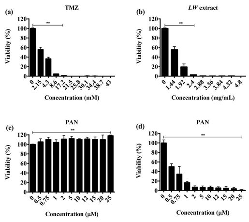
EXCLI Journal 2021;20:614-624 – ISSN 1611-2156
 Received: November 18, 2020, accepted: February 15, 2021, published: March 09, 2021

for 10 min and washed twice with PBS 1X. time- and concentration-dependent manner.
Finally, cells were resuspended in 300 µL of The IC50 value was 8.6 mM for TMZ, and
PBS 1X and inoculated subcutaneously into 1.92 mg/mL for LW extract at 24 h (Figure 1a
the left flank of Wistar rats. The rats were ran- and 1b). In the case of PAN, we observed a
domly divided into five experimental groups: 50 % reduction of viability at the 0.75 µM
1) Control group: without vaccination (n = 5), concentration after 24 h of treatment, and a
2) TMZ group (n = 5), 3) LW extract group (n significant cytotoxic effect (p < 0.001) was
= 5), 4) PAN group (n = 5), and 5) pre-expo- observed after 48 and 72 h with all concentra-
sure treatment with TMZ followed by LW ex- tions of PAN used (Figure 1c). In contrast, the
tract + PAN group (n = 5). After 7 days, rats treatment combination TMZ + LW extract +
were challenged with 5x10⁶ viable C6 cells PAN was not cytotoxic at the IC30, or IC50 val-
resuspended in 300 µL of PBS 1X via subcu- ues (Figure 1d).
taneous injection into the left flank. Tumor
width and length were measured every third Pre-exposure treatment enhanced the
day for 21 days with a digital caliper and tu- cytotoxic effect against C6 cells
mor volume was calculated according to the Pre-exposure with TMZ for 24 h followed
formula: V = (W^2 x L)/2, where V is tumor by treatment with a combination of PAN and
volume, W is tumor width and L is tumor LW extract at IC50 values (0.75 µM and 1.92
length (Santana-Krímskaya et al., 2020). mg/mL, respectively) increased significantly
 The humane endpoint was used to avoid (p < 0.0001) the cytotoxic effect of TMZ
unnecessary suffering, and rats were sacri- against C6 cells (Figure 2a).
ficed 21 days after inoculation. Similarly, LW extract pre-exposure for 24
 h followed by PAN (0.75 µM) and TMZ
Statistical analysis (8.6 mM) combination treatment (Figure 2b),
 The experiments were performed in tripli- and PAN pre-exposure treatment for 24 h, fol-
cate and statistical differences between lowed by LW extract (1.92 mg/mL) and TMZ
groups were analyzed using ANOVA fol- (8.6 mM) (Figure 2c), also increased the cy-
lowed by the Tukey post hoc test. The data are totoxic effect.
presented as mean ± standard deviation (SD)
and differences between groups were consid- TMZ, LW extract, and PAN treatments
ered significant at a p-value ≤ 0.05. Statistical prevent the recovery of C6 cells
analyses were performed with the GraphPad To determine the recuperation capacity of
Prism software version 6 (GraphPad Soft- C6 cells after the treatments, cell viability was
ware, Inc., San Diego, Ca). measured after a recovery period of 5 days
 (Figure 3). Treatment with TMZ and LW ex-
 tract significantly (p < 0.001) decreased cell
 RESULTS
 recovery of C6 cells in a dose-dependent
Treatments with TMZ, LW extract, and manner (Figure 3a and 3b). The PAN treat-
PAN decreased C6 cells viability in a ment significantly (p < 0.001) decreased cell
concentration-dependent manner recovery in a time- and dose-dependent man-
 The TMZ and LW extract treatments sig- ner (Figure 3c and 3d).
nificantly reduced the viability of C6 cells (p
< 0.001) as compared to untreated cells in a

 618
EXCLI Journal 2021;20:614-624 – ISSN 1611-2156
 Received: November 18, 2020, accepted: February 15, 2021, published: March 09, 2021

Figure 1: TMZ, LW extract, and PAN induce cell death in C6 cells as assessed by the resazurin assay.
(a) Representative bar graphs of data obtained of viable cells treated at 24 and 48 h with TMZ. (b)
Representative bar graphs of data obtained of viable cells treated at 24, and 48 h with LW extract. (c)
Representative bar graphs of data obtained of viable cells treated at 24, 48, and 72 h with PAN. (d)
Representative bar graphs of data obtained of viable cells treated with a combination of IC30, and IC50
of all treatments at 24 h. Results were obtained from three independent experiments performed in trip-
licate, and data are presented as the mean ± standard deviation. **p = 0.001 and *p = 0.01 with respect
to control. TMZ, temozolomide; LW extract, Lophophora williamsii extract; PAN, panobinostat

 Figure 2: Pre-ex-
 posure to individual
 treatments for 24 h
 increases the cyto-
 toxic effect over C6
 cells as assessed
 by the resazurin as-
 say. (a) Pre-expo-
 sure with TMZ for
 24 h, followed by
 LW extract (1.92
 mg/mL), and PAN
 (0.75 M) for 24 h.
 (b) Pre-exposure
 with LW extract for
 24 h, followed by
 TMZ (8.6 mM), and
 PAN (0.75 M) for
 24 h.
(c) Pre-exposure with PAN for 24 h, followed by IC50 value of LW extract (1.92 mg/mL), and TMZ (8.6
mM) for 24 h. Results were obtained from three independent experiments performed in triplicate, and
data are presented as the mean ± standard deviation. ***p = 0.0001 with respect to control. TMZ, te-
mozolomide; LW extract, Lophophora williamsii extract; PAN, panobinostat

 619
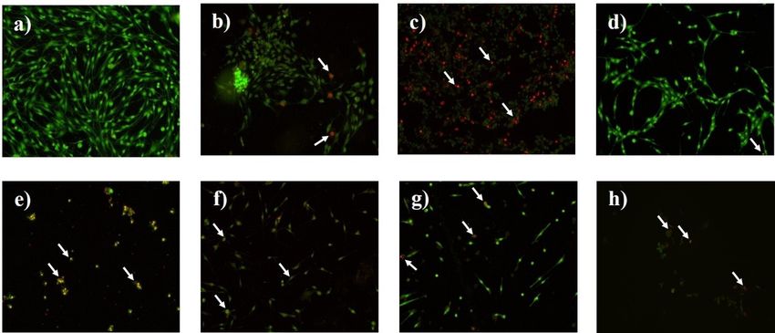
EXCLI Journal 2021;20:614-624 – ISSN 1611-2156
 Received: November 18, 2020, accepted: February 15, 2021, published: March 09, 2021

Figure 3: TMZ, LW extract, and PAN avoid the recovery of C6 cells as assessed by the resazurin assay.
(a) Viability of C6 cells 24 h after removing the TMZ for a recovery period of 5 days. (b) Viability of C6
cells 24 h after removing the LW extract for a recovery period of 5 days. (c) Viability of C6 cells 24 h
after removing the PAN for a recovery period of 5 days. (d) Viability of C6 cells 72 h after removing the
PAN for a recovery period of 5 days. Results were obtained from three independent experiments per-
formed in triplicate, and data are presented as the mean ± standard deviation. **p = 0.001 with respect
to the control. TMZ, temozolomide; LW extract, Lophophora williamsii extract; PAN, panobinostat

TMZ, LW extract, and PAN treatments (bright red cell fluorescence) (Figure 4c). The
induce cell death by apoptosis and necrosis IC50 and IC100 of LW extract (Figure 4d), in-
on C6 cells duced cell death by apoptosis (Figure 4e), as
 Untreated cells presented a bright green did the IC50 and IC100 of PAN (Figure 4f and
nuclear fluorescence and a regular structure 4g). Finally, TMZ pre-exposition followed by
(Figure 4a). Cells treated with the IC50 of LW extract and PAN treatments induced cell
TMZ showed early stages of apoptosis (bright death by necrosis (Figure 4h).
orange cell fluorescence) (Figure 4b), and
cells treated with the IC100 showed necrosis

 620
EXCLI Journal 2021;20:614-624 – ISSN 1611-2156
 Received: November 18, 2020, accepted: February 15, 2021, published: March 09, 2021

Figure 4: AO/EB staining assay shows that treatments TMZ, LW extract, and PAN induce apoptosis
and necrosis on C6 cells. The arrows indicate cell damage. (a) Nontreated cells. (b) Cells treated with
the IC50 of TMZ (8.6 mM) for 24 h. (c) Cells treated with the IC100 of TMZ (43 mM) for 24 h. (d) Cells
treated with the IC50 of LW extract (1.92 mg/mL) for 24 h. (e) Cells treated with the IC100 of LW extract
(4.8 mg/mL) for 24 h. (f) Cells treated with the IC50 of PAN (0.75 M) for 72 h. (g) Cells treated with the
IC100 of PAN (25 M) for 72 h. (h) Cells pre-exposed to TMZ (8.6 mM) for 24 h followed by LW extract
(1.92 mg/mL) and PAN (0.75 M). TMZ, temozolomide; LW extract, Lophophora williamsii extract; PAN,
panobinostat

TMZ, PAN, and pre-exposure to TMZ
followed by LW extract and PAN increase
the release of HMGB1, HSP70, and HSP90
by C6 cells
 There were no significant differences be-
tween the levels of HMGB1, HSP70, and
HSP90 found in cell lysates, however, there
was a significant (p = 0.01) difference in the
HSP90 levels of LW treated cells and the con-
trol (Figure 5a). In the supernatant, TMZ and
PAN treatments increased the release of
HMBG1 and HSP90 as compared to the con-
trol, but this increase was not significant (Fig-
ure 5b). On the other hand, the pre-exposure
to TMZ followed by LW extract and PAN
treatment increased the release of HSP70, in
contrast to the control, but there was no statis-
tical significance (Figure 5b).

Figure 5: TMZ, PAN, and TMZ followed by LW extract and PAN stimulate the release of HMGB1,
HSP70, and HSP90 as measured by ELISA. (a) HMGB1, HSP70, and HSP90 measurement in cell
lysates after 24 h of treatment. (b) HMGB1, HSP70, and HSP90 measurement in the supernatant after
24 h of treatment. Results were obtained from three independent experiments performed in triplicate,
and data are presented as the mean ± standard deviation. *p = 0.01 with respect to the control. TMZ,
temozolomide; LW extract, Lophophora williamsii extract; PAN, panobinostat; HMGB1, high-mobility
group box 1 protein, HSP70 and HSP90, heat shock protein 70 and 90, respectively

 621
EXCLI Journal 2021;20:614-624 – ISSN 1611-2156
 Received: November 18, 2020, accepted: February 15, 2021, published: March 09, 2021

TMZ, LW extract, PAN and TMZ pre- al., 2019). To our knowledge, there are no
exposure followed by LW extract and PAN previous reports of the cytotoxic effect of
do not induce immunogenic cell death in PAN against C6 cells, but it has been reported
vivo that other histone-modifying enzymes
 C6 cells lysed by TMZ, LW extract, PAN, (BIX01294, 3DZNep, VP, TSA, and chae-
or pre-exposure to TMZ followed by LW ex- tocin) do not affect C6 cells viability after 72
tract and PAN treatments were used to vac- hours of exposure (Maleszewska et al., 2014).
cinate rats, which were then challenged with The difference is that this group did not in-
viable C6 cells. Tumor presence was detected clude PAN, for which the apoptotic effect had
at day 6 in all rats (vaccinated and unvac- been previously described, although not
cinated). The tumor volume was measured against glioma cells (Gerson et al., 2018). Re-
until day 13. The group vaccinated with the garding the LW extract cytotoxic effect, our
cell lysed by pre-exposition to TMZ and research group had previously reported that
treated with LW extract and PAN presented LW methanolic extract decreases the viability
the highest tumor volume, compared to the of murine fibrosarcoma L929, murine lym-
other groups (p = 0.001) (Figure 6 and Sup- phoma L5178Y-R, human histiocytic lym-
plementary Tables 1 to 6). phoma U937, and human breast cancer MCF7
 (Franco-Molina et al., 2003) cell lines. How-
 DISCUSSION ever, there are no previous reports of the ef-
 The present study aimed to determine the fect of LW extract over C6 or other glioma cell
cytotoxic and immunogenic potential of lines.
TMZ, PAN, and LW extract in a C6 glioma rat The recovery assay allowed us to discrim-
model. inate cell death from a temporarily inactive
 Our results show that the cytotoxic effect metabolism. TMZ, PAN, and LW extract
of all tested treatments (TMZ, PAN, and LW treated cells were unable to proliferate even
extract) decreased C6 cell viability in a time- after a five-day recovery period, corroborat-
 ing cell death.
and concentration-dependent manner (Ni et

Figure 6: TMZ, LW extract, PAN, and TMZ and followed by LW extract and PAN did not induce immu-
nogenic cell death. The tumor volume was measured every third day for 13 days starting at day 7 post-
inoculation. Results are presented as the mean ± standard deviation. **p = 0.001. TMZ, temozolomide;
LW extract, Lophophora williamsii extract; PAN, panobinostat

 622
EXCLI Journal 2021;20:614-624 – ISSN 1611-2156
 Received: November 18, 2020, accepted: February 15, 2021, published: March 09, 2021

 The single treatments induced apoptosis, tients after conventional treatment; further-
but the simultaneous combination of all treat- more, the use of LW extract should be consid-
ments did not exert any cytotoxic effect. The ered for glioma treatment, alone or sequen-
sequential treatment, which consisted of C6 tially administrated with TMZ and PAN, but
cells pre-exposition to TMZ followed by further in vivo experiments should be per-
treatment with LW extract and PAN resulted formed to determine its antitumor effect.
in necrosis. The combination of cytotoxic
drugs tends to cause antagonism, but this an- Funding
tagonism does not necessarily translate into “Fondo Sectorial de Investigación para la
clinical failure because of other properties, Educación”, grant A1-S-35951, CONACYT,
such as immune response modulation, which México supported this work.
can also inhibit tumor growth (Richards et al.,
2020). The use of multiple agents can result Acknowledgments
in the release of DAMPs from dying cells, and We thank Alejandra Elizabeth Arreola
these molecules interact with immune cells to Triana for article revision; and Universidad
trigger an antitumor adaptive immune re- Autónoma de Nuevo León UANL, Facultad
sponse (Zhou et al., 2019; Asadzadeh et al., de Ciencias Biológicas, Laboratorio de
2020). Inmunología y Virología, P.O. Box 46 “F”,
 In this study, we did not observe a signif- 66455 San Nicolás de los Garza, NL, México
icant increase of HMGB1 and HSP90 in the for facilities provided.
supernatant of C6 cells treated with TMZ (8.6
mM) and PAN (20 M) as compared to the Conflicts of interest
control, but there was an increase of HSP70 The authors declare that there is no con-
in cells pre-exposed to TMZ, followed by LW flict of interest.
extract and PAN.
 Finally, we evaluated ICD induction in REFERENCES
vivo. The treatments did not prevent the im-
plantation of the C6 glioma, suggesting that Alonso-Castro AJ, Juárez-Vázquez M del C, Campos-
 Xolalpa N. Medicinal plants from Mexico, Central
immunogenic death was not the in vivo cell America, and the Caribbean used as immunostimu-
death mechanism. These results correlate with lants. Evid Based Complement Altern Med. 2016;
the recurrence of human glioma after a few 2016:4017676.
months following completion of TMZ treat-
 Asadzadeh Z, Safarzadeh E, Safaei S, Baradaran A,
ment (Daniel et al., 2019), indicating the lack
 Mohammadi A, Hajiasgharzadeh K, et al. Current ap-
of a tumor-specific immune response. Fur- proaches for combination therapy of cancer: The role
thermore, it has been reported that TMZ treat- of immunogenic cell death. Cancers. 2020;12:1047.
ment upregulates programmed cell death-1
ligand-1 (PD-L1) in glioblastoma cells, pro- Bahadur S, Sahu AK, Baghel P, Saha S. Current prom-
 ising treatment strategy for glioblastoma multiforme:
moting immune escape (Wang et al., 2019). If A review. Oncol Rev. 2019;13(2):417.
this is the case, PD-L1 expression could be
antagonizing the effect of DAMPs release. Casado R, Uriarte I, Cavero RY, Calvo MI. LC-PAD
 determination of mescaline in cactus “Peyote” (Lopho-
 phora williamsii). Chroma. 2008;67:665–7.
 CONCLUSIONS
 Daniel P, Sabri S, Chaddad A, Meehan B, Jean-Claude
 In conclusion, this study contributes to the B, Rak J, et al. Temozolomide induced hypermutation
knowledge that TMZ, PAN, or LW extract in glioma: Evolutionary mechanisms and therapeutic
and its combinations do not induce immuno- opportunities. Front Oncol. 2019;9:41.
genic cell death, suggesting an explanation re- Dinis-Oliveira RJ, Pereira CL, da Silva DD. Pharma-
garding the tumor recurrence in glioma pa- cokinetic and pharmacodynamic aspects of peyote and
 mescaline: Clinical and forensic repercussions. CMP.
 2019;12:184–94.

 623
EXCLI Journal 2021;20:614-624 – ISSN 1611-2156
 Received: November 18, 2020, accepted: February 15, 2021, published: March 09, 2021

Du B, Waxman DJ. Medium dose intermittent cyclo- Torres-del-Muro F, et al. IMMUNEPOTENT CRP
phosphamide induces immunogenic cell death and can- plus doxorubicin/cyclophosphamide chemotherapy re-
cer cell autonomous type I interferon production in gli- model the tumor microenvironment in an air pouch tri-
oma models. Cancer Lett. 2020;470:170–80. ple-negative breast cancer murine model. Biomed
 Pharmacother. 2020;126:110062.
Franco-Molina M, Gomez-Flores R, Tamez-Guerra P,
Tamez-Guerra R, Castillo-Leon L, Rodríguez-Padilla Singleton W, Collins A, Bienemann A, Killick-Cole C,
C. In vitro immunopotentiating properties and tumour Haynes H, Asby D, et al. Convection enhanced deliv-
cell toxicity induced by Lophophora williamsii (pe- ery of panobinostat (LBH589)-loaded pluronic nano-
yote) cactus methanolic extract: Immunopotentiation micelles prolongs survival in the F98 rat glioma model.
by Lophophora williamsii extract. Phytother Res. Int J Nanomedicine. 2017;12:1385–99.
2003;17:1076–81.
 Stepanenko AA, Andreieva SV, Korets KV,
Gerson SL, Caimi PF, William BM, Creger RJ. Phar- Mykytenko DO, Baklaushev VP, Huleyuk NL, et al.
macology and molecular mechanisms of antineoplastic Temozolomide promotes genomic and phenotypic
agents for hematologic malignancies. In: Hoffman R, changes in glioblastoma cells. Cancer Cell Int. 2016;
Benz EJ, Silberstein LE, et al. (eds): Hematology. 7th 16:36.
ed. (pp 849–912). Amsterdam: Elsevier, 2018.
 Stiborova M, Eckschlager T, Poljakova J, Hrabeta J,
Lu Q, Ding Y, Li Y, Lu Q. 5-HT receptor agonist Adam V, Kizek R, et al. The synergistic effects of
valerenic acid enhances the innate immunity signal and DNA-targeted chemotherapeutics and histone deacety-
suppresses glioblastoma cell growth and invasion. Int J lase inhibitors as therapeutic strategies for cancer treat-
Biol Sci. 2020;16:2104–15. ment. Curr Med Chem. 2012;19:4218–38.

Maleszewska M, Steranka A, Kaminska B. The effects Turubanova VD, Balalaeva IV, Mishchenko TA,
of selected inhibitors of histone modifying enzyme on Catanzaro E, Alzeibak R, Peskova NN, et al. Immuno-
C6 glioma cells. Pharmacol Rep. 2014;66:107–13. genic cell death induced by a new photodynamic ther-
 apy based on photosens and photodithazine. J Immu-
Ni Q, Fan Y, Zhang X, Fan H, Li Y. In vitro and in vivo nother Cancer. 2019;7:350.
study on glioma treatment enhancement by combining
temozolomide with calycosin and formononetin. J Eth- Van Veggel M, Westerman E, Hamberg P. Clinical
nopharmacol. 2019;242:111699. pharmacokinetics and pharmacodynamics of pano-
 binostat. Clin Pharmacokinet. 2018;57:21–9.
Rapoport B, Anderson R. Realizing the clinical poten-
tial of immunogenic cell death in cancer chemotherapy Wang S, Yao F, Lu X, Li Q, Su Z, Lee J-H, et al. Te-
and radiotherapy. Int J Mol Sci. 2019;20(4):959. mozolomide promotes immune escape of GBM cells
 via upregulating PD-L1. Am J Cancer Res. 2019;9:
Richards R, Schwartz HR, Honeywell ME, Stewart 1161–71.
MS, Cruz-Gordillo P, Joyce AJ, et al. Drug antagonism
and single-agent dominance result from differences in Zhou J, Wang G, Chen Y, Wang H, Hua Y, Cai Z. Im-
death kinetics. Nat Chem Biol. 2020;16:791–800. munogenic cell death in cancer therapy: Present and
 emerging inducers. J Cell Mol Med. 2019;23:4854–65.
Santana-Krímskaya SE, Franco-Molina MA, Zárate-
Triviño DG, Prado-García H, Zapata-Benavides P,

 624
You can also read