Exposição à amônia e alterações de pH desencadeiam danos branquiais e mortalidade em peixes tetras da Amazônia Ammonia exposure and pH alterations ...
←
→
Page content transcription
If your browser does not render page correctly, please read the page content below
Brazilian Journal of Animal and Environmental Research 4070
                                                                                                                 ISSN: 2595-573X

    Exposição à amônia e alterações de pH desencadeiam danos branquiais e
                  mortalidade em peixes tetras da Amazônia

  Ammonia exposure and pH alterations trigger gill damage and mortality in
                         Amazonian tetras fish
DOI: 10.34188/bjaerv4n3-098

Recebimento dos originais: 04/03/2021
Aceitação para publicação: 30/06/2021

                                   Wallice Paxiúba Duncan
         Doutor em Ecologia e Recursos Naturais pela Universidade Federal de São Carlos
            Universidade Federal do Amazonas, Laboratório de Morfologia Funcional
         Endereço: Av. Rodrigo Octávio, 6200, Coroado I, 69080-900. Manaus, AM, Brasil
                                E-mail: wduncan@ufam.edu.br

RESUMO
O objetivo deste estudo foi avaliar os efeitos da toxicidade aguda pela amônia e alterações de pH na
estrutura branquial dos peixes tetras, Paracheirodon axelrodi e Paracheirodon simulans. Estas
espécies são importantes caracídeos neotropicais usados como peixes ornamentais. Os danos
branquiais foram analisados e quantificados. Como esperado, o cardinal (P. axelrodi) foi a espécie
menos tolerantes à toxicidade da amônia e à água ácida. Por outro lado, o neon (P. simulans) foi a
espécie mais resistente à amônia e menos tolerante à água alcalina. Hiperplasia epitelial,
descolamento do epitélio lamelar e fusão das lamelas foram mais frequentemente observadas nas
brânquias de P. simulans expostos aos estressores (amônia e alterações de pH) do que nas brânquias
de P. axelrodi. Tais lesões são inespecíficas, porém podem reduzir a reabsorção de amônia, bem
como proteger o epitélio branquial de danos ainda mais graves causados pelas alterações de pH. Por
outro lado, alterações na estrutura dos vasos sanguíneos, tais como aneurismas, telangiectasias e
necrose focal das células pilares foram comumente observadas no cardinal. A necrose dos
componentes branquiais está relacionada às taxas de mortalidade dos peixes. Embora os caracídeos
pertençam ao mesmo gênero e ocorram na mesma bacia hidrográfica, tais espécies apresentam
respostas morfológicas diferentes aos mesmos agentes estressores.

Palavras-chave: efeito da amônia, alterações branquiais, caracídeos neotropicais, peixes
ornamentais, alterações de pH

ABSTRACT
The goal of this study was to evaluate the effects of acute ammonia toxicity and pH changes on the
gill structure of tetras fish, Paracheirodon axelrodi and Paracheirodon simulans, two important
neotropical characins used as ornamental fish. Fish mortality was recorded, and damage to the gills
was examined and quantified. As expected, cardinal tetras (P. axelrodi) were less tolerant to
ammonia toxicity and acidic water. Conversely, green neon tetras (P. simulans) were more resistant
to acute ammonia levels and less tolerant to alkaline water. Epithelial hyperplasia, lamellar lifting
and lamellar fusion were more frequently observed in the gills of the green neon tetras that were
exposed to stressors (ammonia toxicity and pH alterations) than in the gills of cardinal tetras. These
non-specific lesions may reduce ammonia uptake, as well as protect the gill epithelium from severe
damage caused by pH alterations. In contrast, blood vessel alterations, such as aneurysms and
telangiectasis, and focal necrosis of the pillar cells were commonly observed in the cardinal tetras.
Necrosis in the gill components was related to the mortality rates of these tetra fish. Although these

      Brazilian Journal of Animal and Environmental Research, Curitiba, v.4, n.3, p. 4070-4084 jul./set. 2021.
Brazilian Journal of Animal and Environmental Research 4071
ISSN: 2595-573X
characins belong to the same genus and live in the same hydrological system, they present different
morphological responses to the same stressors.
Keywords: ammonia effects, gill alterations, neotropical characins, ornamental fish, pH alterations
1 INTRODUCTION
Cardinal tetras (Paracheirodon axelrodi Schultz, 1956) and green neon tetras
(Paracheirodon simulans Géry, 1963) are found in the black water that extends throughout the
middle to upper Rio Negro and Orinoco basin (WALKER, 2004). In general, they are sympatric and
syntopic, but they differ in their preferred microhabitats and are rarely found together in same
environment (MARSHALL et al., 2011). Both species are widely exploited as ornamental fish. P.
axelrodi and P. simulans represent 64.6 % and 7.5%, respectively, of total export volume between
2006 and 2015 from the Amazonas state, Brazil. In this period, more than 100 million individuals
were exported (TRIBUZY-NETO et al., 2020).
Detrimental water conditions, such as a concomitant increase in the pH of water and
ammonia, produce higher concentrations of the more toxic un-ionized form of ammonia (MIRON
et al., 2008), which may contribute to high mortality during transport and shipment. In the case of
cardinal tetra, the mortality rates can be as high as 70 % during the capture and transport due to the
poor water quality (WAICHMAN et al., 2001). However, the sensitivity to ammonia appears to be
species-specific, since the cardinal tetras appear to be less tolerant than the green neon tetras
(SOUZA-BASTOS et al., 2017).
The gills are particularly sensitive to changes in the aquatic environment (EVANS et al.,
2005). Structural damage, such as epithelial lifting, lamellar fusion and haemorrhage, can lead to
alterations in those physiological processes (LEASE et al., 2003), such as, reduce respiratory
effectiveness and decrease swimming performance (MIRON ET AL., 2008). Furthermore, toxic
substances and extreme changes in pH tend to promote excessive mucus production due to the
proliferation of mucous cells (PAULINO et al., 2014). However, the proliferation of mucous and
pavement cells results in an increase in the blood-water barrier (EVANS et al., 2005). With this
background, it is possible that extreme pH conditions and high ammonia concentrations will result
in severe gill damage, contributing to the high mortality of tetra fish during management in fisheries
and aquariums.
In this study, the experimental design of the LC50-96h as reported by OLIVEIRA et al.
(2008) was replicated. The goal was to investigate the acute effects of pH stress and ammonia
concentrations on the gill morphology of two ornamental fish. In addition, a correlation and/or a
Brazilian Journal of Animal and Environmental Research, Curitiba, v.4, n.3, p. 4070-4084 jul./set. 2021.Brazilian Journal of Animal and Environmental Research 4072
ISSN: 2595-573X
causal relationship was established between stress-induced alterations and mortality rates in these
fishes.
2 MATERIALS AND METHODS
2.1 ANIMALS AND MAINTENANCE
Cardinal tetras (0.24 ± 0.02 g and 2.74 ± 0.11 cm) and green neon tetras (0.12 ± 0.03 g and
2.05 ± 0.16 cm) collected in the middle sector of the Rio Negro (00º40’S/62º58’W), were purchased
from a local aquarium supply store. Fish were kept in a 1,000 L holding tank with local well water
for two weeks, and the following conditions were maintained: a stabilized temperature (29.2 ± 1.1
o
C), dissolved oxygen 5.4 ± 0.8 mg L-1, conductivity 24.5 ± 2.7 µS cm-1, pH 5.5 ± 1.3 and natural
photoperiod. Fish were fed every morning with commercial feed (38 % crude protein). During this
period, the ammonia concentrations ranged from 0.05 to 0.10 mM, and feeding was stopped 24 h
prior to the beginning of the experiments. All in vivo procedures were approved by Ethical
Committee of Animal Experimentation (CEUA/UFAM) protocol Nº 014/2015 (in accordance with
the guidelines of CONCEA, National Council of Control of Animal Experimentation).
2.2 EXPERIMENTAL DESIGN
In order to evaluate the effects of pH alterations and ammonia concentrations on the gill
morphology of cardinal and green neon tetras, the experimental conditions of LC50-96 h that were
reported by OLIVEIRA et al. (2008) have been replicated. The fish were placed in tanks (40 L) and
kept for 48 hours in tap water (pH 6.4 ± 0.7; temperature 29.4 ± 0.5; electrical conductivity 12.1 ±
0.4 µS/cm; dissolved oxygen 4.7 mg/L, and ammonia was not detected) before the beginning of the
experiments. For ammonia experiments, three groups of ten fish (with three replicates for each
treatment) were exposed to two different levels (1.54 ± 0.06 and 2.75 ± 0.17 mmoles/L of total
ammonia) and tap water without ammonia was used as a control group. The pH monitoring of the
ammonia experiments ranged from 6.7 ± 0.2 and 7.3 ± 0.4 in the tests with 1.54 and 2.75 mmoles/L
of ammonia, respectively. For study with extreme pH conditions, three different levels of acidic
water (with three replicates for each treatment) were used: pH 2.9 ± 0.2 (min – max, 2.5 - 3.2), 4.3
± 0.4 (3.8 - 4.5) and 6.0 ± 0.3 (5.8 - 6.2), as a control group. While three different levels of alkaline
water were tested: pH 7.2 ± 0.5 (6.9 - 7.5), 7.8 ± 0.3 (7.5 - 7.9) and 8.8 ± 0.4 (8.6 - 9.0). The ammonia
levels, acidic and alkaline pH were adjusted as reported by OLIVEIRA et al. (2008). briefly,
adjustment to acidic pH was performed with HCl, while alkaline pH was performed with Tris and
NaOH. Tests with ammonia were done with a NH4Cl solution. In all experiments, solutions were
slowly added every 1-h until reaching the desired value. In addition, the water was partially renewed
Brazilian Journal of Animal and Environmental Research, Curitiba, v.4, n.3, p. 4070-4084 jul./set. 2021.Brazilian Journal of Animal and Environmental Research 4073
ISSN: 2595-573X
daily by 30% of the tank volume. The water quality was analysed twice a day, before the partial
renewal of the tank water. In the ammonia experiments, the physical and chemical variables of the
water were recorded as the fallowing: temperature (29.5 ± 1.2 oC), dissolved oxygen (5.3 ± 1.1
mg/L), and conductivity (92.4 ± 33.7 µS/cm). While in the pH experiments, these variables were:
temperature (30.2 ± 0.8 oC), dissolved oxygen (4.1 ± 2.4 mg/L), and conductivity (70.8 ± 52.1
µS/cm). In all cases, monitoring was recorded using a Consort C535 multiparameter analyser
(CONSORT bvba). Ammonia concentrations were assayed as total ammonia levels (NH3 + NH4+)
using Nessler methods. Animals that lost their equilibrium and died due to the acute toxicity of the
ammonia were immediately necropsied, and haemorrhages were observed in the gills.
2.3 TISSUE PROCESSING
At the end of the exposure period (96 h), the fish were euthanized with a high dose of
anaesthetic 0.1 % benzocaine. The gills were removed and fixed in phosphate-buffered containing
2.5 % glutaraldehyde. Gill arches were washed and then dehydrated through a graded series of
ethanol baths. The right sides of the gills were embedded in 2-hydroxyethyl methacrylate resin. To
quantify the histological changes, thin sections (3 µm) of tissue were cut using a tungsten knife
mounted on a semi-rotary microtome (CUT 5062, Slee Medical GmbH). Slides for quantitative
measurements were stained with 0.12 % toluidine blue. Some slides were stained with periodic acid-
Schiff reagent (PAS) and Alcian blue at pH 2.5 (AB) to quantify the mucous cells with neutral and
acidic mucosubstances, respectively. Changes in cell morphology, such as karyorrhexis, karyolysis,
and pyknosis were also recorded
2.4 GILL MORPHOMETRIC ANALYSIS
The gill components (epithelial lamellar/filament and blood space) were evaluated in random
selected fields of the histological sections. The density of volume of the gill components
(undamaged and damaged) was analysed using a stereological technique. The tissue elements were
quantified from digital images using a square lattice grid of points superimposed by the free software
STEPanizer Stereology Tool version 1 (www.stepanizer.com). The thickness of the water-blood
barrier was estimated using the test line of a semi-circular grid (Merz’s grid) and analysed using
IMAGEJ 1.45s software (www.imagej.nih.gov/ij/).
2.5 STATISTICAL ANALYSIS
The data are expressed as mean values ± standard deviation (SD). In the experiments,
ammonia and pH data are expressed as mean ± SD (minimum - maximum). The volume density
Brazilian Journal of Animal and Environmental Research, Curitiba, v.4, n.3, p. 4070-4084 jul./set. 2021.Brazilian Journal of Animal and Environmental Research 4074
ISSN: 2595-573X
values are proportions that required arcsine-square root transformation before analysis (ZAR, 2010).
The uniformity of the data was tested using the Kolmogorov-Smirnov test. The relationship between
mortality rate and histological changes was examined using Pearson's correlation analysis.
Stereological and morphometric measurements were analysed by parametric one-way ANOVA
followed by Tukey’s test (significance level was set at α < 0.05). The data were analysed using the
Sigma Plot package version 11.0 (Systat software Inc.).
3 RESULTS
The green neon tetras were slightly more sensitive to extreme alkaline water than the cardinal
tetras. In green neon tetras, 100 % mortality occurred when pH 8.8 was reached, while in cardinal
tetras 70 % of mortality was observed at pH 8.8 (Figure 1A). In contrast, the mortality rate reached
100% in the cardinal tetras exposed to pH 2.9; while, at this pH (2.9), 78 % of green neon tetras
died. In both species, the gills were necropsied and exhibited typical haemorrhages, high mucous
content and focal necrosis. In green neon tetras, the high mortality was also positively associated
with the focal necrosis of the gill epithelium (r2 = 0.82; p = 0.011; Figure 1B). Conversely, this
relationship was not observed in cardinal tetras.
Figure 1. Variations in the mortality rate (A) and focal necrosis (B) of the gill components in cardinal tetras and green
neon tetras exposed to different pH values.
As expected, the cardinal tetras were more sensitive to high ammonia concentrations than
the green neon tetras. At an ammonia level of 2.75 mM, 80.3 ± 3.5 % of the cardinal tetras died over
96 hours, whereas only 22.6 ± 1.9 % of the green neon tetras died. Ammonia was toxic to the
cardinal tetras even at low concentrations (1.54 mM), and 24.3 ± 2.1 % of the fish in this group died.
Brazilian Journal of Animal and Environmental Research, Curitiba, v.4, n.3, p. 4070-4084 jul./set. 2021.Brazilian Journal of Animal and Environmental Research 4075
ISSN: 2595-573X
Meanwhile, no cardinal tetras died in this concentration.
The gills of the control fish appeared normal in both species. The density of volume of the
gill components with histological alterations in the green neon and cardinal tetras exposed to the pH
challenges are detailed in the Tables 1 and 2, respectively. Although some of the fish were healthy,
evidence (< 0.7 %) of epithelial hyperplasia on the lamellae and gill filament was still observed.
Complete fusion of all lamellae was also observed. In the healthy cardinal tetras, hyperplasia
occurred more frequently (0.7 % of total tissue alterations) in the lamellar epithelium, while in the
green neon tetras, it occurred in the filament epithelium (0.7 %).
Table 1. Gill damage (as volume density, %) in Paracheirodon axelrodi (cardinal tetra) exposed to the pH challenges.
Different letters on the same line indicate significant differences (p < 0.05) according to the (Tukey test).
Gill alterations Acidic pH Alkaline pH
2.9 4.3 6.0 7.2 7.8 8.8
Hyperplasia of
lamellar epithelium 5.8 ± 0.4a 0.4 ± 0.1b 0.7 ± 0.2b 0.5 ± 0.2b 4.6 ± 1.1a 3.7 ± 1.4a
Hyperplasia of
filament epithelium 0.0a 5.7 ± 1.6b 0.3 ± 0.2c 1.8 ± 1.0d 7.1 ± 5.2b 5.4 ± 1.1b
Epithelial lifting of
lamellae 2.0 ± 0.5a 0.0b 0.0b 0.0b 14.4 ± 8.4c 7.1 ± 2.7d
Dilation of the
marginal channel 0.0a 1.3 ± 1.0b 0.0a 0.2 ± 0.1a 7.7 ± 2.3c 3.6 ± 1.3d
Complete fusion of all
lamellae 0.0a 3.4 ± 2.1b 0.1 ± 0.1a 0.0a 4.3 ± 2.1b 11.4 ± 2.9c
a a b b c
Lamellar aneurysm 1.7 ± 0.6 0.9 ± 0.4 0.0 0.2 ± 0.1 9.5 ± 2.2 2.5 ± 1.2a
a b c b d
Undamaged lamellae 3.4 ± 0.8 52.2 ± 5.0 73.5 ± 9.8 52.4 ± 9.1 19.8 ± 2.5 4.4 ± 1.9a
a b b c a
Undamaged filament 16.5 ± 6.7 22.5 ± 3.9 24.8 ± 6.4 45.4 ± 7.8 16.7 ± 8.4 18.7 ± 5.4a
At extremely low pH levels (e.g., 2.9), hyperplasia due to pavement cell proliferation on the
filament epithelia and chloride cell proliferation on the lamellae was observed. Epithelial lifting of
lamellae, which resulted in the complete fusion of all lamellae, was most frequent in the gills of the
green neon tetras (10.5 %), while this was not observed in cardinal tetras. Conversely, focal necrosis
was most common in the cardinal tetras (70 %) than in the green neon tetras (12.3 %). The neon
tetras exposed to slightly alkaline water (pH 7.8) often had multiple lesions, such as hyperplasia (on
the lamellae and filament epithelia), epithelial lifting of lamellae, dilation of the marginal channel
and fusions between adjacent lamellae. However, these alterations were most frequently observed
in the cardinal tetras. Unlike the cardinal tetras, the green neon tetras had severe blood alterations
(such as lamellar aneurysms) and focal necrosis. Moreover, all fish in the highly alkalized water
(pH 8.8) died during the experiments. The green neon tetras exhibited focal necrosis in almost 80
% of their gill components, whereas the cardinal tetras exhibited focal necrosis in 43.3 % of their
gill components.
Brazilian Journal of Animal and Environmental Research, Curitiba, v.4, n.3, p. 4070-4084 jul./set. 2021.Brazilian Journal of Animal and Environmental Research 4076
ISSN: 2595-573X
Table 2. Gill damage (as volume density, %) in Paracheirodon simulans (green neon tetra) exposed to pH alterations.
Different letters on the same line of data indicate significant differences (Tukey test, p < 0.05).
Gill alterations Acidic pH Alkaline pH
2.9 4.3 6.0 7.2 7.8 8.8
Hyperplasia of
lamellar epithelium 12.1 ± 1.4a 0.1 ± 0.1b 0.0b 1.1 ± 0.5c 1.2 ± 0.2c 2.3 ± 0.9d
Hyperplasia of
filament epithelium 8.7 ± 2.3a 0.0b 0.7 ± 0.4c 0.9 ± 0.7c 4.9 ± 1.4d 1.7 ± 0.6c
Epithelial lifting of
lamellae 9.2 ± 0.7a 0.2 ± 0.1b 0.0b 0.3 ± 0.3b 1.5 ± 0.5c 0.8 ± 0.4c
Dilation of the
marginal channel 5.1 ± 0.4a 4.5 ± 2.7a 0.0b 0.4 ± 0.2b 2.8 ± 1.1c 1.0 ± 0.4d
Complete fusion of all
lamellae 10.5 ± 0.3a 2.4 ± 1.8b 0.3 ± 0.2c 0.5 ± 0.4c 3.9 ± 1.3b 1.2 ± 0.7d
a a b a c
Lamellar aneurysm 2.2 ± 1.4 2.9 ± 0.9 0.0 3.0 ± 1.1 11.9 ± 2.9 4.3 ± 1.5d
a b b b a
Undamaged lamellae 9.2 ± 7.0 60.5 ± 9.8 59.2 ± .5 45.9 ± 8.7 10.3 ± 2.8 3.4 ± 1.2c
a a b b a
Undamaged filament 31.4 ± 8.7 27.6 ± 5.7 39.2 ± 5.2 48.5 ± 7.8 22.8 ± 8.6 7.3 ± 2.1c
Acute ammonia concentrations induced severe gill alterations in both tetra fishes (Table 3).
The most severe types of damage were extensive focal necrosis, complete fusion of all lamellae,
aneurysms, hyperplasia of chloride cells and lamellar fusion. Ammonia also induced cell
proliferation in the gill epithelium. Nevertheless, focal necrosis was more common in the cardinal
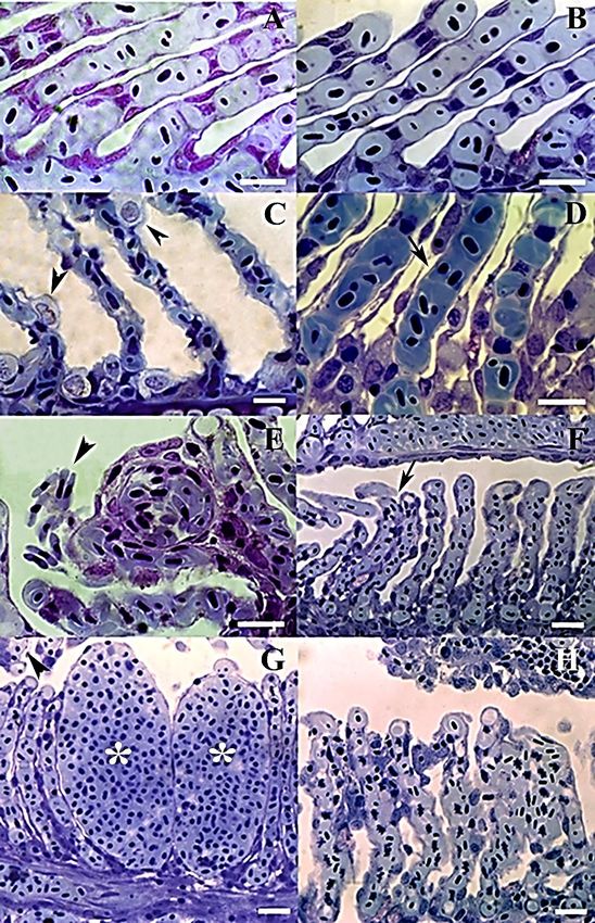
tetras than in the green neon tetras. Some representative histological alterations are showed in the
figure 2. Necrotic cells, both in the gill epithelium and those in support (pillar cells), exhibit
morphological changes such as karyorrhexis, and karyolysis.
Brazilian Journal of Animal and Environmental Research, Curitiba, v.4, n.3, p. 4070-4084 jul./set. 2021.Brazilian Journal of Animal and Environmental Research 4077
ISSN: 2595-573X
Figure 2. Sagittal sections of the gill tissues showing normal aspects of the lamellae of the cardinal (a) and green neon
tetras (b) maintained in tap water (pH 6.0 without ammonia). Representative histological changes in the gills
(arrowheads), such as chloride cell proliferation in cardinal tetras (c), epithelial lifting in green neon tetras (d),
hemorrhages in cardinal tetras (e), complete fusion of lamellae in green neon tetras (f), lamellar aneurysms (g) and focal
necrosis and partial lamellar fusion in green neon tetras (h). Scale bar = 10 µm). Sections were stained with toluidine
blue 0.25 %.
Brazilian Journal of Animal and Environmental Research, Curitiba, v.4, n.3, p. 4070-4084 jul./set. 2021.Brazilian Journal of Animal and Environmental Research 4078
ISSN: 2595-573X
Table 3. Gill damage (as volume density, %) in the gill alterations of Paracheirodon axelrodi and Paracheirodon
simulans after 96 h at different levels of ammonia exposure. Different letters are significantly different according to
one-way ANOVA (Turkey’s test, p < 0.05).
P. axelrodi P. simulans
Histopathologies (total ammonia, mM) (total ammonia, mM)
0 1.54 2.75 0 1.54 2.75
Hyperplasia of
lamellar epithelium 0.3 ± 0.2a 0.9 ± 0.2b 1.9 ± 0.5c 0.0a 12.8 ± 0.5b 3.2 ± 1.1c
Hyperplasia of
filament epithelium 0.2 ± 0.1a 1.0 ± 0.6b 1.8 ± 1.2b 0.5 ± 0.3a 13.0 ± 0.5b 4.7 ± 0.7c
Epithelial lifting of
lamellae 0.0a 2.5 ± 1.7b 3.3 ± 1.7b 0.0a 6.6 ± 1.2b 13.3 ± 1.8c
Dilation of the
marginal channel 0.4 ± 0.2a 12.1 ± 0.1b 9.2 ± 0.4c 0.4 ± 0.3a 6.5 ± 1.5d 7.1 ± 1.3cd
a b b a b
Complete fusion of all 0.0 3.6 ± 0.6 3.5 ± 0.7 0.0 8.8 ± 1.6 6.5 ± 1.7c
lamellae
Lamellar aneurysm 0.0a 15.3 ± 3.8b 8.1 ± 2.4c 0.0a 0.6 ± 0.4b 1.7 ± 0.8c
Necrosis 0.0a 23.2 ± 2.2b 44.8 ± 3.6c 0.0a 9.3 ± 2.8b 19.5 ± 3.8c
Undamaged lamellae 48.3 ± 3.9a 17.4 ± 1.2b 7.5 ± 1.4c 46.9 ± 8.4a 22.1 ± 7.3b 14.6 ± 1.6c
Undamaged filament 51.5 ± 5.2a 22.8 ± 9.8b 18.7 ± 3.6b 51.9 ± 5.8a 20.2 ± 2.7b 29.3 ± 6.5b
Ammonia toxicity and extreme pH conditions also affected the water-blood distance in the
neon tetras (Figure 3). The water-blood barrier became thicker (p < 0.05) in highly acidified or
alkalinized water. At a very low pH, the diffusion barrier increased to 1.45 ± 0.26 µm and 1.34 ±
0.09 µm in the cardinal and green neon tetras, respectively. Moreover, at the highest pH, the water-
blood distance also increased to 1.42 ± 0.13 µm and 1.63 ± 0.35 µm in the cardinal and green neon
tetras, respectively. Similarly, the water-blood distance tended to increase significantly with the
ammonia concentration in both tetra fish.
Figure 3. Effect of pH alterations (A) and total ammonia concentrations (B) on the water-blood distance barrier (2/3 of
the harmonic mean) of cardinal tetras (P. axelrodi) and green neon tetras (P. simulans). Data are presented as the means
± SD. Different letters over bars indicate significant difference among treatments (Tukey test, p < 0.05).
Both stressors (ammonia exposure and pH alterations) induced mucous cell (MC)
proliferation in the leading and trailing edges, as well as in the tips of the gill filaments. In both fish
Brazilian Journal of Animal and Environmental Research, Curitiba, v.4, n.3, p. 4070-4084 jul./set. 2021.Brazilian Journal of Animal and Environmental Research 4079
ISSN: 2595-573X
species, the MCs contained neutral and/or acidic mucosubstances, which were stained with periodic
acid-reagent of Schiff (PAS) and Alcian blue, respectively. Exposure to alkaline pH levels (>pH
7.8) increased the number of MCs containing neutral mucosubstances in the gills of cardinal
tetras (p < 0.05), whereas more MCs with acidic glycoproteins (p < 0.0%) were observed in the
green neon tetras. In contrast, very acidic water (Brazilian Journal of Animal and Environmental Research 4080
ISSN: 2595-573X
4 DISCUSSION
A previous study has described the acute lethal effects of pH alterations and ammonia
toxicity on cardinal tetras (OLIVEIRA et al., 2008). The LC50-96 h for cardinal tetras was estimated
to be pH 2.9 in acidified water, pH 8.8 in alkalinized water and 2.7 mM for ammonia toxicity. More
recently, SOUZA-BASTOS et al. (2017) studied 11 Amazonian fish species and concluded that
cardinal tetras were more sensitive to ammonia toxicity than green neon tetras. Similarly, I observed
the same results in both species. However, I herein provide a histopathological approach to explain
the mortality caused by acute pH variations and ammonia levels. Furthermore, no comparable study
of pH challenges has been carried out to date using green neon tetras, which are a congeneric species
of cardinal tetras and are of equal importance to the ornamental fish market. Extreme pH challenges
(acidic or alkaline) and high ammonia concentrations cause severe gill damage in tetra fish.
However, these two characins exhibited different morphological responses to the same stressor. For
example, green neon tetras submitted to alkaline pH (>8.8) has a greater degree of gill injuries
compared to cardinal tetras; while at acid pH (pH 7.8).
Numerous chloride cells (CCs) were also observed on the filaments (interlamellar space) and
lamellar epithelium of tetras fish. The proliferation of CCs and pavement cells on the lamellar
epithelium was evident in both species under pH challenges. Hyperplasia of the lamellar epithelium
may result in an increase in the water-blood distance (CAMARGO and MARTINEZ, 2007). It is
Brazilian Journal of Animal and Environmental Research, Curitiba, v.4, n.3, p. 4070-4084 jul./set. 2021.Brazilian Journal of Animal and Environmental Research 4081
ISSN: 2595-573X
possible that these alterations reduced the gas-exchange capacity of the gills. The increase in
respiratory epithelium thickness and the associated thickening of the mucus layer due to excessive
secretions from MCs may result in mortality due to hypoxia (EVANS et al., 2005). Moreover, this
can be potentially hazardous to the cardinal tetras, since this species is more sensitive to hypoxia
than the green neon tetras (CAMPOS et al., 2017; SOUZA-BASTOS et al., 2017).
Extreme pH conditions also cause extensive epithelial lifting, lamellae fusion and
aneurysms. In highly acidic water, these lesions were more common in green neon tetras. However,
more deleterious damage, such as necrosis, was observed in the cardinal tetras. Epithelial
hypertrophy and hyperplasia can lead to complete lamellar fusion, thereby reducing the respiratory
surface in neotropical fish (PAULINO et al., 2014). Oedema with epithelial lifting is an
inflammatory process that is initiated as a defence mechanism to reduce the gill surface area of
green neon tetras due to stress at extreme pH levels (pH 7.8). In turn, this response
may increase the water-blood distance, thereby impairing oxygen uptake. The lamellar thickness
and diffusion distance increased in both tetra fish exposed to the extreme environmental conditions.
This can be lethal to the cardinal tetras because hypoxia (15–20 % of oxygen saturation) itself leads
to 50 % mortality in cardinal tetras (SOUZA-BASTOS et al., 2017). Therefore, stress due to pH
alterations may be potentiated by hypoxic conditions. It is important to emphasize that both
conditions occur simultaneously during the handling and transport of tetra fish.
The lethal concentration of ammonia in cardinal tetras was calculated to be 2.78 mM
(OLIVEIRA et al., 2008). A similar value was determined by SOUZA-BASTOS et al. (2017).
According to these authors, cardinal tetras are less tolerant (2.24 mM) than green neon tetras (8.52
mM). In our study, the mortality rate of cardinal tetras was almost 82 % at a total ammonia level of
2.75 mM. As expected, the green neon tetras were more resistant than the cardinal tetras. However,
ammonia caused epithelial lifting and partial or complete fusion of the secondary lamellae even at
low concentrations, possibly due to hyperplasia of the pavement cells. These lesions were similar
to those observed in pH experiments and can reduce the respiratory surface area and interlamellar
space. Again, these gill alterations were observed more frequently in the green neon tetras than in
the cardinal tetras. I suggest that these gill changes can help to prevent ammonia uptake in green
neon tetras. This may explain why green neon tetras are 3.8 times more resistant than cardinal tetras
(SOUZA-BASTOS et al., 2015).
Like extreme pH levels, ammonia toxicity also induced vascular changes in the gills of tetra
fish, and these changes were more common in the cardinal tetras than in the green neon tetras. Gill
telangiectasis and lamellar aneurysms occurred due to the direct action of the ammonia, which
ruptured the pillar cell system of these fish. Blood vessel changes, although reversible, may have
Brazilian Journal of Animal and Environmental Research, Curitiba, v.4, n.3, p. 4070-4084 jul./set. 2021.Brazilian Journal of Animal and Environmental Research 4082
ISSN: 2595-573X
progressed under acute ammonia intoxication and resulted in epithelial rupture and haemorrhages
(MIRON et al., 2008). In fact, the moribund fish showed signs of haemorrhage in their gills. The
tetra fish that were exposed to high ammonia levels exhibited focal necrosis in the pillar cells. These
cells that support the blood space in the lamellae showed karyorrhexis. Additionally, the chloride
cells that proliferated on the lamellae epithelium exhibited karyolysis and nuclear shrinkage
(pyknosis) in the erythrocytes. All these alterations were more severe in the cardinal tetras, which
may explain the high mortality rate of this species when exposed to high ammonia concentrations.
5 CONCLUSIONS
Overall, this study demonstrates that extreme pH conditions and high ammonia levels cause
severe damage to the gill structures and increase water-blood diffusion barrier. The gills of cardinal
tetras exposed to ammonia had 2-fold more necrotic tissue than the gills of the green neon tetras.
These histological alterations explain the different mortality rates reported in these tetra fish. The
present study aimed to reduce mortality and increase the understanding of the effects of common
stressors that occur during the harvest, holding and transport of tetra fish for ornamental purposes.
Brazilian Journal of Animal and Environmental Research, Curitiba, v.4, n.3, p. 4070-4084 jul./set. 2021.Brazilian Journal of Animal and Environmental Research 4083
ISSN: 2595-573X
REFERENCES
CAMARGO, M.M. & MARTINEZ, C.B.R. 2007. Histopathology of gills, kidney and liver of a
Neotropical fish caged in an urban stream. Neotropical ichthyology, 5(3): 327-336.
https://doi.org/10.1590/S1679-62252007000300013.
CAMPOS, D.F.; JESUS, T F.; KOCHHANN, D.; HEINRICHS-CALDAS, W.; COELHO, M.M &;
ALMEIDA-VAL, V.M.F. 2017. Metabolic rate and thermal tolerance in two congeneric Amazon
fishes: Paracheirodon axelrodi Schultz, 1956 and Paracheirodon simulans Géry, 1963
(Characidae). Hydrobiologia, 789: 133-142. https://doi.org/10.1007/s10750-016-2649-2.
DAYE, P.G. & GARSIDE, E.T. 1976. Histopathologic changes in surficial tissues of brook trout,
Salvelinus fontinalis (Mitchill), exposed to acute and chronic levels of pH. Canadian Journal of
Zoology, 54(12): 2140-2155. https://doi.org/10.1139/z76-248
EVANS, D. P.; PIERMARINI, P.M. & CHOE, K.P. 2005. The multifunctional fish gill: dominant
site of gas exchange, osmoregulation, acid-base regulation, and excretion of nitrogenous waste.
Physiological review, 85(1): 97-177. https://doi.org/10.1152/physrev.00050.2003.
FIERTAK, A. & KILARSKI, W.M. 2002. Glycoconjugates of the intestinal goblet cells of four
cyprinids. Cellular and Molecular Life Sciences, 59(10): 1724-1733.
https://doi.org/10.1007/PL00012500.
LEASE, H.M.; HANSEN, J.A.; BERGMAN, H.L. & MEYER, J.S. 2003. Structural changes in gills
of Lost River suckers exposed to elevated pH and ammonia concentrations. Comparative
Biochemistry and Physiology, 134(4): 491-500. https://doi.org/10.1016/S1532-0456(03)00044-9.
MARSHALL, B.G.; FORSBERG, B.R.; HESS, L.L. & FREITAS, C.E.C. 2011. Water temperature
differences in interfluvial palm swamp habitats of Paracheirodon axelrodi and Paracheirodon
simulans (Osteichthyes: Characidae) in the middle Rio Negro, Brazil. Ichthyological Exploration of
Freshwaters, 22(4): 377-383.
MIRON, D.S.; MORAES, B.; BECKER, A.G.; CRESTANI, M.; SPANEVELLA, R.; LORO, V.L.
& BALDISSEROTO, B. 2008. Ammonia and pH effects on some metabolic parameters and gill
histology of silver catfish, Rhamdia quelen (Heptapteridae). Aquaculture, 277(3): 192-196.
https://doi.org/10.1016/j.aquaculture.2008.02.023.
OLIVEIRA, S.R.; SOUZA, R.T.Y. B.; NUNES, E.S.S.; CARVALHO, C.S.M.; MENEZES, G.C.;
MARCON, J. L.; ROUBACH, R.; ONO, E.A. & AFFONSO, E.G. 2008. Tolerance to temperature,
pH, ammonia and nitrite in cardinal tetra, Paracheirodon axelrodi, an Amazonian ornamental fish.
Acta Amazonica, 38(4): 773-780. https://doi.org/10.1590/S0044-59672008000400023.
PAULINO, M.G.; BENZE, T.P.; SADAUSKAS-HENRIQUE, H.; SAKURAGUI, M.M.;
FERNANDES, J.B. & FERNANDES M.N. 2014. The impact of organochlorines and metals on
wild fish living in a tropical hydroelectric reservoir: bioaccumulation and histopathological
biomarkers. Science of the Total Environment, 497-498(1): 293-306.
https://doi.org/10.1016/j.scitotenv.2014.07.122.
RAMOS, C.A.; COSTA, O.T.F.; DUNCAN, W.L.P. & FERNANDES, M.N. 2018.
Morphofunctional description of mucous cells in the gills of the Arapaimidae Arapaima gigas
(Cuvier) during its development. Anatomia, Histologia, Embryologia, 47(4): 330-337.
https://doi.org/ 10.1111/ahe.12358.
Brazilian Journal of Animal and Environmental Research, Curitiba, v.4, n.3, p. 4070-4084 jul./set. 2021.Brazilian Journal of Animal and Environmental Research 4084
ISSN: 2595-573X
SOUZA-BASTOS, L.R.; VAL, A.L. & WOOD, C.M. 2017. Are Amazonian fish more sensitive to
ammonia? Toxicity of ammonia to eleven native species. Hydrobiologia, 789: 143-155.
https://doi.org/10.1007/s10750-015-2623-4.
TRIBUZY-NETO, I.A.; BELTRÃO, H.; BENZAKEN, Z.S. & YAMAMOTO, K.C. 2020. Analysis
of the ornamental fish exports from the Amazon state, Brazil. Boletim do Instituto de Pesca, 46(4):
e554. https://doi.org/10.20950/1678-2305.2020.46.4.554.
WAICHMAN, A.V.; PINHEIRO, M. & MARCON, J.L. 2001. Water quality monitoring during the
commercialization of amazonian ornamental fish. In: CHAO, N.L.; PETRY, P.; PRANG, G.;
SONNESCHEIN, L. & TLUSTY, M. Conservation and Management of Ornamental Fish Resources
of the Rio Negro Basin. Amazonia, Brazil: Project Piaba. Manaus: Editora Universidade do
Amazonas. p. 279-299.
WALKER, I. 2004. The food spectrum of the cardinal - tetra (Paracheirodon axelrodi, Characidae)
in its natural habitat. Acta Amazonica, 34(1): 69-73. https://doi.org/10.1590/S0044-
59672004000100009.
ZAR, J.H. 2010. Biostatistical Analysis. 5th Edition. New Jersey: Prentice-Hall. 944p.
Brazilian Journal of Animal and Environmental Research, Curitiba, v.4, n.3, p. 4070-4084 jul./set. 2021.You can also read