Flowers as Islands: Spatial Distribution of Nectar-Inhabiting Microfungi among Plants of Mimulus aurantiacus, a Hummingbird-Pollinated Shrub
←
→
Page content transcription
If your browser does not render page correctly, please read the page content below
Microb Ecol
DOI 10.1007/s00248-011-9975-8

 NOTES AND SHORT COMMUNICATIONS

Flowers as Islands: Spatial Distribution of Nectar-Inhabiting
Microfungi among Plants of Mimulus aurantiacus,
a Hummingbird-Pollinated Shrub
Melinda Belisle & Kabir G. Peay & Tadashi Fukami

Received: 9 October 2011 / Accepted: 22 October 2011
# Springer Science+Business Media, LLC 2011

Abstract Microfungi that inhabit floral nectar offer unique            selects” [2, 6, 20]. Small propagules and large populations of
opportunities for the study of microbial distribution and the          microorganisms were thought to facilitate unlimited dispers-
role that dispersal limitation may play in generating distribu-        al, promoting microbial ubiquity wherever the environment
tion patterns. Flowers are well-replicated habitat islands,            was suitable. Although some studies corroborated this theory
among which the microbes disperse via pollinators. This                [15, 18–21], others found patterns inconsistent with this
metapopulation system allows for investigation of microbial            theory at large [48] and small scales [39] as well as through
distribution at multiple spatial scales. We examined the               time [3, 29, 36, 47]. With these findings, many authors now
distribution of the yeast, Metschnikowia reukaufii, and other          regard dispersal limitation—the situation in which a species’
fungal species found in the floral nectar of the sticky monkey         limited capability for dispersal prevents it from reaching
flower, Mimulus aurantiacus, a hummingbird-pollinated                  areas of suitable habitat [10]—as a potentially important
shrub, at a California site. We found that the frequency of            factor influencing microbial distribution [41, 47, 48].
nectar-inhabiting microfungi on a given host plant was not             However, microbial dispersal is hard to trace, and microbial
significantly correlated with light availability, nectar volume,       habitat requirements are often unknown, making it difficult
or the percent cover of M. aurantiacus around the plant, but           to decouple effects of environmental factors from those of
was significantly correlated with the location of the host             dispersal limitation [29].
plant and loosely correlated with the density of flowers on                Species of yeast and other microfungi found in floral nectar
the plant. These results suggest that dispersal limitation             offer unique opportunities for the study of microbial distribu-
caused by spatially nonrandom foraging by pollinators may              tion and dispersal limitation. Floral nectar is initially sterile
be a primary factor driving the observed distribution pattern.         and microfungi disperse to flowers on bees, birds, and other
                                                                       pollinators [14], whose movement is more readily traceable
                                                                       than that of the microbes themselves. Flowers are discrete,
Introduction                                                           island-like habitats and microbial metapopulations can be
                                                                       analyzed at multiple scales within and between plants, which
Until recently, the prevailing theory on microbial distribution        function as microbial “archipelagos.” Moreover, only a small
was that “everything is everywhere, but, the environment               number of species can inhabit floral nectar due to high sugar
                                                                       content (about 20–50%) [26, 27] and antimicrobial com-
                                                                       pounds [35], making them relatively simple to analyze
Electronic supplementary material The online version of this article   compared with other microbial communities [11, 35].
(doi:10.1007/s00248-011-9975-8) contains supplementary material,
which is available to authorized users.
                                                                       Despite these advantages of nectar-inhabiting microfungi as
                                                                       a study system, little is known about their distribution
M. Belisle : K. G. Peay : T. Fukami (*)                                patterns or the role that dispersal limitation may play in
Department of Biology, Stanford University,
371 Serra Mall,                                                        generating the patterns.
Stanford, CA 94305-5020, USA                                               In this paper, we investigate distribution patterns of nectar-
e-mail: fukamit@stanford.edu                                           inhabiting microfungi in the flowers of Mimulus aurantiacus,
                                                                       or sticky monkey flower, in central California. We hypoth-
K. G. Peay
Department of Plant Pathology, University of Minnesota,                esize that nectar-inhabiting microfungi show distinct distri-
St. Paul, MN 55108, USA                                                bution patterns possibly attributable to spatially nonrandom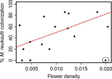
M. Belisle et al.

dispersal by pollinators, which are thought to be the main         number of flowers produced by the plant at the time of
dispersal agent [9]. At our study site, Anna’s hummingbird         sampling. A total of 192 flowers were sampled.
(Calypte anna) is the main pollinator of M. aurantiacus               Immediately after harvesting each flower, nectar was
flowers, although Allen’s hummingbird (Selasphorus sasin),         extracted with a 5-μl microcapillary tube, volume mea-
Rufous hummingbird (Selasphorus rufus), and occasionally           sured, diluted in 60 μl of distilled H2O, and stored at 4°C
bees (Bombus vosnesenskii and Xylocopa micans) have also           until being processed within 1 week of nectar collection in
been noted visiting M. aurantiacus flowers. Since visits by        the field. Dilution plating was used to estimate the density
insect pollinators are not common [45], this study focuses on      of colony forming units (CFUs) in each sample. Briefly,
spatial variation in environmental and floral factors that might   each sample was further diluted in distilled H2O and the
influence nectar availability and hummingbird movement,            nectar–water solution spread on yeast extract–malt extract
including light intensity, flower density, neighboring plant       agar (YM agar) plates [49] using a sterile spreading rod.
density, and nectar volume per flower [7, 44].                     Resulting colonies were counted from plates with dilutions
                                                                   yielding approximately 100 CFUs after 2 days of incuba-
                                                                   tion at 25°C. From each plate, up to 12 colonies were
Methods                                                            randomly chosen for separate DNA extractions (see below).
                                                                   The density of yeast per collected flower was estimated
Study Site                                                         using the dilution factor and the number of colonies
                                                                   recovered after plating. Although this method disregards
The survey was conducted in a 0.25-km2 area at the Jasper          species that may be unculturable, it is commonly used in
Ridge Biological Preserve (JRBP) on the San Francisco              similar studies and has been found to accurately represent
peninsula of California. This area contains several vegetation     the species composition and cell density of nectar-
types, including chaparral, open woodland, and broadleaf           inhabiting microfungi [9].
evergreen forest within a relatively small area (Fig. S1). Our
preliminary survey at JRBP detected nectar-inhabiting micro-       Hummingbird Sampling
fungi in multiple plant species, including M. aurantiacus,
Lepechinia calycina (pitcher sage, Lamiaceae), Pedicularis         Two experiments were carried out to ascertain that
densiflora (Indian warrior, Orobanchaceae) and Eriodictyon         hummingbirds were dispersal agents for microfungi to M.
californicum (yerba santa, Boraginaceae). In this study, we        aurantiacus flowers. In the first, hummingbirds were
focus on M. aurantiacus, a common species at JRBP, to              captured by mist-netting, and their tongues and beaks
standardize host species identity. This species is a shrub         assayed for the presence of microfungi. To this end, after
native to California and Oregon. In JRBP, it is found under a      capture, hummingbirds were fed initially sterile sugar
range of conditions, from relatively moist oak woodland to         water. The remaining water, which had come into contact
dry, open chaparral (Fig. S1), and blooms from approxi-            with birds’ beaks and tongues, was plated on YM agar, and
mately late March to early July [37].                              the resulting colonies were DNA-sequenced for species
                                                                   identification. In the second experiment, we caged M.
Nectar Sampling                                                    aurantiacus plants to experimentally deny access by
                                                                   hummingbirds. Because the mesh size of the cages was
For this study, 16 M. aurantiacus plants under varying             large enough to allow access by potential insect pollinators,
microenvironmental conditions were chosen for sampling             but not hummingbirds, we were able to evaluate the role of
of nectar. Flowers from each plant were collected in June          hummingbirds for dispersal of microfungi through compar-
2010, which coincided with the height of M. aurantiacus            ison of flowers in vs. outside the cages. Yeast abundance in
flowering activity at JRBP. Flower age at the time of              nectar was surveyed by harvesting flowers for nectar
collection was standardized to 6 days by marking flower-           sampling when they were 5 days old.
ing buds before they were open and recording the day of
first flower opening. Although individual M. aurantiacus           Molecular Methods
flowers bloom for up to 10 days in the field, 6 days was
chosen as the time to harvest the flowers because our              Fungal DNA was extracted and amplified using the Sigma
preliminary survey indicated that 6 days would provide a           Extract-N-Amp tissue polymerase chain reaction (PCR) kit
reasonable amount of time for yeast to disperse to the flower      (Sigma-Aldrich, Inc., Saint Louis, MO, USA). PCR
while reducing the number of flowers lost to wilting [42]. Six     reactions were performed in a 20 μl volume using 0.8 μl
to 12 flowers were sampled per plant, depending on the             of extracted DNA, 10 μl of Extract-N-Amp PCR ReadyMix
Distribution of Nectar-Inhabiting Microfungi
(Sigma-Aldrich, Inc., Saint Louis, MO, USA), 0.15 μl of (molecules per square meter per day) was estimated for
each primer at 50 μM and 8.9 μl of H2O. Amplification each of the 16 plants for the year December 2009–
was performed using the D1/D2 domains of the large December 2010 [1, 12], using photographs taken in
subunit nuclear ribosomal RNA with the primers NL1 December 2009 with a digital SLP camera with circular
(5′-GCA TATCAA TAA GCG GAG GAA AAG-3′) and fish eye lens and the Gap Light Analyzer (Simon Fraser
NL4 (5′-GGT CCG TGT TTC AAG GAC GG-3′) [38], University, Institute of Ecosystem Studies, 1999).
which are commonly used for yeast identification [9, 27,
32, 34]. Statistical Analyses
PCR amplification was conducted using a touchdown
PCR protocol with the following settings: an initial To determine which environmental factors were corre-
denaturation step of 94°C for 3 min, denaturation at 94°C lated with microfungal distribution, we performed linear
for 30 s, 10 cycles decreasing in 0.5°C increments from regressions using nectar volume, light intensity, and
56.5°C to 51.5°C for 30 s each, 20 cycles of 51.5°C for flower density as predictors of the percentage of flowers
45 s followed by 72°C for 45 s, and a final elongation step per plant occupied by microfungi using JMP software v.
of 72°C for 10 min. PCR products were separated by gel 8.02 (SAS Institute Inc., Carey, NC, USA). We also
electrophoresis using 1.25% sodium boric acid gel [8] and performed the same analysis to predict the percentage of
visualized using ethidium bromide staining and subsequent flowers per plant occupied by the most common
UV transillumination (Fotodyne Inc., Hartland, WI, USA). microfungal species in our study, Metschnikowia reu-
Samples that produced a visible band during gel electro- kaufii. Other species were too rare to analyze individually.
phoresis were sequenced by Elim Biopharm (Hayward, CA, In addition, we created pairwise similarity matrices of
USA), using an ABI 3730 XL automatic DNA sequencer. geographic distances between plants, differences in flower
density, and the percent of flowers from which microfungi
Sequence Analysis were detected on each plant. Statistical significance of
correlations between these variables was tested using
Forward and reverse sequences were aligned using Clustal Mantel and partial Mantel tests in the programming
W (1.83) software. The consensus sequences were grouped environment R version 2.7.2 (R Core Development Team
into operational taxonomic units (OTUs) using Geneious 2008) and using the packages Ecodist [24] and Vegan
Pro bioinformatics software (Biomatters Ltd., Auckland, [40].
New Zealand). OTUs were defined as groups of sequences
sharing 98% pairwise similarity [32]. A representative
sequence of each OTU was used to perform Basic Local Results
Alignment Search Tool (BLAST) searches against the
National Center for Biotechnology Information’s GenBank. Microfungi were detected from 15 of the 16 plants sampled,
Representative sequences were also placed into a most with up to nine species per plant (Fig. 1a) and from 54 of
likely tree using sequences from other studies of nectar the 97 unwilted flowers sampled, with an average of
yeast and a recent phylogeny of the Saccarhomycetales 4,960 CFUs (min=19.2, max=94,480) per microliter of
[46]. All sequences were aligned in Geneious using the nectar in samples that contained microfungi. Across the
MAFFT [31] plugin and the most likely tree estimated study, we observed a total of nine species using a 98%
using PhyMyl [25]. Species names were assigned using a sequence similarity cutoff and 16 species using a 99%
combination of BLAST match and phylogenetic placement cutoff (Table 1). However, the observed number of species
(Fig. S2, Table 1). per flower was low, with 97% of colonized flowers having
only one fungal species (Fig. 1b, c).
Environmental Factors: Flower Density, Neighboring Plant The proportion of flowers per plant colonized by microfungi
Density, and Light Availability was not significantly correlated with flower density (r2 =0.10,
p=0.22), neighboring plant density (r2 =0.037, p=0.59,
The number of flowers on each sampled plant was recorded Fig. 2b), light availability (r2 =0.06, p=0.36), or average
at the time of flower collection. The percent cover of M. nectar volume per flower (r2 =0.18, p=0.09). However, the
aurantiacus plants within a 3-m radius of each sampled relationship between microfungal frequency and flower
plant was also recorded to estimate neighboring plant density was significant when one outlier (a small plant with
density. The amount of photosynthetically active radiation high flower density but low flower number) was excluded
transmitted through gaps in the overlying tree canopy (r2 =0.35, p=0.01; Fig. 3).M. Belisle et al.
Table 1 Taxonomic assignments of microfungi observed in this study
Species identity Number of sequences Number of sequences Top BLAST match % match Accession
from nectar from birds (accession number) number
Aspergillus fumigatus 0 1 AY660917 100 JN642540
Auerobasidium pullulans 3 2 GQ911488 99.8 JN642535
Beauveria bassiana 1 1 AY283555 99.6 JF906819
Candida albicans 0 3 FJ627953 99.7 JN652537
Candida parapsilosis 0 8 EU660860 100 JN642532
Candida quercitrusa 0 2 DQ466526 100 JN642539
Candida rancensis 20 11 EU523604 100 JN642531
Collophora rubra 0 1 HQ433106 96 JN642541
Cryptococcus albidosimilis 0 1 GU460168 100 JN642543
Cryptococcus flavescens 0 1 AM748548 100 JN642542
Cryptococcus sp. 1 1 DQ513279 100 JF906824
Hanseniaspora uvarum 0 1 EU268654 100 JN642546
Hanseniaspora valbyensis 14 11 U73596 99.8 JG906826
Metschnikowia gruessii 3 0 AF406913 99.8 JF906827
Metschnikowia koreensis 4 0 AB617390 99.5 JN642536
Metschnikowia kunwiensis 0 8 JF809869 100 JN642533
Metschnikowia reukaufii 166 0 JF809868 100 JN642529
Metschnikowia sp. 0 8 JF809868 92.1 JN642530
Penicillium toxacarium 0 1 EF198659 100 JN642544
Pichia fermentans 0 1 EF554827 99.8 JN642545
Rhodotorula sp. 0 3 AF387138 100 JN642538
Starmerella bombicola 2 4 HQ111047 99.7 JN642534
Total 214 69
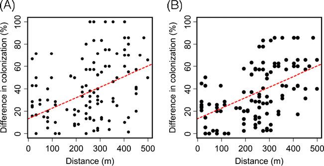
A highly significant positive correlation was found correlation between host plant spatial proximity and micro-
between the spatial proximity of plants and the frequency of fungal colonization frequency was significantly positive even
microfungal colonization (Mantel r=0.32, p=0.008; Fig. 4) after the effect of flower density was controlled for
and M. reukaufii colonization (Mantel r=0.467, p=0.001; (Mantel r=0.32, p=0.008).
Figs. 2a and 4). Although flower density showed spatial The number of flowers analyzed from a given plant was
autocorrelation when the outlier plant was removed (r2 =0.319, not significantly correlated with either the percent of
p=0.003), a partial Mantel test indicated that the positive flowers found with yeast (r2 =0.095, p=0.13), the percent
Figure 1 Histograms summarizing a the number of yeast species by the most commonly detected species, M. reukaufii (% of flowers
detected per plant, b the frequency of yeast colonization (% of flowers from which M. reukaufii was detected) in individual plants of M.
from which yeast was detected), and c the frequency of colonization aurantiacusDistribution of Nectar-Inhabiting Microfungi
Figure 2 Graphical representa-
tion of a flower colonization by
(A) M. reukaufii frequency (B) M. aurantiacus percent cover
the yeast M. reukaufii per plant 37.4085
100 m 100 m
and b estimated percent cover of
M. aurantiacus plants surround-
ing each plant from which nectar
37.4075
was sampled. GPS coordinates
are plotted along the X- and Y-
axis with points representing in-
dividual plants according to lati-
Latitude (o)
37.4065
tude and longitude. Points are
shaded from white to black, with
white indicating 0% flowers col-
37.4055
onized and black indicating
100% flowers colonized in a, and
white indicating 0% estimated 100% 40%
percent cover and black indicat- 37.4045
ing 40% estimated percent cover
by M. aurantiacus plants in a 0% 0%
3 m radius of each sampled plant
37.4035
in b 122.238 122.236 122.234 122.238 122.236 122.234
o
Longitude ( )
of flowers found with the predominant species, M. reukaufii microbes in noncaged flowers than in caged flowers
(r2 =0.023, p=0.26) or the number of yeast species found (number of flowers from which microbes were detected:
per flower (r2 =0.084, p=0.15), indicating that sampling 22 of 36 noncaged flowers vs. 13 of 36 caged flowers;
bias did not affect our results. Pearson’s chi-square test, pM. Belisle et al.
Figure 4 a Correlation between
the percentage of flowers colo-
nized by yeast per plant and the
distance between host plants
(Mantel r=0.319, p=0.008) and
b correlation between the per-
centage of flowers colonized by
M. reukaufii per plant and the
distance between host plants
(Mantel r=0.467, p=0.001).
Matrices used for the Mantel
tests were similarity matrices of
distance in meters between pairs
of plants and pairwise differences
in the percent yeast colonization
It seems feasible that trap-lining and territoriality result in nectar chemistry between plants may not always limit
heavily fungal-inoculated clumps of flowers in some parts colonization of a general nectivorous species like M.
of a given area but not in other parts. As Anna’s reukaufii. Laboratory studies indicate that nectivorous
hummingbird is the main visitor of M. aurantiacus fungal species isolated from M. aurantiacus plants at
flowers [16, 17, 45], their behavior may have created the JRBP can grow on different sugar sources, indicating a
observed spatial patterns. A study conducted at JRBP broad range of tolerance [42].
found that Anna’s hummingbird’s males aggressively Nectar-inhabiting microfungi in Europe, South Africa,
defend their territories [13]. The prime vegetation for and elsewhere seem to be characterized by a similarly low
male territories is flowering chaparral [13], where M. level of species diversity, with M. reukaufii being one of the
aurantiacus are in highest densities (Fig. S1). The core dominant species [9, 14, 27, 28, 33, 43]. These studies,
territory of each male was approximately 1,000 m2 in size, combined with our results, indicate that nectar-inhabiting
and 16 males occupied territories in an area the size of microfungal communities in geographically distant loca-
approximately 1.7 km2, whereas females were nonterrito- tions may consist of similar species, suggesting the
rial and nest in woodlands, feeding on flowers wherever possibility that nectar-inhabiting microfungi have a high
possible [13]. capacity for long-distance dispersal, yet show dispersal
Several microfungal species (e.g., Candida rancensis, limitation within local areas. Sampling more plants in larger
Hanseniaspora valbyensis, Starmerella bombicola) were areas should provide a more comprehensive understanding
detected from both nectar and hummingbird samples of their distribution patterns.
(Table 1), indicating that hummingbirds were indeed their One potential reason for the widespread dominance
dispersal agent. Although M. reukaufii was not detected of M. reukaufii is that this species is superior to others in
from hummingbirds, the sample size was somewhat small local competition. This reason seems unlikely, however, as
(ten birds) and the sampling took place earlier than the time a laboratory study has suggested that other species,
of the nectar sampling. Further research on hummingbird namely C. rancensis and M. koreensis, may be as
behavior along with more microfungal sampling from competitive as M. reukaufii and can completely exclude
hummingbirds is needed to directly test for a connection it if they arrive earlier [42]. Thus, mechanisms other than
between nectar-inhabiting microfungi and pollinator local interactions within flowers may be necessary to
movement. explain the distribution patterns we found. Differences
In this study, we measured nectar quantity, but not between species in dispersal ability, if they exist, may be
quality, which may also have affected microfungal one such explanation.
distribution. Varying concentrations of amino acids Along with findings from previous work [9, 14, 23, 27,
and sugars in nectar are known to affect pollinator 28, 44], the nonrandom distribution patterns we have
visitation [4, 5]. In addition, some yeast species may reported in this paper reinforce the prospect of these
affect the concentrations of amino acids and sugars, thus microfungal species as a useful model system for under-
potentially affecting pollinator visitation as well as the standing the role of dispersal in determining microbial
growth of late-arriving yeast species [26, 42]. Nectar distribution. Future research should more directly investi-
chemistry may provide a strong environmental filter for gate dispersal limitation through both observations and
some microfungal species [28]. However, variation in experiments.Distribution of Nectar-Inhabiting Microfungi
Acknowledgments We thank Nona Chiariello and JRBP staff for ography of stream bacterial communities. Ecology 88:2162–
assistance during field sampling, Chase Mendenhall for his contributions 2173
with hummingbird mist-netting, Trevor Hebert for assistance with Fig. 20. Finlay BJ (2002) Global dispersal of free-living microbial
S1, and the Biology 44Y students in the spring of 2011 for their eukaryote species. Science 296:1061–1063
assistance in collecting the data presented in Fig. S3. We also thank Bill 21. Finlay BJ, Fenchel T (2004) Cosmopolitan metapopulations of
Gomez, Nathan Kim, Christine Kyauk, Katrina Luna, Pat Seawell, free-living microbial eukaryotes. Protist 155:237–244
Sebastian Calderon Bentin, and Diamantis Sellis for field and laboratory 22. Gill FB (1988) Trapline foraging by hermit hummingbirds:
assistance; Annette Golonka and Carlos Herrera for technical advice; competition for an undefended, renewable resource. Ecology
and Paul Ehrlich, Hal Mooney, Karen Nelson, members of the Fukami 69:1933–1942
lab, and several anonymous reviewers for comments. Funding was 23. Golonka AM (2002) Nectar-inhabiting microorganisms (NIMs)
provided by Stanford University. and the dioecious plant species Silene latifolia.. PhD dissertation,
Duke University, Durham, NC
24. Goslee SC, Urban DL (2007) The ecodist package for
dissimilarity-based analysis of ecological data. J Stat Softw
References 22:1–19
25. Guindon S, Gascuel O (2003) A simple, fast, and accurate
algorithm to estimate large phylogenies by maximum likelihood.
1. Anderson MC (1964) Studies of the woodland light climate: I. the Syst Biol 52:696–704
photographic computation of light conditions. J Ecol 52:27–41 26. Herrera CM, García IM, Pérez R (2008) Invisible floral larcenies:
2. Baas Becking LGM (1934) Geobiologie of inleiding tot de microbial communities degrade floral nectar of bumble bee-
milieukunde. W.P. Van Stockum & Zoon, The Hague pollinated plants. Ecology 89:2369–2376
3. Bahl J, Lau MCY, Smith GJD, Vijaykrishna D, Cary SC, Lacap DC, 27. Herrera CM, de Vega C, Canto A, Pozo MI (2009) Yeasts in floral
Lee CK, Papke RT, Warren-Rhodes KA, Wong FKY, McKay CP, nectar: a quantitative survey. Ann Bot 103:1415–1423
Pointing SB (2011) Ancient origins determine global biogeography 28. Herrera CM, Canto A, Pozo MI, Bazaga P (2010) Inhospitable
of hot and cold desert cyanobacteria. Nat Commun 2:163 sweetness: nectar filtering of pollinator-borne inocula leads to
4. Baker HG, Baker I (1986) The occurrence and significance of impoverished, phylogenetically clustered yeast communities. Proc
amino acids in floral nectar. Plant Syst Evol 151:175–186 R Soc B 277:747–754
5. Baker HG, Baker I (1987) The predictability of pollinator type by 29. Hosaka K, Castellano MA, Spatafora JW (2008) Biogeography of
the chemistry of nectar. Am J Bot 74:645 hysterangiales (Phallomycetidae, Basidiomycota). Mycol Res
6. Berkeley MJ (1863) The gardeners' chronicle & agricultural 112:448–462
gazette, London, UK. 30. Janzen DH (1971) Euglossine bees as long-distance pollinators of
7. Boose DL (1997) Sources of variation in floral nectar production tropical plants. Science 171:203–205
rate in Epilobium canum (Onagraceae): implications for natural 31. Katoh K, Misawa K, Kuma K, Miyata T (2002) MAFFT: a novel
selection. Oecologia 110:493–500 method for rapid multiple sequence alignment based on fast
8. Brody JR, Kern SE (2004) Sodium boric acid: a Tris-free, cooler Fourier transform. Nucleic Acids Res 30:3059–3066
conductive medium for DNA electrophoresis. Biotechniques 32. Kurtzman CP, Robnett CJ (1998) Identification and phylogeny of
36:214–216 ascomycetous yeasts from analysis of nuclear large subunit (26S)
9. Brysch-Herzberg M (2004) Ecology of yeasts in plant–bumblebee ribosomal DNA partial sequences. Antonie Van Leeuwenhoek
mutualism in Central Europe. FEMS Microbiol Ecol 50:87–100 73:331–371
10. Cain ML, Bowman WD, Hacker SD (2011) Ecology. Sinauer, 33. Lachance M, Starmer WT, Rosa CA, Bowles JM, Barker JSF,
Sunderland Janzen DH (2001) Biogeography of the yeasts of ephemeral
11. Carter C, Healy R, O’Tool NM, Naqvi SMS, Ren G, Park S, flowers and their insects. FEMS Yeast Res 1:1–8
Beattie GA, Horner HT, Thornburg RW (2007) Tobacco nectaries 34. Lachance MA, Daniel HM, Meyer W, Prasad GS, Gautam SP,
express a novel NADPH oxidase implicated in the defense of Boundy-Mills K (2003) The D1/D2 domain of the large-subunit
floral reproductive tissues against microorganisms. Plant Physiol rDNA of the yeast species Clavispora lusitaniae is unusually
143:389–399 polymorphic. FEMS Yeast Res 4:253–258
12. Chazdon RL, Field CB (1987) Photographic estimation of 35. Lokvam J, Braddock JF (1999) Anti-bacterial function in the
photosynthetically active radiation: evaluation of a computerized sexually dimorphic pollinator rewards of Clusia grandiflora
technique. Oecologia 73:525–532 (Clusiaceae). Oecologia 119:534–540
13. Colwell RR (1994) Breeding territories of the male Anna’s 36. Lumbsch HT, Buchanan PK, May TW, Mueller GM (2008)
hummingbirds at Jasper Ridge Biological Preserve. Biology 96 Phylogeography and biogeography of fungi. Mycol Res 112:423–
project, Jasper Ridge Paper, Stanford University 424
14. de Vega C, Herrera CM, Johnson SD (2009) Yeasts in floral nectar 37. Mooney HA, Ehrlich PA, Lincoln DE, Williams KS (1980)
of some South African plants: quantification and associations with Environmental controls on the seasonality of a drought
pollinator type and sugar concentration. S Afr J Bot 75:798–806 deciduous shrub, Diplacus aurantiacus and its predator, the
15. Fenchel T (2003) Biogeography for bacteria. Science 301:925–926 Checkerspot Butterfly, Euphydryas chalcedona. Oecologia
16. Fetscher AE, Kohn JR (1999) Stigma behavior in Mimulus 45:143–146
aurantiacus (Scrophulariaceae). Am J Bot 86:1130–1135 38. O’Donnell K (1993) Fusarium and its near relatives. In: Reynolds
17. Fetscher AE, Rupert SM, Kohn JR (2002) Hummingbird foraging DR, Taylor JW (eds) The fungal holomorph: mitotic, meiotic and
position is altered by the touch-sensitive stigma of bush monkey- pleomorphic speciation in fungal systematics. CAB International,
flower. Oecologia 133:551–558 Wallingford, pp 225–233
18. Fierer N, Jackson RB (2006) The diversity and biogeography 39. Oda Y, Star B, Huisman LA, Gottschal JC, Forney LJ (2003)
of soil bacterial communities. Proc Natl Acad Sci USA Biogeography of the purple nonsulfur bacterium Rhodopseudo-
103:626–631 monas palustris. Appl Environ Microbiol 69:5186–5191
19. Fierer N, Morse JL, Berthrong ST, Bernhardt ES, Jackson RB 40. Oksanen L, Kindt R, Legendre P, O’Hara B, Simpson GL,
(2007) Environmental controls on the landscape-scale bioge- Solymos P, Henry M, Stevens H, Wagner H (2008) Vegan:M. Belisle et al.
Community Ecology Package. R package version 1.15-1. http:// 45. Streisfeld MA, Kohn JR (2007) Environment and pollinator-
cran.r-project.org/, http://vegan.r-forge.r-project.org/ mediated selection on parapatric floral races of Mimulus aur-
41. Peay KG, Bruns TD, Kennedy PG, Bergemann SE, Garbelotto M antiacus. J Evol Biol 20:122–132
(2007) A strong species–area relationship for eukaryotic soil 46. Suh S, Blackwell M, Kurtzman CP, Lachance M (2006)
microbes: Island size matters for ectomycorrhizal fungi. Ecol Lett Phylogenetics of Saccharomycetales, the ascomycete yeasts.
10:470–480 Mycologia 98:1006–1017
42. Peay KG, Belisle M, Fukami T (2011) Phylogenetic relatedness 47. Telford RJ, Vandvik V, Birks HJB (2006) Dispersal limitations
predicts priority effects in nectar yeast communities. Proc R Soc matter for microbial morphospecies. Science 312:1015
B. doi:10.1098/rspb.2011.1230 48. Whitaker RJ, Grogan DW, Taylor JW (2003) Geographic barriers
43. Pozo MI, Herrera CM, Bazaga P (2011) Species richness of yeast isolate endemic populations of hyperthermophilic archaea. Sci-
communities in floral nectar of southern Spanish plants. Microb ence 301:976–978
Ecol 61:82–91 49. Yarrow D (1998) Methods for the isolation, maintenance
44. Stiles FG (1975) Ecology, flowering phenology, and hummingbird and identification of yeasts. In: Kurtzman CP, Fell JW (eds)
pollination of some Costa Rican Heliconia species. Ecology The yeasts: a taxonomic study. Elsevier, Amsterdam, pp 77–
56:285–301 100You can also read