Glutathione-loaded non-ionic surfactant niosomes: A new approach to improve oral bioavailability and hepatoprotective efficacy of glutathione - De ...
←
→
Page content transcription
If your browser does not render page correctly, please read the page content below
Nanotechnology Reviews 2022; 11: 117–137
Research Article
Esam M. Aboubakr, Hamdoon A. Mohammed*, Abeer S. Hassan, Hebatallah B. Mohamed,
Mahmoud I. El Dosoky, and Adel M. Ahmad*
Glutathione-loaded non-ionic surfactant
niosomes: A new approach to improve oral
bioavailability and hepatoprotective efficacy
of glutathione
https://doi.org/10.1515/ntrev-2022-0010 and Span®60-Tween®80 at a molar ratio of 2:1 of choles-
received October 11, 2021; accepted November 19, 2021 terol/non-ionic surfactant, displaying a particle size of
Abstract: A new formulation (niosomes) was prepared to 688.5 ± 14.52 nm, a zeta potential of −26.47 ± 0.158 mV,
enhance the bioavailability, hepatic tissue uptake, and and encapsulation efficiency (EE) of 66 ± 2.8% was selected
hepatoprotective activity of glutathione (GSH). The GSH- for in vivo testing. The levels of MDA, NO, SOD, NF-κB,
loaded niosomes (nanoform, N-GSH) were formulated by IL-1β, and Bcl-2 were measured. The results demonstrated
the thin-film hydration technique using cholesterol/non- that hepatic tissue damage was ameliorated using N-GSH
ionic surfactants (Span®40, Span®60, and Tween®80) at a as confirmed by the morphological and histopathological
componential ratio of 1:1 and 2:1. The hepatoprotective examination compared to the CCl4 and control groups.
activity of N-GSH, GSH, and the standard silymarin against The N-GSH significantly (p < 0.05) decreased the elevated
CCl4-induced liver damage and oxidative stress were levels of hepatic enzymes, oxidative parameters, and inflam-
tested on the rats’ model. The hepatic morphology and matory mediators, as compared to silymarin and GSH. Also,
histopathological characters were also investigated. The N-GSH significantly (p < 0.05) increased GSH hepatocyte
tissue contents of N-GSH were analysed using a concur- concentrations as compared to the control groups. The pre-
rently validated RP-HPLC method. The optimized nio- sent study demonstrated that N-GSH remarkably improved
somes, composed of glutathione (500 mg), cholesterol, glutathione oral bioavailability and hepatic tissue uptake,
thereby introducing a new glutathione formulation to pro-
tect hepatic tissue from injury and restore its GSH contents.
* Corresponding author: Hamdoon A. Mohammed, Department of Keywords: glutathione, oral bioavailability, niosomes,
Medicinal Chemistry and Pharmacognosy, College of Pharmacy,
RP-HPLC, liver protection, redox status, antioxidants,
Qassim University, Qassim 51452, Saudi Arabia; Department of
Pharmacognosy, Faculty of Pharmacy, Al-Azhar University, anti-inflammatory
Cairo 11371, Egypt, e-mail: ham.mohammed@qu.edu.sa,
tel: +96-6566176074
* Corresponding author: Adel M. Ahmad, Department of
Pharmaceutical Analytical Chemistry, Faculty of Pharmacy, 1 Introduction
South Valley University, Qena 83523, Egypt,
e-mail: adelpharma2004@svu.edu.eg Liver-associated illnesses remain a global health concern.
Esam M. Aboubakr: Department of Pharmacology and Toxicology, These diseases are considered to be the leading cause of
Faculty of Pharmacy, South Valley University, Qena 83523, Egypt,
mortality among populations of low- to middle-income
e-mail: esam_pharma@svu.edu.eg
Abeer S. Hassan: Department of Pharmaceutics, Faculty of Asian and African countries [1]. The liver is highly suscep-
Pharmacy, South Valley University, Qena 83523, Egypt, tible to oxidative stress, inflammation, and degeneration
e-mail: abeer.saad@svu.edu.eg since it is the main organ responsible for the metabolism
Hebatallah B. Mohamed: Department of Pharmaceutics, Faculty of and detoxification of nutrients/drugs [2]. Several physio-
Pharmacy, South Valley University, Qena 83523, Egypt,
logical and pathological processes in human and animal
e-mail: dochoba2014@svu.edu.eg
Mahmoud I. El Dosoky: Department of Pathology, Faculty of
cells, e.g. gene expression, signal transduction, growth,
Medicine, South Valley University, Qena 83523, Egypt, and even death, are associated with reactive oxygen spe-
e-mail: mahmouddosoky@med.svu.edu.eg cies (ROS) production [3]. However, excessive ROS are
Open Access. © 2022 Esam M. Aboubakr et al., published by De Gruyter. This work is licensed under the Creative Commons Attribution 4.0
International License.
118 Esam M. Aboubakr et al.
implicated in soft-tissue degeneration, cancer, neuro- ∼2.5 min in the blood [16]. Therefore, the oral formula-
degenerative, cardiovascular diseases, cellular damage, tions of glutathione that withstand the gastrointestinal
and death [4]. environment and degradation are imperative in nature
The protective antioxidant mechanisms and a number and are in demand to overcome these shortcomings. Dif-
of antioxidant systems are conspicuous in the biological ferent approaches have been adopted to improve the GSH
systems that are involved in the free radicals capture/neu- bioavailability through the oral route by different novel
tralization and thereby performing cellular protection [5]. formulations, and in this context, the nanoformulations
Two antioxidant protective systems are available in the have taken the lead. The GSH encapsulating chitosan
human body, i.e. enzymatic antioxidants and non-enzymatic nanoparticles [17] and Eudragit VR RS 100/cyclodextrin
antioxidants. The first system includes several enzymes, e.g. material-based formulations [16] have been prepared.
glutathione peroxidase (GPx) and catalase (CAT), which Also, the mucoadhesive polymer-based film for GSH was
combats free radical formation and neutralize their harmful formulated [18]. The appropriate liposomal nanovesicles
effects [6]. The second antioxidant system, non-enzymatic formulation that may enhance the bioavailability and
antioxidants, includes compounds such as glutathione, stability of the GSH component and reduce the dosing
coenzyme Q10, melatonin, thioredoxin, glutaredoxin, and frequency of GSH has also been proposed. The phospho-
lipoic acid, produced as endogenous metabolic products, lipid bilayer composition of liposomes has a similar struc-
contributes to cellular protection against excessive ROS ture to that of the cell membranes, which enhanced the
[5]. In addition, nutrients such as greens, fruits, vitamin entrapped/core drugs, e.g. the bioavailabilities of peptides
E, and vitamin C-rich foods are also supportive materials and proteins [19]. However, drug leakage, phospholipid
used as protective antioxidant nutraceuticals [7]. hydrolysis, phospholipid oxidation, sedimentation, and
Glutathione (GSH) is the predominant non-enzymatic particles aggregation are common drawbacks and stability
thiol antioxidant in cells with a primary role in scaven- shortcomings of the liposomal preparations [20,21]. There-
ging the free radicals. GSH is present in the cytosol and fore, alternative non-ionic surfactant nanovesicles (nio-
other cell structures, such as mitochondria, peroxisomes, somes) have been proposed and are currently developed
and nuclear matrices [8]. Also, all the cells can biosynthe- to overcome the stability shortcomings of the liposomes.
size GSH; however, hepatocytes are the main exporting Niosomes are vesicular bilayer structures of non-ionic sur-
source [8]. The oxidized glutathione, glutathione disul- factants and cholesterol that self-assemble spontaneously
phide (GS-SG), produced from the reaction of GSH with on hydration with aqueous media. Niosomes are also
free radicals, further undergoes the enzymatic reduction better candidates for drug delivery than liposomes owing
process by nicotinamide adenine dinucleotide phosphate to their lower costs and higher stability. In addition, the
(NADPH)-dependent glutathione reductase to regenerate niosomes have better resistance to hydrolysis and oxida-
the glutathione (GSH) in a reduced form [9]. Other roles tion during storage. Drug solubility and stability enhance-
of GSH, i.e. in the regeneration of disulphide bonds of ments are also added advantages for the niosomes formu-
proteins, as co-factor for detoxifying the enzymes, and lations. The drug’s permeation and bioavailability have
lessening a load of electrophiles and oxidants as a result also been widely explored as advantages of the niosomes
of the presence of cysteine in the GSH structure, have also formulations when delivered through various routes of
been also reported [10]. Therefore, the cells’ ability to administration [22]. These nanovesicles, the niosomes,
combat free radicals is directly affected by the GSH/GS- are comparatively thermodynamically more stable than
SG, and to a lesser extent, to the ratio of other redox cou- the liposomes, thereby adjusting the process temperature
ples, e.g. NADPH/NADP+ and thioredoxinred/thioredoxinox above the gel/liquid transition of the main lipid composi-
[9,11]. The factors affecting GSH levels in the plasma have tion through the use of suitable mixtures of the surfactants
also been reported in the literature. For instance, the levels and a proper stabilizer, i.e. cholesterol, in the formulation
of GSH normally decrease with age and could be patholo- of niosomes. By considering the parameters of componen-
gically depleted due to several physiological malfunc- tial ratios of the ingredients, the nature of the surfactant,
tioning in response to diseases, e.g. neurodegenerative temperature, and the stabilizer, the prepared nanovesicles
[12], pulmonary, hepatic [13], immune, and cardiovascular are considered to be suitable for use as a targeted carrier
disorders [14]. for controlled release of the dosage form for the lipophilic
The GSH bioavailability through the oral route is poor and hydrophilic drugs [23].
as it is degraded by the intestinal enzyme, γ-glutamyl The current study was designed to develop and eval-
transpeptidase, leading to its lowered absorption from uate various formulations of the GSH-loaded niosomes
gout [15]. In addition, GSH also has a short half-life of prepared with different non-ionic surfactants to enhance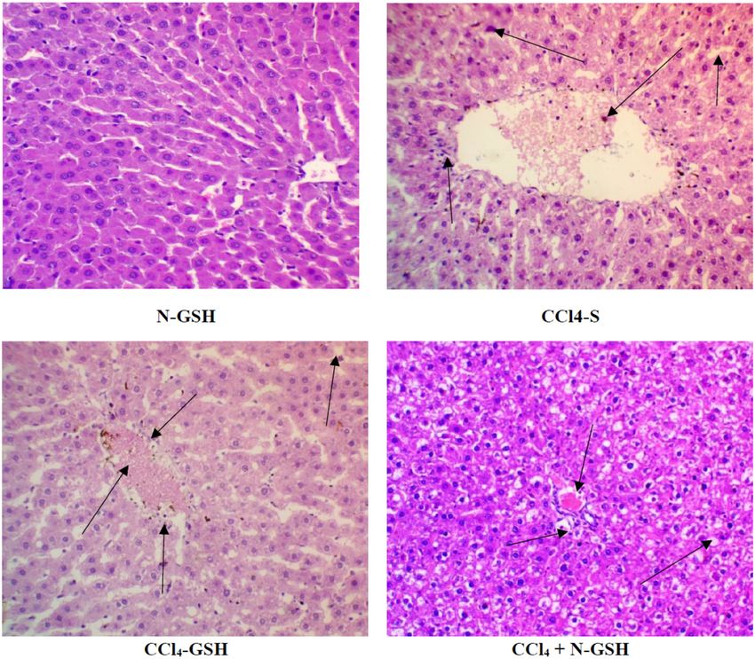
Improved oral-bioavailability and hepatoprotective efficacy of glutathione 119
the stability of the GSH, and bioavailability of the GSH- purification system (Millipore, Milford, MA, USA). The
loaded formulations when administered through the oral standard solution of GSH was prepared by dissolving
route. The GSH-nanovesicles (n-GSH) formulated through 50 mg/L GSH in 10 mM ethylenediamine tetraacetic acid
the thin-film hydration method and characterized in terms (EDTA) to prevent oxidation by trace metals. The standard
of their particle size, zeta potential, and entrapment effi- working solution was prepared through dilutions with the
ciency (EE). Furthermore, the biological evaluations of the HPLC mobile phase.
N-GSH as a liver-protecting agent against carbon tetra-
chloride (CCl4)-induced liver injury in comparison to sily-
marin and GSH were performed. GSH concentrations in the
liver tissue homogenates of the injured and treated animals 2.2 Preparation of glutathione-loaded
were assayed. The intracellular redox status and oxidative niosomes
stress conditions were also evaluated by measuring the
malondialdehyde (MDA) levels, anti-oxidant enzyme Glutathione-loaded-niosomes were prepared using the
activity, superoxide dismutase (SOD) reactivity, and thin-film hydration method with different mixtures of
the serum levels of the inflammatory cytokines, tumor cholesterol and non-ionic surfactants at molar ratios of
necrosis factor TNF-α, IL-6, aspartate aminotransferase 1:1 and 2:1 [24]. A 500 µmol of non-ionic surfactants mix-
(AST), alanine aminotransferase (ALT), and nitric oxide ture (Span® 40 and Span® 60: Tween® 80) and choles-
(NO) status. A histopathological examination for hepatic terol were dissolved in a chloroform/methanol mixture
tissues of the normal, injured, and treated animals were (2:1 v/v) in a round bottom flask. Organic solvents were
also performed. evaporated (15 min at 55°C, 900 rpm) using a rotary eva-
porator (Buchi 200, BU¨CHI Labortechnik AG, Flawil,
Switzerland). The resultant film that formed in the bottom
of the rounded flask was hydrated by gently shaking it for
2 Materials and methods 45 min at 55°C with distilled water containing 250 mg and
500 mg of GSH. The developed niosomal dispersion was
stored at 4°C for further study. The compositions of dif-
2.1 Chemicals and reagents ferent niosomal formulations are summarized in Table 1.
Cholesterol, silymarin, and glutathione were purchased
from Sigma Chemical Co, St. Louis, MO, USA. Span®20,
Span® 40, Span® 60, Span® 80, Tween® 20, Tween® 40, 2.3 RP-HPLC analytical procedure
Tween® 60, Tween® 80, and propylene glycol were pur-
chased from Adwic, El-Naser Chemical Co., Egypt. The Agilent 1260 Infinity® HPLC system (Agilent Technologies,
HPLC-grade chloroform, methanol, acetonitrile, and ana- Germany), used for the method development, consisted of
lytical-grade sodium acetate, disodium hydrogen phos- an Agilent 1260 Infinity® binary solvent pump, thermostat
phate, CCl4, thiobarbituric acid (TBA), and diethyl ether column, autosampler, and Agilent 1260 Infinity® Diode
were purchased from Merck, Darmstadt, Germany. Ultra- array detector. The output signals were monitored and
pure water was obtained from the Milli-Q Plus® water processed using OpenLAB CDS ChemStation® software.
Table 1: Composition of glutathione-loaded niosomes prepared with 1:1 and 2:1 molar ratios of cholesterol and non-ionic surfactant mixture
(Span 40® and span 60®: Tween 80®)
Formulation GSH (mg) Cholesterol (µmol) Span 40® (µmol) Span 60® (µmol) Tween 80® (µmol) EE (%)
GTN1 250 250 125 — 125 35 ± 3.6
GTN2 250 250 — 125 125 45 ± 1.6
GTN3 250 335 82.5 — 82.5 53 ± 1.6
GTN4 250 335 — 82.5 82.5 65 ± 2.5
GTN5 500 250 125 — 125 47 ± 2.2
GTN6 500 250 — 125 125 54 ± 2.7
GTN7 500 335 82.5 — 82.5 60 ± 1.4
GTN8 500 335 — 82.5 — 66 ± 2.8
120 Esam M. Aboubakr et al.
All the solutions were degassed by ultrasonication (Power- where T is the initial total amount of drug added and C is
Sonic 420, Labtech, Korea) and filtered through a 0.45 µm the free unentrapped drug in the supernatant. Each
Nylon filter (PALL Life Sciences, USA). GSH concentrations experiment was performed in triplicate.
were determined using the method explained by Tsiasioti
et al. [25], with certain modifications. At 30°C, isocratic
2.4.3 Transmission electron microscopy (TEM)
separation was carried out on Pursuit-3® C18 column
(150 mm × 4.6 mm i.d. and 3 µm particle size, Agilent Tech-
The niosomal dispersion of GSH was diluted (10×) using
nologies, Netherland) using a mobile phase prepared by
distilled water. A diluted GSH vesicle dispersion drop was
mixing 10% methanol and 90% 0.02 M phosphate buffer
applied to a carbon-coated 300 mesh copper grid and left
(pH 2.5) at a flow rate of 1.0 mL/min. The injected volume
for 1 min to allow some vesicles to adhere to the carbon
of the standard solution was 20 µL, and detected with a UV
substrate. Excess dispersion was removed by a piece of
spectrophotometer at 210 nm. Various chromatographic
filter paper followed by rising the grid twice in deionized
conditions were tested, optimized, and validated according
water for 3–5 s. Next, a drop of 2% aqueous solution of
to ICH guidelines for use [26].
uranyl acetate was applied for 1 s. The remaining solution
was removed using filter paper, and the sample was air-
dried. Afterward, the sample was viewed under the micro-
scope at 10–100k magnification power using an acceler-
2.4 Characterization of glutathione-loaded
ating voltage of 100 kV using the JEOL TEM (Model 100 CX
niosomes II; Tokyo, Japan).
2.4.1 Evaluation of the vesicle size and zeta potential
2.4.4 In vitro drug release study
An average diameter of N-GSH (GSH-loaded niosomes,
z-average) and polydispersity index (PDI) of all the for- The in vitro drug release from the N-GSH niosomal for-
mulations were measured at 25°C by the dynamic light mulation as compared to the free GSH was determined at
scattering (DLS) method utilizing the Malvern Zetasizer 37°C similar to the pH of the stomach (pH 1.2) and small
Nano-ZS® instrument (Malvern Instruments, UK) equipped intestine (pH 6.8). The pH was adjusted to 6.8 with 0.5 M
with a backscattered light detector operating at 173°. Before HCl and 0.05 M phosphate buffer. These were used to
each measurement, the niosomal dispersion was diluted mimic the pH conditions of the stomach and small intes-
(20×) with double-distilled water. The zeta potential of the tine. About 1 mL of N-GSH formulation, equivalent to
vesicles was estimated by laser Doppler anemometry using 50 mg of GSH, was placed over a previously soaked cel-
a Malvern Zetasizer ZS®. All the measurements were per- lulose membrane (Spectro/Por membranes, molecular
formed in triplicate. weight cut-off: 12–14 kDa) fitted at the lower end of a
glass cylinder, and the glass cylinder was then dipped
in a beaker containing 100 mL of phosphate buffer (pH
2.4.2 Entrapment efficiency 1.2 and 6.8, 100 mL) at 37 ± 0.5°C, and shaken at 50 rpm
using a thermostatically controlled water bath (Gesellschaft
The EE of the GSH into the N-GSH niosomes was esti- Labor Technik MBH & Co., GFL. Germany). Aliquots (5 mL)
mated indirectly by measuring the free drug (GSH) in were removed and substituted with a freshly prepared
the supernatant of the hydrated N-GSH preparation using buffer medium. The drug content was estimated by the
the developed RP-HPLC method. The unentrapped drug RP-HPLC method at the predefined time points for 24 h.
was separated from drug-loaded niosomes by centrifuga- The in vitro release experiment was repeated in triplicate.
tion at 4°C, 14,000 rpm, for 60 min, using a bench-top
refrigerated centrifuge (Centurion Scientific Ltd, Sussex,
2.4.5 Kinetic release study
UK). The obtained niosomes pellets were reconstituted in
distilled water and washed twice using the same proce-
The data obtained from the in vitro release studies were ana-
dures. The EE (in percentage) was calculated according to
lysed using the linear regression method (r2). The release data
the following equation:
were analysed according to the zero-order kinetic model,
EE(%) = [T − C ]/ T × 100, (1) Higuchi diffusion model, and Korsmeyer–Peppas model.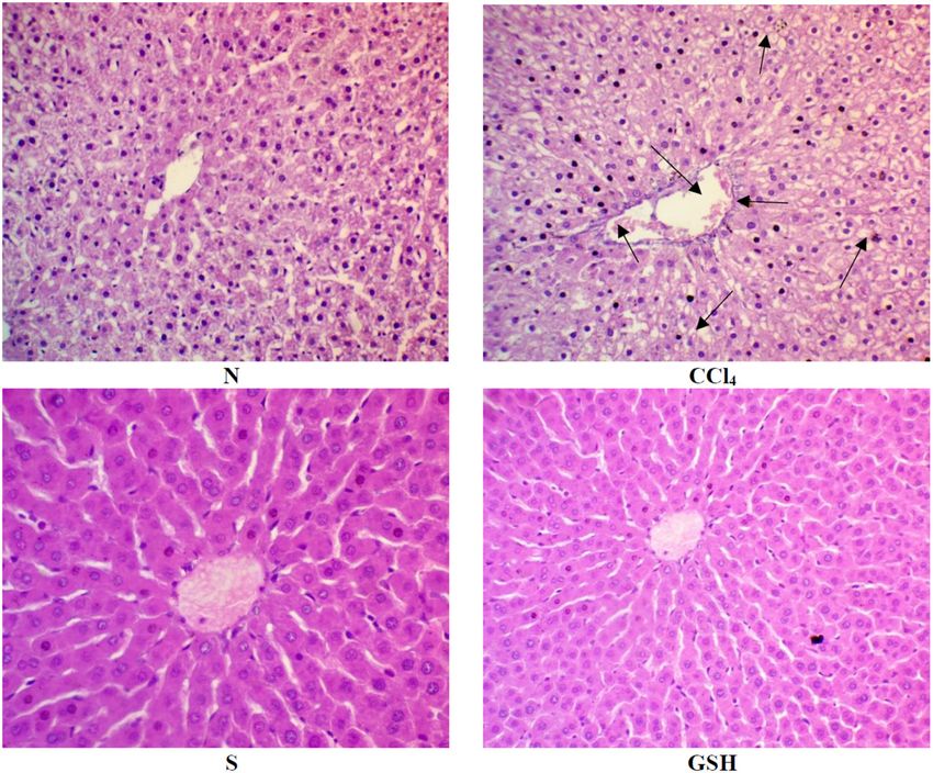
Improved oral-bioavailability and hepatoprotective efficacy of glutathione 121
Zero-order kinetics: animals received corn oil (1 mL/kg, twice a week) by intra-
Q = k 0t, peritoneal injection (i.p.) and distilled water (0.5 mL/day,
orally) for 8 consecutive weeks; G2 (CCl4 group), animals
where Q is the drug released at time t and k0 is the zero-
were i.p. injected with CCl4 as 50% solution in corn oil
order release constant. (1 mL/kg, twice a week) for 8 consecutive weeks and
Higuchi model: 0.5 mL normal saline (0.5 mL/day, orally) for 8 consecutive
Q = KHt 1 / 2, weeks; G3 (silymarin group), rats were orally administered
where Q is the amount of drug released at time t per unit 100 mg/kg/day silymarin for 8 consecutive weeks; G4
area, and KH is the Higuchi release rate constant. (GSH group), rats were orally administered 100 mg/kg/day
Korsmeyer–Peppas equation: of reduced glutathione for 8 consecutive weeks; G5 (N-GSH
group), rats were orally administered 100 mg/kg/day
Mt /M ∞ = ktn, reduced N-GSH (nano-glutathione) for 8 consecutive
where Mt/M∞ is the fraction of drug released at time t weeks; G6 (CCl4 + silymarin), rats were administered
and n is the release exponent. The n value is indicative of 1 mL of CCl4 (50% solution in corn oil, i.p., 1 mL/kg, twice
the drug release mechanism, where n ≤0.5 indicates a a week) + 100 mg/kg/day silymarin orally for 8 consecu-
Fickian diffusion mechanism, while 0.5 < n < 1 indicates tive weeks; G7 (CCl4 + GSH), rats were administered 1 mL
a non-Fickian mechanism (anomalous diffusion). If n = 1, CCl4 (50% solution in corn oil, i.p., 1 mL/kg, twice a
it indicates a zero-order mechanism (case II relaxation). In week) + 100 mg/kg/day reduced glutathione (GSH) orally
the case of n > 1, it indicates a super case II transport. The for 8 consecutive weeks; and G8 (CCl4 + N-GSH), rats
anomalous diffusion or non-Fickian diffusion refers to a com- were administered 1 mL of CCl4 (50% solution in corn
bination of both diffusion and erosion controlled release rate, oil, i.p, 1 mL/kg, twice a week) + 100 mg/kg reduced
while case II relaxation and super case II transport refer to the N-GSH orally for 8 consecutive weeks.
erosion of the polymeric matrix.
2.5.2 Blood and liver sampling
2.4.6 Stability studies
Twenty-four hours after the last administered dose, under
The stability of all formulations was determined by storing light diethyl ether anaesthesia, blood samples were col-
them at 4°C and room temperature in a sealed 20 mL glass vial. lected from inferior vena cava in clean, dry test tubes, and
The size, PDI, and zeta-potential values were recorded at pre- centrifuged at 3,000 rpm for 10 min; serum was separated
defined time intervals (fresh preparation and 4 weeks after and refrigerated at −20°C for further analysis. The liver of
storage). All the measurements were repeated in triplicate. each animal was immediately dissected out, washed with
ice-cold saline. The liver samples were macroscopically
examined to determine the apparent intensity of the damage,
2.5 In vivo experiment
and its relative weight to the animal’s total weight was
recorded. Parts of liver samples were collected and homoge-
All approvals were obtained by the ethical committee at the
nized using 100 mmol KH2PO4 buffer containing 1 mmol
faculty of Pharmacy, South Valley University (Approval #
EDTA (pH 7.4) and centrifuged at 12,000×g for 30 min at
P1001). Animals were purchased from Helwan Animal Breed-
4°C to produce the 10% homogenate. The supernatants
ing House, Cairo, Egypt. Sixty-four adult male Sprague–
were collected and refrigerated at −80°C for further analysis
Dawley rats weighing between 130 and 150 g were used.
through RP-HPLC. Other parts of the hepatic tissues
Animals were kept under optimized conditions of 12 h
were also preserved in 10% formalin for histopatholo-
light–12 h dark cycle, 25 ± 2°C temperature, and were fed
gical examinations.
standard rat chow and water ad libitum. The animals were
treated according to international and national ethical
guidelines.
2.5.3 Histopathological examination
2.5.1 Experimental design The hepatic tissue samples were fixed in 10% formalin at
room temperature for 24 h, and samples were dehydrated
Animals were randomly divided into 8 groups (n = 8 per using graded ethanol and embedded in paraffin wax. Tissue
group) as follows: G1 (Normal group), healthy control sections with a thickness of 5 µm were deparaffinized using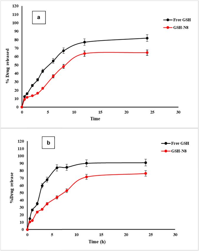
122 Esam M. Aboubakr et al.
xylene and stained with haematoxylin and eosin (H&E) [27]. 2.5.9 Assessment of MDA
Hepatic tissue staining for NF-KB [28] and Bcl-2 [29] were
performed using a standard immune-histochemical proce- MDA levels in the hepatic tissue homogenates were deter-
dure. Histopathological analysis was performed in the mined using the spectrophotometric method based on
Pathology Department at the faculty of Medicine, South the reaction between MDA (the product of lipid peroxida-
Valley University, and visualized using an Olympus micro- tion) and TBA. The pink color produced was measured
scope with 200× magnification. spectrophotometrically at 532 nm [33].
2.5.4 Determination of total protein
2.6 Statistical analysis
Protein concentrations in the hepatic tissue homogenates
were determined using the Bradford technique [30]. Statistical analysis was performed using graph-pad prism
version 9.2.0 (332). Values were presented as mean ±
standard error of the mean (SEM), n = 8. A one-way ana-
2.5.5 Determination of liver enzymes lysis of variance was used to compare groups’ results,
followed by the Tukey–Kramer test. The difference was
Levels of serum AST and ALT were determined calorime- considered significant for p < 0.05.
trically [31] using commercial kits obtained from Biodiag-
nostic Company, Egypt.
3 Results
2.5.6 Assessment of NO concentration
NO levels in the hepatic tissue were determined using a 3.1 Development and characterization of
commercial kit (Biodiagnostics, Cairo, Egypt) following GSH-loaded niosomes
the manufacturer's instructions. NO is converted to nitrous
acid followed by a reaction with sulfanilamide and N-(1- 3.1.1 Vesicle size, zeta potential, and EE measurements
naphthyl)ethylenediamine producing azo dye. The absor-
bances of samples were measured spectrophotometrically The results in Table 2 showed the vesicle sizes of all the formula-
at 540 nm. tions that were in the range of 476 ± 10.32 to 688.5 ± 14.52 nm.
Formulations prepared with varying molar ratios of cho-
lesterol under similar experimental conditions showed a
2.5.7 Assessment of SOD slight increase in the vesicle size. The optimized formula-
tion, GTN-8, was evaluated for its zeta potential, which
The hepatic tissue SOD activity was measured using a was at −26.47 ± 0.158 mV. The negative zeta-potential
standard kit (Biodiagnostics, Cairo, Egypt) according to values might be due to the presence of hydroxyl groups
the manufacturer’s protocol. Serial dilutions of the stan- of the cholesterol molecule.
dard SOD and samples were added to each well, followed Table 2 illustrates the EE (%) of GSH of the niosomes
by the radical detector and xanthine oxidase. The plate was containing cholesterol and non-ionic surfactants in ratios
shaken and incubated at room temperature for 30 min. of 1:1 and 2:1. It was observed that by increasing the
Samples absorbances were recorded at 440–460 nm using cholesterol contents from a 1:1 to 2:1 molar ratio of both
the ELISA microplate reader. the Span60® and Span40®, the EE of GSH niosomes
increased significantly (p < 0.05). Formulations GTN-4
and GTN-8 (Table 1) with the composition of cholesterol
2.5.8 Assessment of inflammatory marker (IL-Iβ) and Span60® in the ratio of 2:1 showed a maximum EE
percentage at 65 ± 3.6 and 66 ± 2.8%, respectively. Also,
The total hepatic IL-1β was measured in the hepatic tissue the EE of the GSH increased significantly by increasing
supernatant obtained using the corresponding rat-spe- the drug contents from 250 to 500 mg (p < 0.05) in the
cific ELISA kit by following the manufacturer’s protocols prepared niosomes formulations. Based on the above-
[32]. mentioned results of the EE, the optimized formulation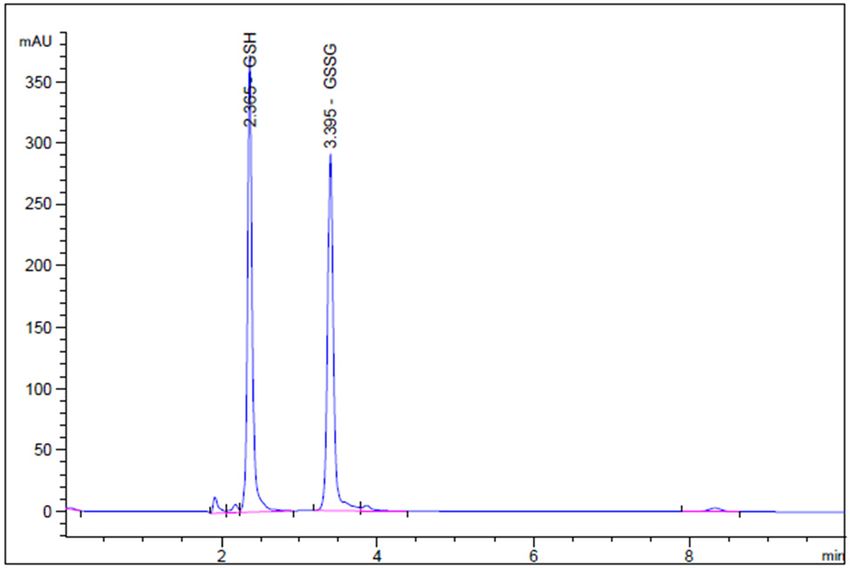
Improved oral-bioavailability and hepatoprotective efficacy of glutathione 123
Table 2: Physicochemical characteristics of the prepared Consequently, 90.9% of the drug was released within
glutathione-loaded niosomes 24 h at the pH of the intestine. Also, it was noticed that
in the stomach environment (Figure 2a), only 35.5% of
Formulation Particle size (nm) PDI GSH was released from the niosomes after 6 h of incuba-
GTN-1 476 ± 10.32 0.501 ± 0.10 tion, which represented the integrity of the nanovesicles
GTN-2 658.28 ± 15.32 0.433 ± 0.04 maintained in this simulated condition, and their ability
GTN-3 500.5 ± 7.887 0.426 ± 0.02 to restrict GSH release in an acidic environment. How-
GTN-4 686.0 ± 20.38 0.506 ± 0.19
ever, in terms of intestinal pH (pH 6.8) (Figure 2b), the
GTN-5 482.9 ± 7.75 0.491 ± 0.08
GTN-6 662.5 ± 2.23 0.462 ± 0.28
release of GSH showed a higher release rate than at pH
GTN-7 506.9 ± 12.73 0.470 ± 0.28 1.2, and the release of GSH reached 45% after 6 h of time.
GTN-8 688.5 ± 14.52 0.509 ± 0.08
3.2.1 Release kinetics of glutathione (GSH)
GTN-8 that showed higher EE% was selected for further
The kinetic parameters of GSH release from GTN-8 for-
in vitro release and in vivo studies.
mulation that followed the zero-order kinetics model,
Higuchi model, and Korsmeyer–Peppas model are sum-
marized in Table 3. It was found out that the best-fit
3.1.2 TEM and microscopic examination
model for the GSH release from the GTN-8 formulation
was the Higuchi diffusion model. According to all the
Figure 1 shows the TEM image of the optimized glu-
applied models, the release speed (k) was less at pH 1.2
tathione-loaded niosomes (prepared formulation, GTN-8).
as compared to that at pH 6.8. The calculated n value was
The vesicles had uniform spherical shapes and were free
between 0.5 and 1, which corresponded to the anomalous
of aggregation.
non-Fickian transport, whereas the mechanism of drug
release seemed to be governed by matrix erosion and diffu-
sion [34]. These results agreed with the previous investiga-
3.2 In vitro drug release studies tions that reported similar release patterns for niosomes [35].
The GSH release in pure form and from niosomes formu-
lation (GTN-8) at similar pH of the gastrointestinal tract
(stomach: pH 1.2, and intestine: pH 6.8) is exhibited in 3.3 Stability studies
Figure 2. The release profile of the free drug at pH of the
stomach and intestine demonstrated a maximum drug Stability studies were conducted at 4°C and at room tem-
release of 33 and 59.7%, respectively, in a 3 h period. perature for the optimized niosomal formulation GTN-8,
and the results are summarized in Table 4. It was found
out that no considerable variations occurred. After 4 weeks
of storage, the formulation showed a slight increase in size
at 4°C, and at room temperature, respectively, as 710 ± 12.45
and 760 ± 14.32 nm, which were within the acceptable
range. Further, the values of PDI were found to be 0.509
and 0.580. In addition, the zeta-potential values remained
near −20 mV, which indicated the homogenous nature of
the vesicles distribution and the niosomes stability. As
shown in Table 4, there are also no significant changes in
the EE values of the GSH-loaded niosomes.
3.4 RP-HPLC analysis validation
Figure 1: Transmission electron micrographs (TEMs) of glutathione- Mobile phase parameters for HPLC separations were opti-
loaded niosomes, N-GSH (formulation, GTN-8). mized to produce symmetric, sharp, and well-resolved124 Esam M. Aboubakr et al.
Figure 2: Release profiles of GSH (free and selected niosomes (formulation, GTN-8)) at pH 1.2 (a) and pH 6.8 (b) (n = 3, mean ± SD).
peaks. After several trials, the optimized mobile phase phosphate buffer (pH 2.5) containing 1 mM EDTA,
was found to be a mixture of 10% methanol/90% wherein the column temperature was set at 30°C, and a
Table 3: Modelling of glutathione release kinetics from the selected formulation GTN8 in the stomach (pH 1.2) and intestine (pH 6.8)
pH Zero-order Higuchi-model Korsmeyer–Peppas
K0 (mg/mL/h) R2 Kh (h0.5) R2 K R2 n
1.2 2.65 0.88899 16.03209 0.947581 5.91 0.948971031 0.7
6.8 2.903418 0.903418 17.66243 0.971605 6.79 0.986886917 0.6Improved oral-bioavailability and hepatoprotective efficacy of glutathione 125
Table 4: Vesicle size, zeta potential, PDI, and EE% for the selected Table 5: Summary of the development and validation of the RP-
formulation GTN8 stored at 4°C and at room temperature for four HPLC method
weeks (n = 3)
Parameter Value
Storage temperature 4°C Room temperature
Retention time (min) 2.36
(25°C)
Linearity range (µg/mL) 2.0–7.5
Size (nm) 710 ± 12.45 760 ± 14.32 Regression equation Y = 247.04 + 601.45x
PDI 0.509 ± 0.08 0.580 ± 0.08 Correlation coefficient 0.998
Zeta potential (mV) −19.6 ± 0.08 −12.6 ± 0.23 LOD (µg/mL) 0.642
EE% 62.5 ± 2.4 59.5 ± 1.5 LOQ (µg/mL) 1.94
Accuracy (n = 3)
Mean recovery (%) 98.36–102.03
% RSD 0.853–1.44
Precision (n = 3)
Intra-day (% RDS) 1.05
retention time of 2.35 min for GSH and 3.39 min for GS-SG
Inter-day (% RDS) 1.08
(Figure 3) at 210 nm were obtained. The validation para- Assay of N-GSH
meters, such as linearity, accuracy, precision, limit of Mean recovery% 100.25 ± 0.58
detection (LOD), and limit of quantitation (LOQ) were % RSD 0.58
recorded (Table 5). The system suitability parameters,
LOD: limit of detection; LOQ: limit of quantitation; RSD: relative
such as the tailing factor, asymmetry factor, number of standard deviation.
theoretical plates, and the height equivalent to a theore-
tical plate (HETP) were also calculated (Table 6).
The validated RP-HPLC method was developed to quan- respectively, indicating that the developed method has
tify the loaded GSH in the prepared niosomes. Different good sensitivity. The method showed high recovery
mobile phase compositions and ratios were evaluated; the (100.25 ± 0.58%) and an RSD% value of less than 2, which
final optimized mobile phase selected was composed of 10% were under the accepted criteria for the study. Also, the
methanol and 90% phosphate buffer (pH 2.5) containing precision study showed that the method was found to be
1 mM EDTA where the column temperature was set at precise and accurate. The developed method utilized UV
30°C. The acidic pH of the mobile phase was used to prevent absorbance detectors, which are relatively inexpensive
oxidation of GSH and to suppress the ionization of the resi- and are already widely employed in pharmaceutical
dual silanols, or other active sites on the stationary phase laboratories. Moreover, the developed method does not
material [36]. This composition showed a retention time of require the test material to be derivatized prior to ana-
2.36 min at 210 nm. The high linearity range (2.5–75.0 µg/mL) lysis [37].
showed LOD and LOQ values of 0.64 and 1.94 µg/mL, The tailing and asymmetry factors of the 5 µg/mL
peaks were 1.16 and 0.820, respectively. The theoretical
plate number was determined to be greater than 2,000,
and the HETP was 0.00186 cm. These values are within
acceptable limits and demonstrate the system’s suit-
ability for the proposed test (Table 5). The well-shaped
peaks in the chromatograms confirmed that the method
has satisfactory specificity.
Table 6: System suitability parameters for the determination of GSH
by the HPLC method
HPLC parameter GSH Acceptable limits
Asymmetry factor 0.820 >1.5
Theoretical plates (m) 10,939 2.0
mixture of GSH (2.35 min) and GSSG (3.39 min) (5.0 µg/mL) under
HETP (cm) 0.00186
optimized conditions.126 Esam M. Aboubakr et al.
3.5 Hepatic contents of the reduced levels as compared to the normal group, while this effect
glutathione (GSH) in animal groups was moderately suppressed by co-administration with
silymarin and GSH (Figure 4). However, a marked reduc-
Hepatic GSH contents were measured by RP-HPLC. The tion (p < 0.05) in both the inflammatory mediators, IL-1β
i.p. administration of CCl4 to the rats significantly (p < 0.05) (126.5 pg/mL) and NO (8.27 µM/g protein), was demon-
decreased the GSH hepatic tissue contents (8.15 ± 0.66 µg/g strated by co-administration of CCl4 with N-GSH as com-
protein) as compared to its contents in the untreated normal pared to other groups.
group of animals (15.83 ± 2.16 µg/g protein). Oral adminis-
tration of silymarin partially restored the GSH contents
(12.33 ± 0.63 µg/g protein) and ameliorated the CCl4 effects.
However, a modest protective effect was observed by oral 3.8 Histopathology of the liver in the injured
administration of GSH against CCl4 reducing effects (9.91 ± and treated rats
1.07 µg/g protein). These results also revealed that N-GSH
was significantly (p < 0.05) protecting the hepatocytes The untreated rat’s liver cross-section (Figure 5, plate N)
against the CCl4 depleting effects and normalized the GSH showed a normal central vein with hepatocyte cords arranged
levels in the hepatocytes (15.90 ± 1.02 µg/g protein). around the central vein. The section also showed no fatty
changes or degeneration, and hepatocytes were separated
with non-dilated and non-congested sinusoids. The CCl4
3.6 Levels of oxidative stress and administered rat’s liver (Figure 5, plate CCl4) showed
biochemical parameters of hepatic markedly dilated congested central vein, and the sur-
tissues in the injured and treated rats rounding hepatocytes showed moderate vacuolar clear
cytoplasm with hepatocytes degeneration (arrow-head)
As presented in Figure 4, the i.p. administration of CCl4 and were separated by dilated and congested sinusoids
significantly (p < 0.05) resulted in the hepatic tissue (arrow). The animals treated with silymarin (Figure 5,
damage that was represented by the extremely elevated plate S) showed the hepatic tissue of normal architecture,
MDA concentrations (3.25 µmol/mg protein) in the hepatic wherein hepatocytes were normally radiating from the
tissue and the hepatic enzymes ALT (58 U/L) and AST central vein and sinusoidal space. In the section obtained
(84 U/L) concentration in the serum levels, while the from glutathione-treated rat’s liver (Figure 5, plate GSH),
SOD concentration was significantly reduced (21 µg/g normal central veins with normal hepatic architecture
protein). Moreover, the CCl4 animal group, which was were observed (Figure 5).
concomitantly treated by silymarin, showed a moderate The tissue section obtained from the N-GSH treated
decrease in that damage as shown by the reduced levels rat’s liver showed the normal central vein with hepato-
of MDA (2.5 µmol/mg protein), ALT (42.5 U/L), AST (72 U/L), cyte cords arranged around the central vein and regular
and elevated SOD (24.8 µg/g protein) levels as compared marginal disruption in the normal liver histology. In the
with the CCl4 group. In contrast, the GSH + CCl4 co-admin- group of animals administered with CCl4 and silymarin
istration produced marginal changes in these parameters. (CCl4-S), the liver sections showed dilated congested
However, co-administration of N-GSH + CCl4 significantly central vein with surrounding hepatocytes showing
(pImproved oral-bioavailability and hepatoprotective efficacy of glutathione 127 Figure 4: Variations in the levels of biochemical parameters in the normal, injured, and treated rats. (N = significantly different compared to the normal group, C = significantly different compared to the CCl4 group, S = significantly different compared to the CCl4 + silymarin group, G = significantly different compared to the CCl4 + glutathione group).
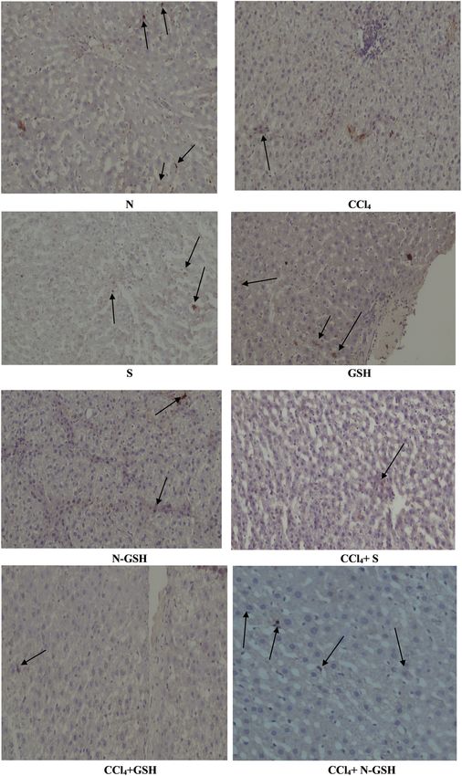
128 Esam M. Aboubakr et al. Figure 5: Histopathology of rats’ liver (H&E stain). N = normal rats; CCl4 = injured rats; S = silymarin-treated rats; GSH = glutathione- treated rats. Figure 6: Histopathology of rats’ liver (H&E stain). N-GSH = nano-glutathione-treated rats; CCl4-S = CCl4 + silymarin-treated rats; CCl4-GSH = CCl4 + glutathione-treated rats; CCl4 + N-GSH = CCl4 + nanoglutathione-treated rats.
Improved oral-bioavailability and hepatoprotective efficacy of glutathione 129 Figure 7: Immunohistochemistry results of Bcl-2 on hepatic tissues of N, normal rats; CCl4, treated rats; S, silymarin-treated rats; GSH, glutathione-treated rats; N-GSH, nano-glutathione-treated rats; CCl4 + S, CCl4 + silymarin-treated rats; CCl4 + GSH, CCl4 + glutathione- treated rats; and CCl4 + N-GSH, CCl4 + nanoglutathione-treated rats.
130 Esam M. Aboubakr et al.
3.9 Effects of Bcl-2 protein on the injured 4 Discussion
and treated rats
Hepatic damage represents one of the most serious health
Bcl-2 protein was detected in the hepatic tissue of normal, problems owing to its high prevalence and limited treat-
silymarin, GSH, and N-GSH administered animal groups. ment options [38]. When injuries with different etiologies
However, Bcl-2 protein was absent in the CCl4 group affect hepatic tissues, it triggers fibrogenesis and damage
stained hepatocytes with Bcl-2 antibody detection. The with major clinical implications and fatality at the end-
CCl4 + silymarin group showed a small number of mild stage liver disease. The molecular processes underlying
and intense hepatocytes, whereas the Bcl-2 protein in the pathogenesis of hepatic injury involve complex inter-
the CCl4 + GSH group was nearly absent. On the contrary, changes of oxidative stress, inflammation, apoptosis, and
the CCl4 group treated by N-GSH showed a remarkable necrosis [39].
presence of Bcl-2 protein, also represented by multiple In this context of liver damage, when the GSH level
stained hepatocytes (Figure 7). decreases, the excessive oxidative stress provokes severe
health complications, including hepatocyte damage [40].
GSH plays a crucial role in the hepatocytes’ protection
against free radicals, thereupon resulting in protecting
the DNA, lipids, proteins, encountered toxins’ neutraliza-
3.10 Effect of NF-Kβ on the injured and
tions, and regulation of cell cycle progression and apop-
treated rats tosis [41]. Moreover, GSH is also conjugated to a wide
range of endogenous hepatotoxic compounds and xeno-
Positive NF-Kβ hepatocytes were not observed in normal, biotics, making them safer for the liver through enhan-
silymarin, GSH, and N-GSH administered animal groups, cing their excretion [41].
while it was highly upregulated by CCl4 treatment (CCl4 GSH administration is therapeutically beneficial.
group). The GSH treatment (CCl4 + GSH group) nearly did Unfortunately, GSH cannot be used orally, as oral GSH
not decrease the NF-Kβ-positive hepatocytes as com- undergoes intestinal hydrolysis by γ-glutamyltransferase
pared to the CCl4 non-treated group. However, silymarin that consequently diminishes its bioavailability. Also, the
treatment (CCl4 + silymarin group) moderately decreased reports on GSH systemic availability revealed that the oral
the NF-Kβ-positive hepatocytes. A remarkable decrease administration of GSH in high doses is also not helpful to
in the number of NF-Kβ-positive hepatocytes was observed increase the GSH levels inside the hepatic tissue to a clini-
in the CCl4 group treated by nano-glutathione (CCl4 + N- cally beneficial level [42]. Therefore, there is a need to
GSH) (Figure 8). increase the systemic bioavailability and hepatic tissue
accumulation of GSH by oral administration. For this pur-
pose, an appropriate pharmaceutical dosage form of GSH
is required that provides the GSH as an effective hepato-
3.11 Macroscopic examination of the liver in protective agent [15].
the injured and treated rats Recently, nano-medicines-based approaches have been
introduced as a promising alternative to conventional
Normal gross morphology with the normal architecture of therapy. Nanoparticles (NPs) were prepared to deliver drugs
the liver was observed in normal, silymarin, GSH, and in a site-directed manner that were achieved through
N-GSH-administered animal groups, while the CCl4- amending the drugs’ physicochemical properties, use of
treated animal groups showed irregular, pale, and gross tissue-specific homing devices, allowing specific organ tar-
surfaces, with multiple macro- and micronodules, while geting with minimal side effects, and utilization of the
the silymarin oral administration attenuated CCl4 dama- encapsulating material, mostly polymers of synthetic and
ging effect, showed lower numbers of macro- and micro- natural origins [43]. The formulated NPs protect the drug,
nodules as compared to the CCl4-administered animal especially macromolecular entities, such as proteins against
group with retraction of the liver capsule. In contrast, the inactivation until they reach their target organ. Further-
CCl4 + GSH group showed many micro- and macronodules more, the liver is the major organ for the accumulation of
with irregular hepatic surfaces. On the other hand, the nanomedicines owing to its rich blood supply [44].
CCl4 + N-GSH-administered animal group liver tissue showed The current work presents the preparation of nio-
soft texture and smooth surface, and nearly preserved the somes formulations with several non-ionic surfactants
liver’s normal anatomy and appearance (Figure 9). with a view to enhance the orally routed GSH’sImproved oral-bioavailability and hepatoprotective efficacy of glutathione 131 Figure 8: Immunohistochemistry results of NF-Kβ on hepatic tissued of N, normal rats; CCl4, treated rats; S, silymarin-treated rats; GSH, glutathione-treated rats; N-GSH, nano-glutathione-treated rats; CCl4 + S, CCl4 + silymarin-treated rats; CCl4 + GSH, CCl4 + glutathione- treated rats; and CCl4 + N-GSH, CCl4 + nanoglutathione-treated rats.
132 Esam M. Aboubakr et al. Figure 9: Macroscopic examination of the liver tissues from N, normal rats; CCl4, treated rats; S, silymarin-treated rats; GSH, glutathione- treated rats; N-GSH, nano-glutathione-treated rats; CCl4 + S, CCl4 + silymarin-treated rats; CCl4 + GSH, CCl4 + glutathione-treated rats; and CCl4 + N-GSH, CCl4 + nanoglutathione-treated rats.
Improved oral-bioavailability and hepatoprotective efficacy of glutathione 133
bioavailability. The thin-film hydration method has been niosomes for 1 month at 4°C. The results obtained from
used efficiently to formulate nanovesicles [34]. Several the formulation also revealed that the presence of different
non-ionic surfactants were utilized in the niosomes pre- molar ratios of cholesterol and different types of non-ionic
paration to understand their effects on niosomes proper- surfactants in the fabricated niosomes could affect the
ties and stability. Span40® and Span60® were selected GSH encapsulation into nanovesicles. The encapsulation
because of their better stability and biocompatibility, com- of GSH into niosomes may avoid metabolic degradation
pared with other surfactants. Also, Span40® and Span60® and provide a small particle size, which enhances oral
have lower irritability and toxicity as compared to other glutathione bioavailability. Glutathione release from nio-
surfactants. These surfactants also have low toxic effects somes in the stomach environment (pH 1.2) was less as
due to their tendency to degrade in vivo to triglycerides compared with the intestine pH (6.8). Therefore, smaller
and fatty acids [45]. Two other factors also affect the encap- amounts of GSH will supposedly be degraded in the sto-
sulation of glutathione as a hydrophilic drug within the mach during the digestion time (about 2 h), and thus,
niosomes, first the permeability of bilayer membranes and higher quantities of glutathione will be made available
second the structural consistency of the hydrocarbon chain for absorption in the intestine. The release of GSH depends
of the surfactant [46]. Hence, the formulation, GTN-8, con- on the pH and the ionic strength of the dispersing medium.
taining high cholesterol and Span60® showed the highest Niosomes containing cholesterol (anionic moiety) are cap-
EE (66 ± 2.8%) as Span60® strongly interacts with choles- able of swelling and shrinking, which are used to trigger
terol molecules and forms large core space for hydrophilic its release. Responding to changes in the pH or ionic con-
drug entrapments [47]. In addition, the presence of centrations, the anionic cholesterol has the tendency to
Tween80® in the formulation enhances the encapsula- shrink in the acidic media [51]. So, it was anticipated
tion of hydrophilic glutathione into the aqueous core of that the GSH niosomes would shrink in an acidic medium
the niosomes vesicles. These observations have been and minimize the glutathione release in the stomach
reported earlier and helped us to conclude that the sur- environment.
factant type and cholesterol concentrations are important The CCl4-induced liver injury in rats is a widely used
and primary factors affecting the niosomes stability and model to investigate the potential therapeutic effect of
drug entrapments efficiency [46]. A high amount of cho- new agents due to their similarities with chemical liver
lesterol inhibited the gel to the liquid phase transition of injury in humans [50]. CCl4 is metabolized in the liver by
the surfactant by firming itself in the bilayer and thereby cytochrome P450 enzymes to produce reactive intermedi-
providing rigidity to the vesicles and preventing the ates, such as trichloromethyl free radicals and peroxyl
entrapped drugs leakage. This also explained the increase free radicals, which initiate the peroxidation of proteins
in the EE% of GSH and its sustained release over time and lipids, leading to hepatocellular damages [52]. In the
[48,49]. present study, 50% solution of CCl4 (1 mL/kg) was given
The loading of 500 mg of GSH in niosomes formula- intraperitoneally (i.p.) for eight weeks, which was used as
tion, GTN-8, significantly increased the EE as compared an inducer of hepatic injury and resulted in severe hepatic
with the formulation GTN-4, which contained 250 mg of tissue damage as noticed by both macro- and microscopic
GSH. The results may be attributed to increased EE, examinations of the hepatic tissue, which is in parallel to
which provided the most drug loading into the currently other studies utilizing the same inducer [53].
formulated GTN-8 preparation of the niosomes [24]. The In the present study, the CCl4 i.p. injection resulted in
niosomes provided a promising oral delivery module for irregular and nodular hepatic surfaces with fatty deposi-
GSH, partly also owing to their small particle size. All the tions, but when CCl4 was co-administered with silymarin
formulations (GTN-1 to GTN-8) were found in the nano- (100 mg/kg, orally), there were moderate reductions in
sized range with low values of PDI. The PDI value of ≤0.5 the damage that were observed macroscopically; hence,
is considered appropriate for drug delivery applications silymarin was also used as a standard hepatoprotective
that represent a relatively homogenous distribution of the agent to compare the results with GSH and the formu-
nanocarriers. According to the stability studies, it was lated N-GSH. The oral administration of GSH did not
found that the optimized formulation, GTN-8, was a stable reduce the hepatic damage by CCl4, which is attributable
niosomes formulation with the acceptable smallest vesicle to the poor oral bioavailability of GSH and its diminished
size and PDI value. The presence of cholesterol and the ability to reach hepatocytes. On the other hand, the for-
negative zeta potential resulted in improved stability of the mulated N-GSH niosomes significantly protected the rats’
developed niosomes formulation. This result provided evi- liver from CCl4 damage, as the macroscopic examination
dence for the maintenance of the structural integrity of of the rat liver showed a smooth surface with a few small134 Esam M. Aboubakr et al.
nodules and sporadic fat deposition. These results were MDA levels in the hepatic tissue homogenate is a clear
also supported by the histo-microscopic examinations, marker for the severity of oxidative stress, causing tissue
proposing that the N-GSH was more potent than sily- damage and depletion of the antioxidant defences [59].
marin in protecting the liver from hepatic injury. The present study revealed a significant increase in the
Inflammation, a commonly associated condition, with hepatic MDA concentrations in the CCl4-administered ani-
hepatic injury, is an outcome of free radical metabolites mals’ group tissue homogenate compared to the normal
of the CCl4 attack on the hepatocytes, which cause the group of animals’ tissue, which is consistent with the pre-
damage of parenchymal cells, promote the hepatocyte’s viously reported findings [60]. However, the elevated
inflammatory responses, and upregulate the inflammatory hepatic MDA concentrations were attenuated significantly
mediators, such as TNF-α IL-1β, and IL-6 [54]. IL-1β is one by oral administration of N-GSH, silymarin, and GSH;
of the major players in hepatic inflammation, and its con- however, the MDA attenuation produced through induced
centration is extremely increased in the case of hepatic N-GSH was more potent than silymarin and GSH. Conse-
injury. It was also found out that IL-1β induces hepatic quently, it is suggested that N-GSH-protective effects
inflammation and collagen synthesis [55]. Additionally, against CCl4-induced hepatic damage by inhibiting lipid
IL-1β is produced by activated hepatic stellate cells (HSCs) peroxidation that is produced by oxidative stress are
and enhances the hepatic production of extracellular noteworthy.
matrix, including type I collagen, which leads to hepatic The CCl4 i.p. administration to the experimental animal
fibrosis and necrosis [55]. The present study also found out resulted in GSH depletion GSH 8.15 ± 0.66 µg/g protein in
that the N-GSH significantly ameliorated the serum IL-1β the CCl4 group as compared to 15.83 ± 2.16 µg/g levels of
levels when co-administered with CCl4 as compared to the protein in the normal group of animals, which was in agree-
CCl4 group results in a similar context. Silymarin oral ment with the reported result [61]. On the other hand,
administration also attenuated the CCl4 upregulatory N-GSH oral administration significantly increased the GSH
effects on IL-1β, which is consistent with and is supported concentration inside the hepatic tissue of CCl4-injured ani-
by prior studies [56]. However, N-GSH was significantly mals (15.90 ± 1.02 µg/g protein levels) to about two-fold of
more active than silymarin, while oral GSH showed negli- its concentration in the untreated injured animals, and it
gible inhibitory effect for the upregulation of IL-1β as was significantly higher than the silymarin and GSH groups.
induced by CCl4. Additionally, the N-GSH, when administered to normal ani-
Moreover, Bcl-2 is an apoptosis regulator present in mals, increased the hepatic GSH contents than the normal
outer mitochondria and plays an important role in liver group animals.
fibrosis. It was found out that Bcl-2 overexpression in CCl4 induces hepatic injury by generating chloro-
mice livers causes liver fibrosis, indicating that Bcl-2 methyl free radicals (–CCl3), which enables the peroxida-
could be pro-fibrogenic in nature. It was also reported tion of membrane lipids, and other lipidic contents as
that animals exposed to liver injury inducer, CCl4, not well as subcellular structures of the hepatocytes, thereby
only demonstrated the increase in Bcl-2 expression but causing the increase of membrane permeability and
also, surprisingly, decreased its expression [57]. In the resulting in the release of large amounts of the hepatic
present study, hepatic tissue immunostaining showed a enzymes, ALT and AST, from the cytoplasm into the blood
decline of the Bcl-2 protein expression in the CCl4-admi- [62]. For the current study, the CCl4 + N-GSH-administered
nistered animal group as compared to the normal group. group showed significantly lowered concentrations of the
Our findings are consistent with the previous study where hepatic enzymes among the CCl4-treated groups, although
CCl4 reduces Bcl-2 protein expression leading to hepatic less than the reference (silymarin) group, indicating the
cell apoptosis [58]. On the contrary, the N-GSH oral outstanding capability of N-GSH to preserve the hepato-
administration exhibited a remarkable increase in Bcl-2 cytes’ integrity even under highly stressful conditions. On
protein expression as compared to other CCl4 groups the other hand, GSH also ameliorated the increased AST
treated with silymarin and GSH. The increased Bcl-2 pro- and ALT levels but it was much less than those in the
tein expression suggested that the N-GSH may protect N-GSH-administered group.
hepatic tissues against CCl4-induced hepatotoxicity by Drugs and different substances and materials’ accu-
keeping Bcl-2 from being downregulated. mulative studies have demonstrated that hepatocytes
Lipid peroxide is the main parameter, considered a secrete SOD after exposure to injurious pro-oxidants
good indicator, of oxidative injury severity. Also, the compounds, such as CCl4, which leads to depletion of
hepatic tissue MDA concentration is commonly used as SOD stores and its dramatically decreased concentrations
an indicator for hepatic tissue damage. The increase of [63], which was in line with the present findings. SODImproved oral-bioavailability and hepatoprotective efficacy of glutathione 135
catalyses the conversion of superoxide to H2O2, which is (as N-GSH) with physicochemical properties that enabled it to
further converted into water and oxygen by CAT and per- accumulate inside the hepatic tissue and overcome the phar-
oxidase. Thus, SOD is a major defence and protects macokinetic barriers, which limits the GSH use as one of the
hepatic tissues from oxidative stress [64]. In the current most potent and easily available hepatoprotective agents.
study, the N-GSH administration protected the hepatic
SOD stores from depletion by CCl4, which could be attri- Acknowledgements: The authors thank the technical sup-
buted to its ability to highly upregulate the GSH hepatic port of Qassim University, Saudi Arabia, and South Valley
concentrations, which seemingly is among the major University, Egypt.
defensive mediator against oxidative stress in the given
situations. Funding information: The authors state no funding involved.
Nonetheless, the increased NO tissue concentration
is believed to be involved in the pathogenesis of hepatic Author contributions: All authors have accepted respon-
injury, including oxidative stress and inflammatory reac- sibility for the entire content of this manuscript and
tions. The CCl4 exposure generates excessive amounts of approved its submission.
NO by activating the iNOS, contributing to hepatic tissue
damages [64], an observation circumstantially consistent Conflict of interest: The authors declare no conflict of
with the present study’s findings. Furthermore, these interest.
results also revealed that the oral administration of
N-GSH also dramatically decreased NO concentrations Ethical approval: The research related to animals’ use has
in the hepatic tissue, thereby contributing to its anti- been complied with all the relevant national regulations
oxidant and anti-inflammatory actions. and institutional policies for the care and use of animals.
Numerous studies have demonstrated that excessive
free radical production induced by CCl4 can induce upre-
gulation of NF-κB and its nuclear translocation, which
is considered responsible for hepatic tissue injury by References
increasing the inflammatory cytokine production [65,66].
These results also exhibited that N-GSH significantly [1] Younossi ZM. Non-alcoholic fatty liver disease–a global public
inhibits the NF-κB tissue expression, which was also con- health perspective. J Hepatol. 2019;70(3):531–44.
sistent with the current findings of the decreased levels of [2] Reyes-Gordillo K, Shah R, Muriel P. Oxidative stress and
the NO hepatic tissue levels. In contrast, silymarin and inflammation in hepatic diseases: current and future therapy.
Oxid Med Cell Longev. 2017;2017:3140673.
GSH oral administration did not significantly affect the
[3] Fang YZ, Yang S, Wu G. Free radicals, antioxidants, and
NF-κB tissue expressions.
nutrition. Nutrition. 2002;18(10):872–9.
[4] Battin EE, Brumaghim JL. Antioxidant activity of sulfur and
selenium: a review of reactive oxygen species scavenging,
glutathione peroxidase, and metal-binding antioxidant
5 Conclusion mechanisms. Cell Biochem Biophys. 2009;55(1):1–23.
[5] Pham-Huy LA, He H, Pham-Huy C. Free radicals, anti-
oxidants in disease and health. Int J Biomed Sci IJBS.
A designated, novel formulation of niosomes incorpor- 2008;4(2):89–96.
ating GSH was successfully prepared, characterized, and [6] Armstrong D, editor. Oxidative stress in applied basic research
biologically tested for their potential hepatoprotective and clinical practice. New York: Springer; 2014.
activity. The concentrations of GSH in the liver tissue [7] Mohammed HA. The valuable impacts of halophytic genus
Suaeda; nutritional, chemical, and biological values. Med
homogenates obtained from the CCl4-injured animal’s
Chem (Los Angeles). 2020;16(8):1044–57.
group and the treated animals with silymarin, GSH,
[8] Franco R, Cidlowski JA. Apoptosis and glutathione: beyond an
and N-GSH, indicated that N-GSH was prominent in antioxidant. Cell Death Differ. 2009;16(10):1303–14.
restoring glutathione in the liver to its normal levels as [9] Cnubben NHP, Rietjens IMCM, Wortelboer H, van Zanden J, van
compared to the normal group of animals. These results Bladeren PJ. The interplay of glutathione-related processes in
also proved the successfulness and utility of the N-GSH antioxidant defense. Environ Toxicol Pharmacol.
2001;10(4):141–52.
niosomes formulation in enhancing the absorption, pro-
[10] Scirè A, Cianfruglia L, Minnelli C, Bartolini D, Torquato P,
tection, liver tissue uptake, and subsequently, the bioac- Principato G, et al. Glutathione compartmentalization and its
tivity of GSH. Therefore, the present study introduced an role in glutathionylation and other regulatory processes of
improved, novel formulation for reduced glutathione GSH cellular pathways. Biofactors. 2019;45(2):152–68.You can also read