Histological evidence for secretory bioluminescence from pectoral pockets of the American Pocket Shark (Mollisquama mississippiensis) - Nature
←
→
Page content transcription
If your browser does not render page correctly, please read the page content below
www.nature.com/scientificreports
OPEN Histological evidence for secretory
bioluminescence from pectoral
pockets of the American
Pocket Shark (Mollisquama
mississippiensis)
Julien M. Claes 1*, Jérôme Delroisse 2, Mark A. Grace 3
, Michael H. Doosey 4
,
Laurent Duchatelet 1 & Jérôme Mallefet 1
The function of pocket shark pectoral pockets has puzzled scientists over decades. Here, we show that
the pockets of the American Pocket Shark (Mollisquama mississippiensis) contain a brightly fluorescent
stratified cubic epithelium enclosed in a pigmented sheath and in close contact with the basal cartilage
of the pectoral fins; cells of this epithelium display a centripetal gradient in size and a centrifuge
gradient in fluorescence. These results strongly support the idea that pocket shark’s pockets are
exocrine holocrine glands capable of discharging a bioluminescent fluid, potentially upon a given
movement of the pectoral fin. Such capability has been reported in many other marine organisms and
is typically used as a close-range defensive trick. In situ observations would be required to confirm this
hypothesis.
Pocket sharks (Mollisquama spp., Squaliformes) are among the most enigmatic cartilaginous fish species.
Extremely rare —only one mature female and one immature male have been collected so far, from the Nazca
Submarine Ridge in the southeast Pacific Ocean (M. parini discovered in 1984)1 and the central Gulf of Mexico
(M. mississippiensis, discovered in 2010 and described in 2019)2,3, respectively—, these oceanic deepwater sharks
also display an extraordinary combination of morphological traits including several cranial synapomorphies4, a
strong dignathic heterodonty possibly reflecting an ectoparasitic feeding strategy, a peculiar denticle morphology,
putative photogenic organ (photophore) aggregations on the ventral side and a lateral conspicuous slit above
each of the pectoral fin bases2. These slits form the opening of the ‘pockets’ from which pocket sharks received
their name. Representing a unique feature among sharks, these side pockets have puzzled scientists since their
discovery, which led to many speculations about their functional significance. While Dolganov considered these
glands to produce sexual p heromones1, more recent work suggests they might be analogous to the abdominal
pouch of the Taillight Shark (Euprotomicroides zantedeschia), another shark species related to Mollisquama spe-
cies, that has been observed to secrete and eject a blue bioluminescent fluid upon mechanical s timulation2,5–7.
This pouch on the taillight shark, located immediately in front of the cloaca of both male and female specimens,
shares indeed several morphological similarities with pocket shark pectoral pockets, including the presence of
voluminous unscaled skinfolds raised above surrounding tissue along the slit margin and internal tightly packed
villiform projections. Besides, the inner cavity of this pouch is also dark grey with a lighter bluish-grey color
close to the opening. The secretory tissue of the Taillight Shark pouch is pseudostratified, made of tall columnar
cells (containing small cytoplasmic granules and a large distal inclusion) separated and topped by flattened cells5.
In this work, we used light and fluorescence microscopy to analyze several histological sections through the
right pocket of the holotype specimen of the American Pocket Shark (M. mississippiensis, male, 142 mm TL) as
an attempt to elucidate their biological function.
1
Laboratoire de Biologie Marine, Earth and Life Institute, Université Catholique de Louvain,
1348 Louvain‑la‑Neuve, Belgium. 2Biology of Marine Organisms and Biomimetics, Biosciences Institute, University
of Mons, 7000 Mons, Belgium. 3NOAA/NMFS, SEFSC/Mississippi Laboratories, 3209 Fredric St., Pascagoula,
MS 39564, USA. 4Department of Biological Sciences, University of New Orleans, 2000 Lakeshore Dr., New Orleans,
Louisiana 70148, USA. *email: julien.claes@gmail.com
Scientific Reports | (2020) 10:18762 | https://doi.org/10.1038/s41598-020-75656-8 1
Vol.:(0123456789)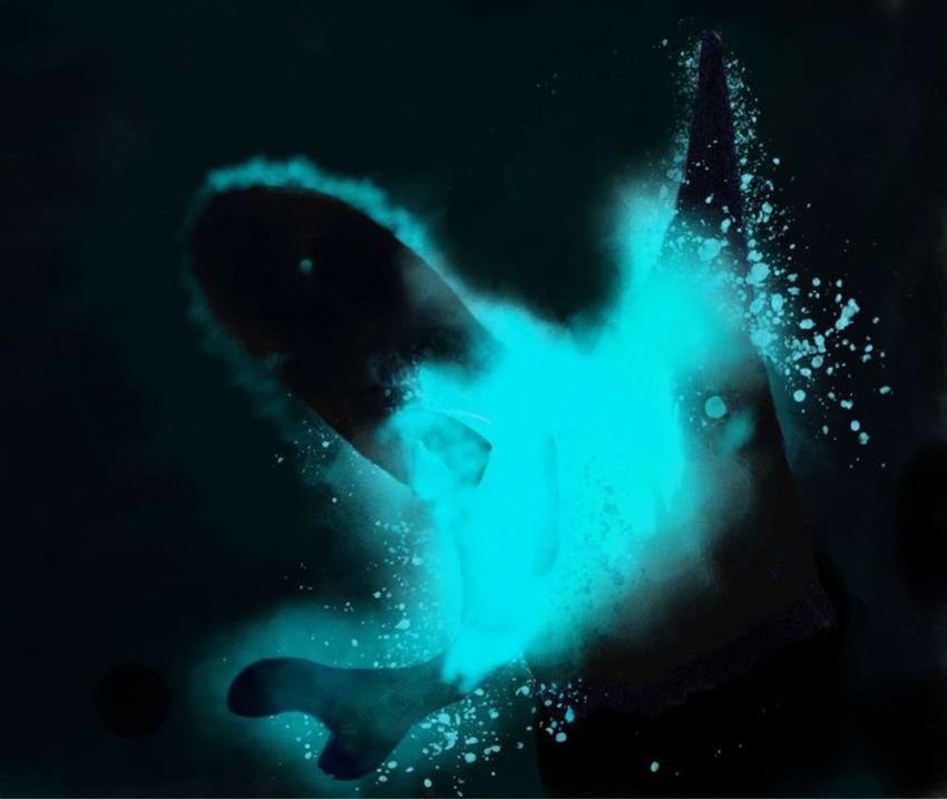
www.nature.com/scientificreports/
Results
Sloping ~ 45° posteriorly to just below right pectoral fin base, the right pocket gland’s orifice had a length
of ~ 4 mm (Fig. 1a) and was bordered with a series of bluish dermal folds. The interior of the gland was made of
numerous darker villiform structures, which encompasses 95% of the gland volume (~ 16 m m3 or 0.1% of total
shark volume) and its blind end was pointing anterodorsally (towards the head). No fluid was observed into the
lumen of the gland but a frozen milky white fluid, potentially coming from the pocket, was observed surround-
ing the specimen before fixation.
Histological sections demonstrated that villiform structures reached a length of 50–150 μm and consisted in
a stratified cuboidal epithelium composed of over 50 cell layers–colored in red by Masson’s trichrome (Fig. 1b,c).
This epithelium was in close contact with the basal cartilage of the pectoral fin (Fig. 1b) and enclosed in a pig-
mented sheath surrounded by connective tissue (Fig. 1b,c). Fluorescence microscopy revealed intense green
autofluorescence from villiform structures (Fig. 1d) and highlighted two cell populations: (i) small cells (2−4 μm)
present close to the basal lamina and displaying a bright, homogeneously fluorescent cytoplasm (Fig. 1e) and
larger cells (4−8 μm) showing only peripheral fluorescence (but with an overall larger quantity of fluorescent
material) and situated between basal cells and the gland’s lumen (Fig. 1f). Due to the presence of a continuum
between these two cell populations, cells composing the villiform structures displayed a centripetal gradient in
size (Fig. 1g, Supplementary Spreadsheet S1) and a centrifuge gradient in fluorescence (Fig. 1h, Supplementary
Spreadsheet S1).
Discussion
Bioluminescence—e.g., the ability for an organism to produce a visible light thanks to a chemical reaction– is
common in marine organisms, and especially in the permanent darkness of the deep-sea where most taxa use
light to evade predators, acquire food or communicate with conspecifics8,9. In sharks, only two families are
known to be endowed with bioluminescence capability: the Etmopteridae (Squaliformes; 53 species) and the
Dalatiidae (Squaliformes; 10 species including pocket sharks and the Taillight Shark)10,11; phylogenetic analyses
and putative photogenic organs found in Zameus squamulosus suggests that Somniosidae (Squaliformes) might
also contain bioluminescent species11 but observation of light-emission displays would be needed to confirm this
hypothesis. Catsharks (Scyliorhinidae, Carcharhiniformes) and carpet sharks (Orectolobidae, Orectolobiformes),
on the other hand, show biofluorescence, a phenomenon distinct from bioluminescence, which results from
the absorbance of the ambient down-welling light and its re-emittance at longer, lower-energy wavelengths12.
The pocket shark pockets are made of brightly fluorescent cells enclosed in a pigmented sheath connected
to the exterior via a folded aperture and closely associated with the basal cartilage of the pectoral fin. This
structure supports the hypothesis that these peculiar organs secrete and discharge a light-producing substance
to the exterior in response to a specific movement of the pectoral fins (upon a given stimulation). Indeed, (1)
bioluminescent compounds often display autofluorescence8,13–16, (2) photogenic cells (including secretory glands)
from mesopelagic animals are often enclosed in light-tight pigmented sheaths to avoid unwanted biolumines-
cence detection17–19 and (3) secretory photogenic glands are sometimes associated with structures allowing fast
discharge of luminous compounds20,21.
The ring-shaped fluorescent pattern of the cells situated at the apex of the villiform structures, which contrasts
with the homogeneously fluorescent cytoplasm of basal cells, might result from a progressive accumulation of
cytoplasmic inclusions in the center of the cell. Cytoplasmic inclusions have been observed in both the photo-
cytes and secretory photogenic cells of dalatiid sharks and might be linked to luminescence c ompetence5,22. The
histological organisation of the villiform structures is consistent with a holocrine secretion mode, where small
basal cells, accumulating fluorescent material and developing a cytoplasmic inclusion, would progressively turn
into larger apical cells when approaching the lumen of the gland, finally becoming the product of the secretion23.
Holocrine secretions are rare among secretory photophores from marine animals, which appear—based on the
morphology of secretory cells and/or the examination of secretion products—to be mostly apocrine (secretion
via the breaking up of the apical part of the secretory cell, resulting in the formation of extracellular membrane-
bound vesicles)21,24–27 or merocrine (secretion via exocytosis from secretory cells)20,28 glands. Interestingly, the
only other reported case of holocrine photogenic glands is found in tubeshoulder fishes (Searsidae), which
also possess two light-producing glands opening in the close vicinity of pectoral fins29; such similar structural
organization likely represents a case of convergent evolution. Furthermore, piscine venom glands—which are also
holocrine structures lacking associated musculature—, rely on a mechanical coupling with skeleton structures
(e.g., grooved spines) to discharge their secretion30–33, potentially representing another convergent evolution
case. Surprisingly, however, the stratified cubic and putatively holocrine epithelium of the pocket shark pockets
appears dramatically different from the pseudostratified columnar and putatively apocrine (based on morphol-
ogy) epithelium of the pelvic photogenic pouch of the Taillight S hark5, suggesting an independent origin of the
two organs in these closely related species. Reaching close to 10 μm in diameter, apical cells of the villiform struc-
tures reach a similar size as photocytes of juvenile dalatiid (Squaliolus aliae; J. M. Claes, personal observation)
and etmopterid sharks (Etmopterus spinax)34. In the Slendertail Lantern Shark (Etmopterus molleri), photophores
are known to form enlarged photogenic structure at the base of the dorsal fin35. Similarly, one can suppose the
pocket shark pockets to be modified photophores, that merged and invaginated themselves to become a secre-
tory gland. The phylogenetic position of M. mississippiensis among Dalatiidae2,3 supports the pocket glands to
be a derived character, posterior to the presence of epidermal ventral photophores.
Secretory luminescence is relatively uncommon among marine organisms, i.e., concerning ~ 10% of marine
bioluminescent genera. However, ‘bioluminescent cloud’ emitters are found in most marine taxonomic group-
ings (Fig. 2a), which probably results from multiple independent origins of the cloud-emitting capability and
highlights its paramount ecological significance. Although secretory luminescence is involved in intraspecific
Scientific Reports | (2020) 10:18762 | https://doi.org/10.1038/s41598-020-75656-8 2
Vol:.(1234567890)www.nature.com/scientificreports/
a
Op Co
Lu
Pi b Pi
Vi Co
Vi
Ca
Ca Ca
c d e
Co
Pi
f
Vi
g h
10 120
100
Cell fluorescence (%)
Cell diameter (µm)
8
80
6
60
4
40
2 20
0 0
0 20 40 60 0 20 40 60
Distance from basal lamina (µm) Distance from basal lamina (µm)
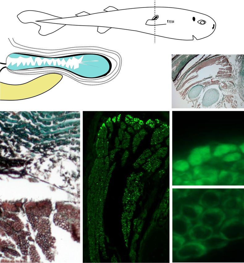
Figure 1. Right pocket histology of American Pocket Shark (Mollisquama mississippiensis). (a) Schematic
drawing of right pocket histology in transversal view (illustration by J. M. Claes, adapted from Grace et al.3). Ca
basal cartilage from the right pectoral fin, Co, connective tissue, Lu pocket lumen, Pi pigmented layer, Op pocket
opening, Vi villiform structure. (b) Transversal section (Masson’s trichrome) through pocket and underlying
basal cartilage from the right pectoral fin; arrows highlight the intimate connection between the cartilage
elements and the villiform structures. (c) Transversal section (Masson’s trichrome) of pocket’s upper wall.
(d) Green autofluorescence from a selected villiform structure in transversal section. (e) Diffuse bright green
autofluorescence from small basal cells of the villiform structure. (f) Marginal weak green autofluorescence from
large apical cells of the villiform structure. (g) Plot of villiform structure cell diameter according to the distance
to basal lamina, with associated regression line (cell diameter = 0.10 distance to basal lamina + 4.00, r2 = 0.69,
n = 100). (h) Plot of villiform structure cell relative fluorescence intensity according to the distance to the basal
lamina, with associated regression line (cell relative fluorescence intensity = 101.80 (distance to basal lamina)−0.29,
r2 = 0.81, n = 100). Indicative scale bars represent 2 cm in (a) (top); 1 mm in (a) (bottom); 10 μm in (b–f).
stracods36 and in Odontosyllis worms37,38, probably because of the energy costs associated
sexual courtship in o
Scientific Reports | (2020) 10:18762 | https://doi.org/10.1038/s41598-020-75656-8 3
Vol.:(0123456789)www.nature.com/scientificreports/
a CTENOPHORA
Anthozoa
CNIDARIA
Other Cnidaria
NEMERTEA
ANNELIDA
Gastropoda
Lamellibranchia MOLLUSCA
Cephalopoda
Ostracoda
Copepoda
Amphipoda CRUSTACEA
Mysid shrimps
Euphausiacea
Decapod shrimps
HEMICHORDATA
ECHINODERMATA
CHAETOGNATHA
TUNICATA
Tubeshoulder fishes
Other teleost fishes
VERTEBRATA
Etmopterid sharks
Dalatiid sharks
b
Figure 2. (a) Systematic distribution of ‘bioluminescent cloud’ emitters among 566 bioluminescent marine
genera. Names in capital letters represent “phylum-level” groups or broader. Circles scale to the number of
marine bioluminescent genera in a given taxonomic grouping while blue color indicates the proportion of
genera with ‘bioluminescent cloud’ emitters. Blue taxonomic grouping contains at least one genus known
to discharge intrinsic bioluminescent secretions outside of their body. (b) Artistic illustration of a putative
defensive cloud-emitting behavior in an American Pocket Shark (left) following the attack of a Goblin Shark
(Mitsukurina owstoni; right) in the darkness of the deep-sea (illustration by J. M. Claes).
with the production of the secretory products, the primary function of bioluminescent cloud emission appears
to be last-minute short-distance defensive behaviors to facilitate escape8. This and the fact that the investigated
specimen is not sexually mature let us hypothesize that the pocket shark pockets (and the Taillight Shark pouch)
are used in similar ‘smoke screen’ predation avoidance contexts, potentially blinding approaching predators
(Fig. 2b). In situ observations and additional biological material would be needed to confirm this hypothesis,
even if this might require some patience given the extreme rarity of pocket sharks.
Methods
Histology. The holotype (TU 203676; 142.0 mm in total length and 14.6 g in total weight) of M. mississip-
piensis (which represents the unique specimen of this species) was collected in the central Gulf of Mexico on 4
February 2010 by the NOAA Ship PISCES (survey 7620101001, station 053) and immediately frozen in water
after capture for about 3 years before being preserved in 20% formalin for 3 weeks and gradually transferred to
a final solution of 70% ethanol for storage. The right pocket of this specimen was excised, cut in several subsam-
ples and dehydrated in a graded ethanol series, with some of the subsamples fixed in Bouin’s fluid beforehand.
Subsamples were then embedded in paraffin using a routine method39. Sections (10 µm in thickness) were cut
using a Microm HM340E microtome and mounted on clean glass slides (SuperFrost Ultra Plus, Menzel GmbH
& Co. KG, Germany). Sections obtained from ethanol-fixed samples were analyzed using the FITC-filter of a
Zeiss Axioscope A1 microscope equipped with an AxioCam ICc3 camera (Zeiss, Oberkochen, Germany) for
Scientific Reports | (2020) 10:18762 | https://doi.org/10.1038/s41598-020-75656-8 4
Vol:.(1234567890)www.nature.com/scientificreports/
autofluorescence detection while sections from samples fixed in Bouin’s fluid were stained with the Masson’s tri-
chrome39. Digital pictures of sections were acquired using the Zeiss Zen Blue 2.5 software and finally analyzed
in Image J v. 1.46 (National Institutes of Health, Bethesda, USA) to estimate the diameter (defined as square root
of total 2D area) and relative fluorescent compound concentration (defined as relative fluorescence intensity)
of putative light-producing cells (n = 100) constituting the pocket’s villiform structures. All experimental meth-
ods were conducted in accordance with relevant guidelines and regulations, and were approved by the NOAA/
NMFS Southeast Regional Office Committee in St. Petersburg, Florida (Scientific Research Permit No. 779-1633;
granting date: 9 January 2009).
Phylogenetic tree building. A phylogenetic tree of marine bioluminescent organisms was built based
on NCBI taxonomy using PhyloT40 and a series of representatives of each marine taxonomic grouping: Bathy-
ctena chuni (Ctenophora), Anthoptilum murrayi (Anthozoa), Aurelia labiata (‘Other Cnidarias’, which encom-
passes the remaining bioluminescent cnidarian genera), Hubrechtella juliae (Nermetea), Odontosyllis phosphorea
(Annelida), Doris kergelenensis (Gastropoda), Pholas dactylus (Lamellibranchia), Heteroteuthis dispar (Ceph-
alopoda), Vargula tsujii (Ostracoda), Metridia lucens (Copepoda), Scina borealis (Amphipoda), Mysis relicta
(‘Mysid shrimps’), Meganyctiphanes norvegica (Euphausiacea), Acanthephyra purpurea (‘Decapod shrimps’),
Balanoglossus carnosus (Hemichordata), Ophiocomina nigra (Echinodermata), Caecosagitta macrocephala
(Chaetognatha), Didemnum granulatum (Tunicata), Searsia koefoedi (‘Tubeshoulder fishes’), Porichthys notatus
(‘Other teleost fishes’, which encompasses the remaining bioluminescent teleost fish genera), Etmopterus spinax
(‘Etmopterid sharks’) and Isistius brasiliensis (‘Dalatiid sharks’). The number of marine bioluminescent genera –
and the proportion of these latter endowed with the capability to discharge ‘bioluminescent clouds’, e.g., intrinsic
bioluminescent secretions– was determined for each taxonomic grouping by compiling information from sci-
entific literature8,13,24–29,36,41–55. External bioluminescent displays resulting from autotomy (e.g., in the pelagic sea
cucumber Enypniastes eximia56) were not considered as bioluminescent clouds given their non-secretory nature.
Pocket shark’s cloud‑emitting behavior illustration. The artistic illustration of the defensive cloud-
emitting behavior of the American Pocket Shark’s was drawn using Adobe Photoshop Sketch (version 4.9.0).
Given that both internal57 and external shark luminescent d
isplays7 were always found to fall in the blue wave-
length range (450–495 nm), we used a blue color to represent American Pocket Shark’s bioluminescent cloud.
We selected the Goblin Shark (Mitsukurina owstoni) as the attacking predator given this species is large (up to
6 m), piscivorous and also occurs in the Gulf of Mexico’s mesopelagic z one58.
Received: 20 July 2020; Accepted: 8 October 2020
References
1. Dolganov, V. N. A new shark from the family Squalidae caught on the Naska Submarine Ridge. Zool. Zh. 63, 1589–1591 (1984).
2. Grace, M. A., Doosey, M. H, Bart, H. L., Naylor, G. J. First record of Mollisquama sp. (Chondrichthyes: Squaliformes: Dalatiidae)
from the Gulf of Mexico, with a morphological comparison to the holotype description of Mollisquama parini Dolganov. Zootaxa
3948,587–600 (2015).
3. Grace, M. A. et al. A new Western North Atlantic Ocean kitefin shark (Squaliformes: Dalatiidae) from the Gulf of Mexico. Zootaxa
4619, 109–120 (2019).
4. Denton, J. S. et al. Cranial morphology in Mollisquama sp. (Squaliformes; Dalatiidae) and patterns of cranial evolution in dalatiid
sharks. J. Anat. 233, 15–32 (2018).
5. Munk, O. & Jorgensen, J. M. Putatively luminous tissue in the abdominal pouch of a male dalatiine shark, Euprotomicroides zant-
edeschia Hulley & Penrith, 1966. Acta Zool. (Stockh) 69, 247–251 (1988).
6. Stehmann, M. & Krefft, G. Results of the research cruises of FRV "Walter Herwig" to South America. LXVIII. Complementary
redescription of the dalatiine shark Euprotomicroides zantedeschia Hulley & Penrith, 1966 (Chondrichthyes, Squalidae), based on
a second specimen from the western south Atlantic. Arch. Fisch. Wiss. 30, 1–30 (1988).
7. Stehmann, M. F. W., Van Oijen, M. & Kamminga, P. Re-description of the rare taillight shark Euprotomicroides zantedeschia
(Squaliformes, Dalatiidae), based on third and fourth record from off Chile. Cybium 40, 187–197 (2016).
8. Haddock, S. H. D., Moline, M. A. & Case, J. F. Bioluminescence in the sea. Annu. Rev. Mar. Sci. 2, 443–493 (2009).
9. Widder, E. A. Bioluminescence in the ocean: origins of biological, chemical, and ecological diversity. Science 328, 704–708 (2010).
10. Pollerspöck, J. & Straube, N. Bibliography Database|Shark-References. www.shark-references.com (2015).
11. Straube, N., Li, C., Claes, J. M., Corrigan, S. & Naylor, G. J. Molecular phylogeny of Squaliformes and first occurrence of biolumi-
nescence in sharks. BMC Evol. Biol. 15, 162 (2015).
12. Gruber, D. F. et al. Biofluorescence in catsharks (Scyliorhinidae): fundamental description and relevance for elasmobranch visual
ecology. Sci. Rep. 6, 24751 (2016).
13. Bowlby, M. R. & Case, J. F. Ultrastructure and neuronal control of luminous cells in the copepod Gaussia princeps. Biol. Bull. 180,
440–446 (1991).
14. Robison, B. H., Reisenbichler, K. R., Hunt, J. C. & Haddock, S. H. Light production by the arm tips of the deep-sea cephalopod
Vampyroteuthis infernalis. Biol. Bull. 205, 102–109 (2003).
15. Haddock, S. H. D., Dunn, C. W., Pugh, P. R. & Schnitzler, C. E. Bioluminescent and red-fluorescent lures in a deep-sea siphono-
phore. Science 309, 263 (2005).
16. Claes, J. M., Krönström, J., Holmgren, S. & Mallefet, J. Nitric oxide in the control of luminescence from lantern shark (Etmopterus
spinax) photophores. J. Exp. Biol. 17, 3005–3011 (2010).
17. Denton, E. J., Herring, P. J., Widder, E. A., Latz, M. F. & Case, J. F. The roles of filters in the photophores of oceanic animals and
their relation to vision in the oceanic environment. Proc. R. Soc. B 225, 63–97 (1985).
18. Herring, P. J. Depth distribution of the carotenoid pigments and lipids of some oceanic animals. 2. Decapod crustaceans. J. Mar.
Biol. Assoc. UK 53, 539–562 (1973).
19. Herring, P. J. Bioluminescent signals and the role of reflectors. J. Opt. A Pure Appl. Op. 2, R29 (2000).
Scientific Reports | (2020) 10:18762 | https://doi.org/10.1038/s41598-020-75656-8 5
Vol.:(0123456789)www.nature.com/scientificreports/
20. Anctil, M. The epithelial luminescent system of Chaetopterus variopedatus. Can. J. Zool. 57, 1290–1310 (1979).
21. Huvard, A. L. Ultrastructure of the light organ and immunocytochemical localization of luciferase in luminescent marine ostracods
(Crustacea: Ostracoda: Cypridinidae). J. Morphol. 218, 181–193 (1993).
22. Hubbs, C. L., Iwai, T. & Matsubara, K. External and internal characters, horizontal and vertical distribution, luminescence, and
food of the dwarf pelagic shark Euprotomicrurus bispinatus. Bull. Scripps Inst. Ocenogr. 10, 1–81 (1967).
23. Schorno, S. Biogenesis of Hagfish Slime: Timing and Process of Slime Gland Refilling in Hagfishes (Eptatretus stoutii and Myxine
glutinosa). (Doctoral dissertation, University of Guelph, USA, 2018).
24. Clarke, G. L., Conover, R. J., David, C. N. & Nicol, J. A. C. Comparative studies of luminescence in copepods and other pelagic
marine animals. J. Mar. Biol. Assoc. UK 42, 541–564 (1962).
25. Dilly, P. N. & Herring, P. J. The light organ and ink sac of Heteroteuthis dispar (Mollusca: Cephalopoda). J. Zool. 186, 47–59 (1978).
26. Bowlby, M. R., Widder, E. A. & Case, J. F. Disparate forms of bioluminescence from the amphipods Cyphocaris faurei, Scina cras-
sicornis and S. borealis. Mar. Biol. 108, 247−253 (1991).
27. Gosliner, T. M. & Vallès, Y. Shedding light onto the genera (Mollusca: Nudibranchia) Kaloplocamus and Plocamopherus with
description of new species belonging to these unique bioluminescent dorids. Veliger 48, 178–205 (2006).
28. Nicol, J. A. C. Histology of the light organs of Pholas dactylus (Lamellibranchia). J. Mar. Biol. Assoc. UK 39, 109–115 (1960).
29. Nicol, J. A. C. Observations on luminescence in pelagic animals. J. Mar. Biol. Assoc. UK 37, 705–752 (1958).
30. Sivan, G. Fish venom: pharmacological features and biological significance. Fish Fish. 10, 159–172 (2009).
31. Ziegman, R. & Alewood, P. Bioactive components in fish venoms. Toxins 7, 1497–1531 (2015).
32. Borges, M. H. et al. Combined proteomic and functional analysis reveals rich sources of protein diversity in skin mucus and venom
from the Scorpaena plumieri fish. J. Proteom. 187, 200–211 (2018).
33. Gorman, L. M. et al. The venoms of the lesser (Echiichthys vipera) and greater (Trachinus draco) weever fish—a review. Toxicon 6,
100025 (2020).
34. Duchatelet, L., Claes, J. M. & Mallefet, J. Embryonic expression of encephalopsin supports bioluminescence perception in lantern-
shark photophores. Mar. Biol. 166, 21 (2019).
35. Duchatelet, L., Pinte, N., Tomita, T., Sato, K. & Mallefet, J. Etmopteridae bioluminescence: dorsal pattern specificity and aposematic
use. Zool. Lett. 5, 9 (2019).
36. Morin, J. G. Luminaries of the reef: The history of luminescent ostracods and their courtship displays in the Caribbean. J. Crust.
Biol. 39, 227–243 (2019).
37. Galloway, T. W. & Welch, P. S. Studies on a phosphorescent bermudian annelid, Odontosyllis enopla Verill. Trans. Am. Microsc.
Soc. 30, 13–39 (1911).
38. Markert, R. E., Markert, B. J. & Vertrees, N. J. Lunar periodicity in spawning and luminescence in Odontosyllis enopla. Ecology 42,
414–415 (1961).
39. Gabe, M. Techniques histologiques (Masson et Cie Editeurs, Paris, 1968).
40. Letunic, I. PhyloT. https://phlot.biobyte.de (2015).
41. Harvey, E. N. Studies on bioluminescence: VI. Light production by a Japanese Pennatulid, Cavernularia haberi. Am. J. Physiol. 42,
349–358 (1917).
42. Haneda, Y. Luminosity in Rocellaria grandis (Deshayes) (Lamellibranchia). Kagaku Nanyo 2, 36–39 (1939).
43. Herring, P. J. Bioluminescence in decapod crustacea. J. Mar. Biol. Assoc. UK 56, 1029–1047 (1976).
44. Herring, P. J. Studies on bioluminescent marine amphipods. J. Mar. Biol. Assoc. UK 61, 161–176 (1981).
45. Herring, P. J. Bioluminescence in the Crustacea. J. Crust. Biol. 5, 557–573 (1985).
46. Herring, P. J. Systematic distribution of bioluminescence in living organisms. J. Biol. Chem. 1, 147–163 (1987).
47. Herring, P. J. Copepod luminescence. Hydrobiologia 167, 183–195 (1988).
48. Haddock, S. H. & Case, J. F. Bioluminescence spectra of shallow and deep-sea gelatinous zooplankton: ctenophores, medusae and
siphonophores. Mar. Biol. 133, 571–582 (1999).
49. Deheyn, D. D. & Latz, M. I. Internal and secreted bioluminescence of the marine polychaete Odontosyllis phosphorea (Syllidae).
Invert. Biol. 128, 31–45 (2009).
50. Thuesen, E. V., Goetz, F. E. & Haddock, S. H. Bioluminescent organs of two deep-sea arrow worms, Eukrohnia fowleri and Cae-
cosagitta macrocephala, with further observations on bioluminescence in chaetognaths. Biol. Bull. 219, 100–111 (2010).
51. Jones, A. & Mallefet, J. Study of the luminescence in the black brittle-star Ophiocomina nigra: toward a new pattern of light emis-
sion in ophiuroids. Zoosymposia 7, 139–145 (2012).
52. Gouveneaux, A. Bioluminescence of Tomopteridae species (Annelida): multidisciplinary approach. (Doctoral dissertation, Centre
National de la Recherche Scientifique, Université catholique de Louvain, Belgium, 2016).
53. Paitio, J., Oba, Y. & Meyer-Rochow, V. B. Bioluminescent fishes and their eyes. In Luminescence—An Outlook on the Phenomena
and Their Applications. (Thirumalai, J., Ed.) (Intech, London, 2016).
54. Verdes, A. & Gruber, D. F. Glowing worms: Biological, chemical, and functional diversity of bioluminescent annelids. Integr. Comp.
Biol. 57, 18–32 (2017).
55. Poulsen, J. Y. New observations and ontogenetic transformation of photogenic tissues in the tubeshoulder Sagamichthys schnaken-
becki (Platytroctidae, Alepocephaliformes). J. Fish Biol. 94, 62–76 (2019).
56. Robison, B. H. Bioluminescence in the benthopelagic holothurian Enypniastes eximia. J. Mar. Biol. Assoc. UK 72, 463–472 (1992).
57. Claes, J. M., Nilsson, D. E., Straube, N., Collin, S. P. & Mallefet, J. Iso-luminance counterillumination drove bioluminescent shark
radiation. Sci. Rep. 4, 4328 (2014).
58. Parsons, G. R., Ingram, G. W. & Havard, R. First record of the goblin shark Mitsukurina owstoni, Jordan (Family Mitsukurinidae)
in the Gulf of Mexico. Southeast. Nat. 1, 189–192 (2002).
Acknowledgements
J.M.C. is a Scientific Collaborator of the Catholic University of Louvain (Marine Biology Laboratory). J.D. is
postdoctoral researcher from the F.R.S.–FNRS (Ref n°34761044). L.D. is a postdoctoral researcher from the
Catholic University of Louvain. J.M. is Research Associate to F.R.S.–FNRS. This paper is a contribution to the
Earth and Life Institute – Biodiversity (ELIB) and the Centre Interuniversitaire de Biologie Marine (CIBIM).
We deeply thank Henry L. Bart from Tulane University Biodiversity Research Institute for giving us access to
the holotype of M. mississippiensis, as well as the two anonymous reviewers whose valuable suggestions allowed
to significantly improve the manuscript.
Author contributions
M.A.G. and M.H.D. collected the specimen and provided the pocket tissue for histological analysis. J.M.C., J.D.
and L.D. performed the histological analyses. J.D. took light and fluorescent microscopy images. J.M.C. analyzed
the results, wrote the article and created the exhibits. All authors reviewed the final version of the manuscript.
Scientific Reports | (2020) 10:18762 | https://doi.org/10.1038/s41598-020-75656-8 6
Vol:.(1234567890)www.nature.com/scientificreports/
Competing interests
The authors declare no competing interests.
Additional information
Supplementary information is available for this paper at https://doi.org/10.1038/s41598-020-75656-8.
Correspondence and requests for materials should be addressed to J.M.C.
Reprints and permissions information is available at www.nature.com/reprints.
Publisher’s note Springer Nature remains neutral with regard to jurisdictional claims in published maps and
institutional affiliations.
Open Access This article is licensed under a Creative Commons Attribution 4.0 International
License, which permits use, sharing, adaptation, distribution and reproduction in any medium or
format, as long as you give appropriate credit to the original author(s) and the source, provide a link to the
Creative Commons licence, and indicate if changes were made. The images or other third party material in this
article are included in the article’s Creative Commons licence, unless indicated otherwise in a credit line to the
material. If material is not included in the article’s Creative Commons licence and your intended use is not
permitted by statutory regulation or exceeds the permitted use, you will need to obtain permission directly from
the copyright holder. To view a copy of this licence, visit http://creativecommons.org/licenses/by/4.0/.
© The Author(s) 2020
Scientific Reports | (2020) 10:18762 | https://doi.org/10.1038/s41598-020-75656-8 7
Vol.:(0123456789)You can also read