Imaging of Developmental Anomalies of the Eye and the Orbit
←
→
Page content transcription
If your browser does not render page correctly, please read the page content below
Imaging of Developmental Anomalies of the Eye and the Orbit

                           1
Larissa T. Bilaniuk and Martha Farber

   From the Children's Hospital of Philadelphia, Philadelphia, PA (LTB) and the Scheie Eye Institute ,
                          University of Pennsylvania, Philadelphia, PA (MF)

   The development of the eye and the orbit                                             supports growth of the eye (2, 4). The mesenchy-
encompasses a series of stages that take place                                          mal tissue that gives rise to the eye and to the
during the embryonic and fetal periods (1-4).                                           adnexal structures originates from neural crest
These stages overlap each other temporally , and                                        cells that themselves arise from ectoderm located
are interrelated. Multiple diverse genetic and ac-                                      at the junction of surface ectoderm and neuroec-
quired conditions may derange this normal pat-                                          toderm . Neural crest cells also give rise to the
tern of development. The specific type of anom-                                         bony orbit, the fat , and nerve sheaths. At a
aly that results, however, depends more heavily                                         specific time during the fetal development, the
on the time that the insult or the developmental                                        crest cells migrate to surround the optic cup and
arrest occurred than on the specific etiology of                                        optic stalk (8) . The development of the globe and
the derangement (5). Familiarity with the se-                                           the development of the orbit are independent of
quence of developmental events is a prerequisite                                        each other. In the developing eye and orbit, the
for understanding the various anomalies of the                                          paraxial mesoderm contributes only to the vas-
visual organ and for correct interpretation of                                          cular endothelium and to the striated extraocular
diagnostic studies. Imaging modalities now allow                                        muscles. The origin, belly , and insertion of the
precise delineation of the ocular and orbital (6)                                       extraocular muscles develop concurrently, and
anomalies and of any associated central nervous                                         each individual muscle develops at the same time
system, facial (7), and systemic abnormalities.                                         (9).
Such information may be critical in planning the                                            During the period of embryogenesis (first 3
treatment and management of the patient, as well                                        weeks after conception), the neuroectoderm
as in establishing a prognosis.                                                         forms the neural plate with a central groove and
                                                                                        paired neural folds that flank the groove on each
                                                                                        side. During this period , a group of cells within
Normal Development
                                                                                        the neural ectoderm is induced by the underlying
   The eye develops from the neuroectoderm                                              paraxial mesoderm to become the eye-forming
(neural plate), the surface ectoderm, and neural                                        tissue. The neural groove deepens and begins to
crest cells (1, 3, 4). Recent experimental embry-                                       close. At the 22nd day , before the anterior neu-
ologic studies have disproved the long held theory                                      ropore closes , the optic primordium can be iden-
that mesoderm contributes significantly to the                                          tified at either side of the still-open anterior neural
structures of the eye and the orbit. However, it is                                     groove. Eventually, the neural folds fuse to form
believed that the paraxial mesoderm that lies                                           a tube; the eye and the chiasm form from that
beneath the neuroectoderm induces it to become                                          portion of the neural tube destined to become the
the eye-forming tissue , and also nourishes and                                         diencephalon.
                                                                                            By the 24th day , while the neural tube is still
                                                                                        open anteriorly , an ocular pit forms in each of
    1
     Address reprint req uests to Dr Bilaniuk , Children 's Hospita l of Phila-         the paired neural fold s (Fig. 1A) and extends
delphia, 34th St. and Center Blvd .• Philadelphia , PA 19104.                           laterally to touch the surface ectoderm on its own
Index terms: Eyes, abnormalities and anoma lies; Orbits , abnormalities                 s ide . As the optic pits deepen into sulci, neural
and anomalies; Pedia tric neuroradiology                                                crest cells insinuate themselves between the sur-
AJNR 13:793-803 , Mar/ A pr 1992 0195-6 108/ 92/ 1302-0793                              face ectoderm and the neural ectoderm of the
© A m erica n Society of Neuroradiology                                                 optic vesicles.
                                                                                  793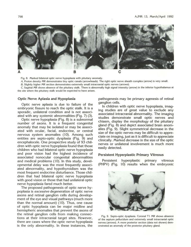
794                                                                                                         AJNR: 13, March/ April 1992

                                                                        ~--4                                                     --9

                          r----2

                                  --3                                                 --6
                                                                                   \.__ -1
                                                                               \
                                                                                   \._ __ 8                        "'----8
A                                              8                                                 c
                                                                                                      --13
                                                                -12                                    --14
                                                                                                          -11
                                                   -----8
                         D                                                 E
   Fig. 1. A-D, Schematic drawings of early stages of eye development. £,Cross section of optic stalk with optic fissure . I , optic pits;
2, neural ectoderm ; 3, surface ectoderm ; 4, forebrain cavity; 5, optic vesicle; 6, lens placode; 7, neural crest; 8, optic stalk; 9, optic
cup; 70, lens vesicle; I 1, optic fissure with hyaloid vessels ; I2, lens; I3, optic stalk lumen ; I4, axons from ganglion cells of the retina .

   By the 26th day, the neural tube is closed at                          tory vascular system (the hyaloid artery and
the anterior neuropore. The optic pits have deep-                         branches) (Fig. 1D) that forms the primary vitre-
ened and expanded to form prominent outpouch-                             ous and nourishes the structures of the eye. This
ings, the optic vesicles, that now project from the                       primitive vascular system involutes by the 35th
neural tube on short necks (the primitive optic                           week of gestation. The vascular primary vitreous
stalks) (Fig. 18). The surface ectoderm overlying                         also atrophies, and secondary vitreous is formed.
the vesicles thickens and forms a lenticular plac-                           The optic nerve develops in the optic stalk,
ode which then invaginates and , through multiple                         which connects the optic vesicle to the forebrain
steps, eventually forms the lens. At the same                             cavity (Fig. 18). A single layer of undifferentiated
time, the optic vesicle invaginates to form a cup                         neuroectodermal cells that lines the cavity of the
with an outer layer of cells destined to become                           forebrain is continuous with the retinal pigment
the retinal pigment epithelium, an inner layer                            epithelium of the outer cup (4). During the invag-
destined to form the neurosensory retina, and an                          ination of the optic vesicle, there is also invagi-
intervening cavity called the subretinal space.                           nation of the optic stalk (Fig. 1E). At the sixth
This process can be compared to a hollow rubber                           week, axons from ganglion cells of the retina
ball where one side has been pushed in against                            grow posteriorly into the optic stalk, toward the
the opposite side (Fig. 1C) . However; the optic                          central nervous system. There is also proliferation
cup is not completely continuous; ventrally it                            of glial cells to form a supporting framework and
contains a fetal (embryonic) fissure that permits                         mantle for the optic nerve. During normal embry-
mesenchymal and vascular tissues (the hyaloid                             ogenesis, an excess of ganglion cells is generated.
artery) to enter the globe (Fig. 1D). This fissure                        All these ganglion cells send axons into the optic
extends along the optic stalk. In normal devel-                           stalk toward the brain. Those axons that do not
opment, the fissure is eventually closed at its                           make appropriate connections or do not reach
margins by multipotential cells.                                          their target centrally undergo degeneration. Thus,
   During its development, the eye has a transi-                          normally there is massive retrograde degenera-
AJNR: 13, March/ April 1992                                                                                                            795

                                                                          Fig. 3 . Coloboma of the left posterior globe (morning glory
                                                                       syndrome). Axial CT demonstrates a smaller left globe with
                                                                       prominent outpouching posteriorly . (Case courtesy of Thomas P.
                                                                       Naidich, MD , Miami, FL.)

                                                                          Fig. 4. Microphthalmia with cyst. Axial CT shows a small
                                                                       deformed right globe with a centrally located lens (open arrow)
                                                                       and calcified or ossified tissue (arrow) at the junction of the globe
                                                                       with the cyst (arrowheads). Extreme microphthalmia is present
                                                                       on the opposite (left) side.

                                                                       the process of invagination and by the subse-
                                                                       quent growth of axons, the direct communication
                                                                       between the cavity of the optic vesicle and the
                                                                       third ventricle is lost. The optic recess on the
                                                                       floor of the third ventricle represents the site of
                                                                       the original connection (4) and is the only residual
        B                                                              of the previous direct communication between
                                                                       the third ventricle and the eye.
    Fig. 2. Right secondary anophthalmia and left congenital cystic
eye.
                                                                          Initially, the chiasm is formed only from
    A, Axial CT shows absence of the right globe. Somewhat             crossed optic nerve fibers. Later, between the
thickened rectus muscles lying in the center of the orbit extend       11th and 13th weeks, uncrossed fibers enter the
to irregular soft tissue that does not have any characteristics of a   chiasm. Myelination begins posteriorly in the re-
globe. The left orbit contains a congenital cystic eye (arrow) with    gion of the geniculate bodies at about the fifth
a centrally located malformed lens and a small calcific density at
                                                                       month and reaches the globe by the eighth month
its posterior aspect.
    8 , Histologic section of the congenitally cystic eye demon-       (4).
strates the abnormal , centrally located lens (open arrow) , disor-
ganized retinal tissue (closed arrows) that extends into the optic
stalk, and bony tissue (arrowhead) adjacent to the cystic eye.
                                                                       Anophthalmia and Microphthalmia
                                                                         Anophthalmia results when there is failure of
tion of optic nerve fibers and retinal ganglion cells                  the optic pit to form a vesicle (primary anophthal-
(10).                                                                  mia) or when the optic vesicle forms and then
   Once the lumen of the stalk is obliterated by                       degenerates (secondary anophthalmia) (Fig. 2A).
796                                                                                    AJNR : 13, March/ April 1992

Complete failure of eye development is extremely
 rare. Only histologic examination can differentiate
 between true anophthalmia and severe micro-
 phthalmia. In cases of microphthalmia, ocular
structures are identified , whereas they are absent
in primary anophthalmia .
    Microphthalmos is considered simple or pure
when the eye is small but anatomically correct,
and complex when the eye is malformed (11).
Some use the term nanophthalmos to refer to the                 A
simple microphthalmos, but others reserve this
term for those cases where there is marked de-
crease in the axial length of the eye (less than 18
mm) , microcornea, and absence of systemic ab-
normalities (12) . Microphthalmos, whether simple
or complex, can be unilateral or bilateral; it can
be the only abnormality or it can be as·s ociated
with systemic disorders that may be due to ge-
netic causes (eg, microphthalmos is present in
75 % of Trisomy 13 patients), environmental
causes (viral prenatal infections), or unknown
causes (13). Weiss et al (11, 12) proposed that
the pathogenetic mechanism for complex micro-
phthalmos is inadequate production of secondary
vitreous. They also stated that small optic cup,
low intraocular pressure, altered vitreous proteo-
                                                                8
glycans, and abnormal release of growth factors
play a role in the development of simple micro-            Fig. 5 . Microphthalmia with cyst.
phthalmos. The complex microphthalmos is fur-              A, Photograph of child with right microphthalmia and a huge
                                                        cyst that obscures the small eye.
ther subdivided into two categories: noncolobom-           B, Histologic section from another case demonstrates a smaller
atous and colobomatous forms.                           malformed globe with an abnormal, centrally located lens (open
                                                        arrow) and neuroectodermal tissue (solid arrow) prolapsing
                                                        through a coloboma into a much larger cyst. (Case courtesy of
Congenital Cystic Eye                                   Dr Morton Smith, Washington University, Seattle, WA.)

   If the invagination of the optic vesicle does not
occur or is incomplete, then a congenital cystic
eye results (Fig. 2). This is an extremely rare         choroidal fissure to close properly are classified
condition . Microscopic examination reveals the         as typical colobomas (15). Colobomas are com-
cyst to be lined by malformed retina that shows         mon malformations, constituting about 2% of all
an inverted arrangement of its layers, because          congenital abnormalities. They are usually bilat-
there has been no invagination. That is, the outer      eral (16).
layer lies on the side towards the cavity of the           A variant of typical coloboma of the disc is
cyst (14) (Fig. 28). Tendons of extraocular mus-        designated the morning glory syndrome, because
cles insert into the outer fibrous wall of the cystic   in this malformation the optic disc is posteriorly
eye and there is an abnormal lens (Fig. 2). Imaging     displaced at the apex of a funnel-shaped staphy-
studies may not be able to differentiate this entity    lomatous excavation (Fig. 3). This configuration
from a very disorganized severe microphthalmia.         is thought to resemble the flower ( 17). Apple and
                                                        colleagues ( 17) explain this on the basis of the
                                                        fact that, during development, just as the cells at
Colobomas                                               the margins of the embryonic fissure remain
   The term coloboma is of Greek origin meaning         undifferentiated for a longer time and thus are
curtailment and is used in ophthalmology to refer       more sensitive to insults, so do the cells of the
to defects in ocular structures (14). Those colo-       future optic nerve head that may be considered
bomas that result from failure of the embryonic         to be one huge embryonic fissure.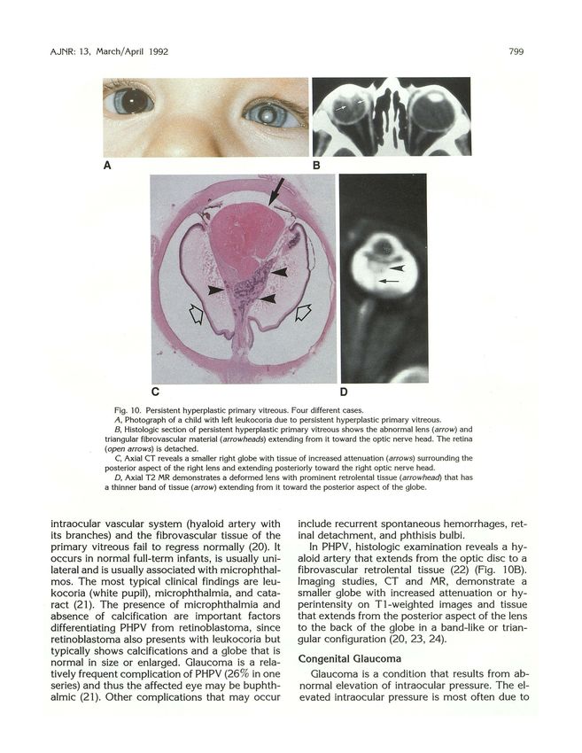
AJNR: 13, March/ April 1992                                                                                                          797

A                                            8                                              c

                        D                                             E
   Fig. 6. Microphthalmos with a cyst.
   A, Axial CT reveals a very small malformed globe (arrow) with centrally located lens lying medial to a huge cyst which has expanded
the bony orbit.
   8, Axial MR at the same level reveals a small deformed medially· located globe and a huge cyst that contains a smaller cyst
(arrowheads) adjacent to the lateral aspect of the abnormal globe. The second centrally located cyst was not identified on CT.
   C, Axial CT section slightly superior to A reveals extraocular muscles and an optic nerve sheath complex (arrow) that extends to the
abnormally small globe.
   D, Coronal MR demonstrates the small deformed globe containing a lens (arrow). These lie inferomedial to the huge cyst.
   E , Coronal MR slightly posterior to D reveals the smaller centrally located cyst whose wall is continuous with abnormal tissue (arrow)
that is continuous with the abnormally small globe. (Case courtesy of Richard Boyer, MD, Primary Children's Medical Center, Salt Lake
City, UT.)

                                                                          Microphthalmia with Cyst
                                                                          Microphthalmos with cyst is a severe colobom-
                                                                       atous malformation that results when neuroec-
                                                                       todermal tissue (the inner layer of the retina)
                                                                       proliferates at the margins of the unclosed fetal
                                                                       fissure and protrudes outward forming a cyst or
                                                                       cysts (14, 18). The sizes of the eye and the cyst
                                                                       vary: the globe may be near normal size or small
                                                                       and the cyst small (Fig. 4), or there may be a tiny
                                                                       malformed eye with a huge cyst (Figs. 5 and 6).
                                                                       This entity may be associated with numerous
                                                                       systemic abnormalities.
                                                                          Imaging modalities play an important role in
                                                                       the diagnosis of microphthalmos with a cyst for
                                                                       they demonstrate and characterize the eye and
                                                                       the cyst , and frequently demonstrate continuity
                                                                       between the globe and the cyst (19) (Figs. 4 and
                                                                       6). Thus, this entity can be differentiated from
   Fig. 7. Aplasia of the right optic nerve. Axial Tl MR shows
only the intracranial segment of the left optic nerve (arrow). No
                                                                       other eye anomalies. In addition , computed to-
right optic nerve cou ld be identified intracranially or within the    mography (CT) or magnetic resonance (MR) can
right orbit.                                                           demonstrate associated brain abnormalities.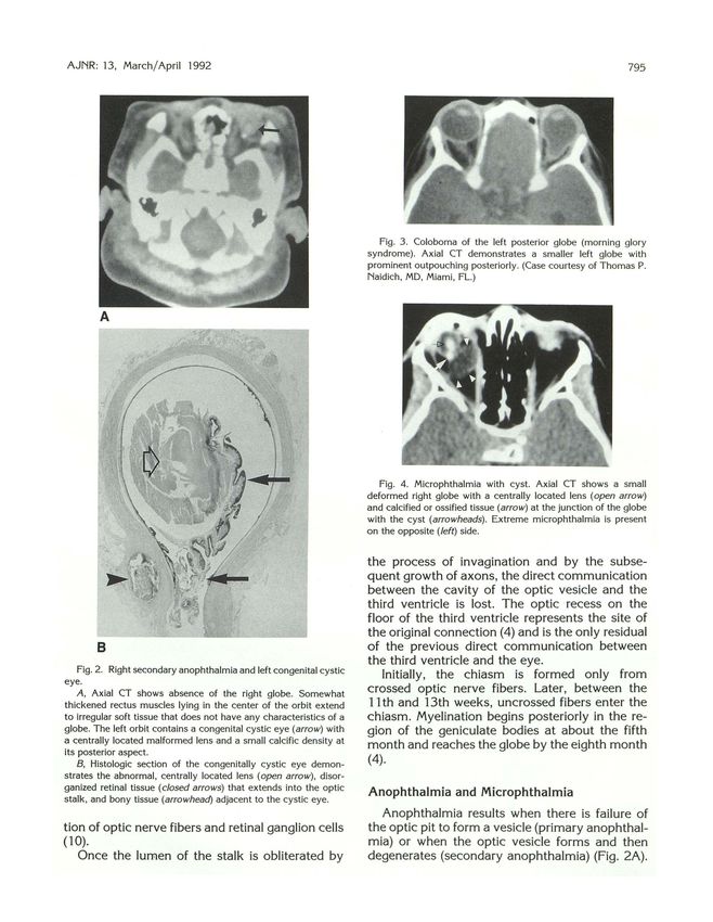
798 AJNR : 13, March/April 1992
A B c
Fig. 8. Marked bilateral optic nerve hypoplasia with pituitary anomaly .
A , Proton density MR demonstrates tiny optic canals (arrowheads) . The right optic nerve sheath complex (arrow) is very small.
8 , Slightly higher MR section dem onstrates extremely small intracranial optic nerves (arrows) .
C, Sagittal MR shows absence of the pituitary stalk. There is abnormally high signal intensity (arrow) in the inferior hypothalamus at
the site where the pituitary stalk would be expected to have arisen.
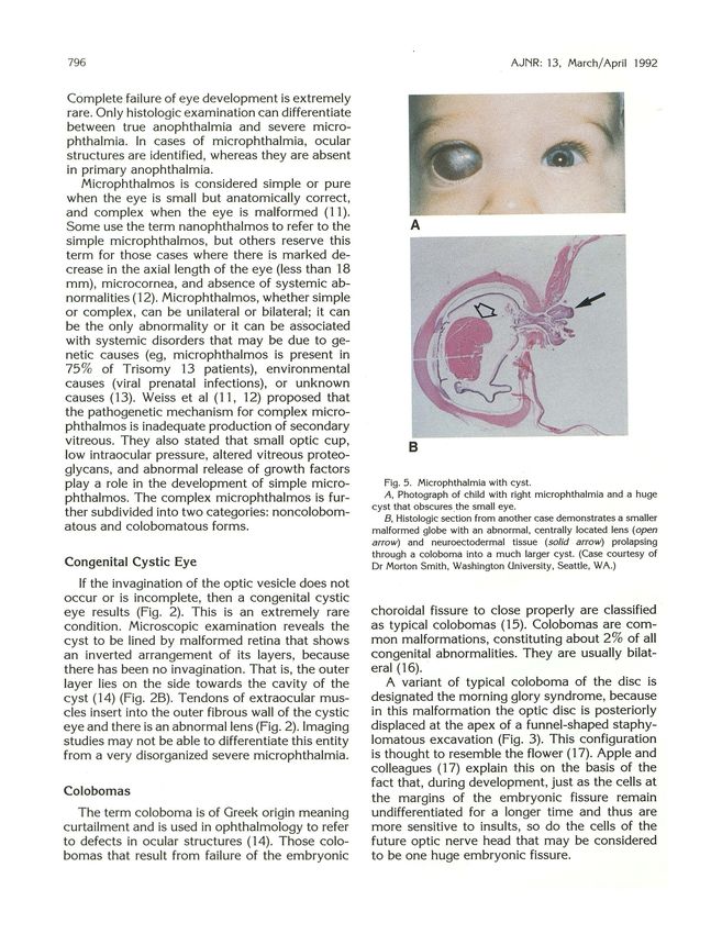
Optic Nerve Aplasia and Hypoplasia pathogenesis may be primary agenesis of retinal
ganglion cells.
Optic nerve aplasia is due to failure of the
In children with optic nerve hypoplasia, imag-
embryonic fissure to reach the optic stalk. It is a
ing studies are of great value to exclude any
sporadic, unilateral condition and is not associ-
associated intracranial abnormality. The imaging
ated with any systemic abnormalities (Fig. 7) (3).
studies demonstrate small optic nerves and
Optic nerve hypoplasia (Fig. 8) is a subnormal
chiasm, display the morphology of the pituitary
number of axons. It is a frequent congenital
gland (Fig. 8) and depict associated brain anom-
anomaly that may be isolated or may be associ-
alies (Fig. 9). Slight symmetrical decrease in the
ated with ocular, facial, endocrine, or central
size of the optic nerves may be difficult to appre-
nervous system anomalies (10). Among such
ciate on imaging , just as it is difficult to appreciate
entities are septo-optic dysplasia (Fig. 9) and clinically. Marked decrease in the size of the optic
encephalocele. One prospective study of 93 chil-
nerves or unilateral involvement is much more
dren with optic nerve hypoplasia found that those
easily detected.
children who had bilateral optic nerve hypoplasia
and poor vision had the highest incidence of Persistent Hyperplastic Primary Vitreous
associated nonocular congenital abnormalities
and medical problems (10). In this study, devel- Persistent hyperplastic primary vitreous
opmental delay was the most frequently associ- (PHPV) (Fig. 10) results when the embryonic
ated abnormality , and hypothyroidism was the
most frequent endocrine disturbance. Those chil-
dren that had bilateral optic nerve hypoplasia
with good vision or those that had unilateral optic
nerve hypoplasia fared much better.
The proposed pathogenesis of optic nerve hy-
poplasia is excessive degeneration of optic nerve
axons and retinal ganglion cells during develop-
ment of the eye and visual pathways (much more
than the normal amount) (10). Thus , one cause
of optic hypoplasia can be major· midline and
hemispheric anomalies that prevent the axons of
the retinal ganglion cells from making connec-
Fig. 9. Septo-optic dysplasia. Coronal Tl MR shows absence
tions at their intracranial target sites. However, o f the septum pellucidum and ex trem ely small intracranial optic
there are cases where the optic nerve hypoplasia nerves (arro ws) . A more posterior section (data not shown) dem-
is the only abnormality. In these instances, the on strated an anomal y of the posterior pituitary gland.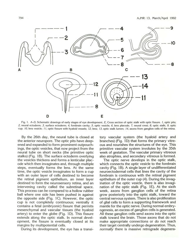
AJNR : 13, March/April 1992                                                                                               799

            A                                                           B

                Fig. 10. Persistent hyperplastic primary vitreous. Four different cases.
                A, Photograph of a child with left leukocoria due to persistent hyperplastic primary vitreous.
                B, Histologic section of persistent hyperplastic primary vitreous shows the abnormal lens (arrow) and
            triangular fibrovascular material (arrowheads) extending from it toward the optic nerve head . The retina
            (open arrows) is detached.
                C, Axial CT reveals a smaller right globe with tissue of increased attenuation (arrows) surrounding the
            posterior aspect of the right lens and extending posteriorly toward the right optic nerve head .
                D, Axial T2 MR demonstrates a deformed lens with prominent retrolental tissue (arrowhead) that has
            a thinner band of tissue (arrow) extending from it toward the posterior aspect of the globe.

intraocufar vascular system (hyaloid artery with                    include recurrent spontaneous hemorrhages, ret-
its branches) and the fibrovascular tissue of the                   inal detachment, and phthisis bulbi.
primary vitreous fail to regress normally (20). It                     In PHPV, histologic examination reveals a hy-
occurs in normal full-term infants, is usually uni-                 aloid artery that extends from the optic disc to a
lateral and is usually associated with microphthal-                 fibrovascular retrolental tissue (22) (Fig. 1OB).
mos. The most typical clinical findings are leu-                    Imaging studies, CT and MR , demonstrate a
kocoria (white pupil), microphthalmia, and cata-                    smaller globe with increased attenuation or hy-
ract (21). The presence of microphthalmia and                       perintensity on T1-weighted images and tissue
absence of calcification are important factors                      that extends from the posterior aspect of the lens
differentiating PHPV from retinoblastoma, since                     to the back of the globe in a band-like or trian-
retinoblastoma also presents with leukocoria but                    gular configuration (20, 23, 24).
typically shows calcifications and a globe that is
normal in size or enlarged. Glaucoma is a rela-                     Congenital Glaucoma
tively frequent complication of PHPV (26 % in one                     Glaucoma is a condition that results from ab-
series) and thus the affected eye may be buphth-                    normal elevation of intraocular pressure. The el-
almic (21 ). Other complications that may occur                     evated intraocular pressure is most often due to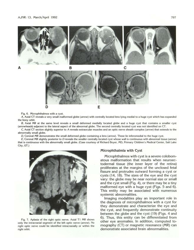
800                                                                                                 AJNR : 13, March/ April 1992

        A

                                                                             A

        B

                                                                             B
       c
    Fig. 11 . Severe primary congenital glaucoma of the left eye.      Fig. 12. Cryptophthalmia.
    A, Three-dimensional soft tissue CT shows marked enlarge-          A, Photograph of the infant shows total absence of the eyelids
ment of the left eye that protrudes between the lids.               and continuous skin from forehead across the orbits to the cheeks.
    8 , Three-dimensional bone CT shows enlargement of the bony     (Case courtesy of Dr. James Katowitz, The Children's Hospital of
left orbit.                                                         Philadelphia, PA.)
    C. Axial CT demonstrates prominent diffuse enlargement of          8, Axial CT shows malformed anterior structures of each globe
the left eye, particularly the anterior chamber. The left lens is   and continuity of the skin (arrow) with the tissue of the orbits.
flattened. There is slight indentation (arrow) at the optic nerve   There is absence of the cornea, anterior chamber, and lens on
head consistent with cupping.                                       each side. (Reprinted with permission from Levine et al (28).)

increased resistance to normal outflow of                           it may occur in association with other develop-
aqueous humor from the eye. Primary congenital                      mental disorders including phakomatoses. The
glaucoma is most frequently due to abnormal                         glaucoma may minifest itself at birth or during
development of the trabecular meshwork . Ander-                     the first few years of life. Increase in intraocular
son's investigations (25) suggest that the primary                  pressure causes enlargement of the entire eye
defect of congenital glaucoma may be premature                      (Fig. 11).
or excessive formation of collagenous beams                            Secondary congenital glaucoma refers to those
within the trabecular meshwork. These beams                         cases of glaucoma that result from intrauterine
are thought to generate traction forces that cause                  eye inflammation (eg, rubella), trauma, or ocular
compaction of the trabecular sheets with conse-                     tumor (eg, retinoblastoma). Insults or maldevel-
quent obstruction to the outflow of aqueous hu-                     opments that result in glaucoma occur anywhere
mor (25).                                                           from the fourth lunar month of gestation onward,
   Congenital glaucoma is bilateral in up to 80 %                   at a time when the anterior structures of the eye
of cases (26). It may be the only abnormality, or                   are developing .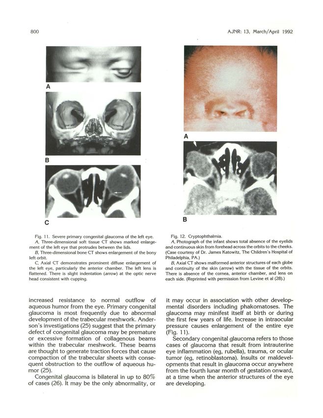
AJNR: 13, March/April 1992                                                                                                        801

Cryptophthalmos                                                        (28). Imaging studies are needed to demonstrate
                                                                       the globes and the rest of the intraorbital struc-
   Cryptophthalmos ("hidden eye") results when
                                                                       tures prior to surgical intervention.
the lid folds fail to develop. Cryptophthalmos is
considered typical when there is complete ab-                          Obstruction of the Nasal Lacrimal Duct
sence of the lids (complete ablepharia) and the
skin is continuous from the forehead, across the                          The nasal lacrimal duct forms in the invagin-
orbits, to the cheeks (27) (Fig. 12). The skin over                    ated surface ectoderm between the inner canthus
the globes is shown pathologically to be corneal                       and the inferior turbinate. Obstruction of this duct
epithelium that has undergone metaplastic                              is a frequent anomaly and is usually due to an
change (2). There is absence of eyelashes, mei-                        imperforate membrane at the lower end of the
bomian glands, lacrimal glands, lacrimal puncta,                       duct (29). The membrane consists of epithelium
and usually also eyebrows (27). Cryptophthalmos                        of the lacrimal duct and the mucosa of the nose.
is not an isolated anomaly but is part of a sys-                       The obstruction is manifested by overflow of
temic syndrome that often includes anomalies of                        tears (epiphora), or less commonly by a dacry-
face, cranium, extremities, and urogenital system                      ocystocele. Imaging studies are helpful in differ-
                                                                       entiating this entity from other masses that may
                                                                       present in the newborn.

                                                                       Congenital Tumors of the Orbit
                                                                          Congenital tumors of the orbit may be derived
                                                                       from one germ cell layer, such as dermoid cysts,
                                                                       from two germ cell layers (teratoid tumors) , or
                                                                       from three germ cell layers (teratoma) (30). These
                                                                       congenital tumors are not true neoplasms but
                                                                       developmental choristomas-that is , malformed
                                                                       tissue found at a site that normally does not
                                                                       contain that tissue .
   Fig. 13. Dermoid cyst. Axial CT demonstrates a heterogeneous .
mass with a wall of higher attenuation (arrowhead) . The content       Dermoid and Epidermoid Cysts
of the mass is heterogeneous with some fatty tissue (low atten-
uation). In addition, the dermoid extends into the frontal zygomatic     Dermoid and epidermoid cysts originate from
suture (arrow) .                                                       sequestered embryonic surface ectoderm and oc-

A                                                               B                                   c
    Fig. 14. Left orbital teratoma .
    A, Photograph of a child shows marked protrusion of the globe by a large posteriorl y loca ted mass that has expanded the orbit.
    8 , and C, Contrast-enhanced axial CT scans demonstrate the large cystic lesion that markedly expands the bony orbit and displ aces
the globe (arrows) anteriorly . (Case courtesy of Thomas P. Naidich, MD , Miami, FL.)
802 AJNR : 13, March/ April 1992
A B
c D
Fig. 15. Orbital teratoma .
A, Frontal and B, Left lateral views of the child show protrusion of the markedly deformed globe anteriorly through the palpebral
fissure.
C, Coronal CT shows heterogeneous irregular tissue within the left orbit.
D, Contrast-enhanced axial CT reveals multiloculated cystic tissue that extends from the orbit into the left temporal fossa. At surgery,
the teratoma lay entirely anterior to the temporal dura . Histologic evaluation of the tumor revealed tissues with pulmonary ,
gastrointestinal, muscular, and renal characteristics. Histologic examination of the globe revealed severely dysplastic microphthalmia
with absent lens, iris, and anterior charnber segments; persistent hyperplastic primary vitreous; and retinal dysplasia. (Case courtesy of
Lucy Rorke, MD , Philadelphia , PA.)
cur frequently in the orbit. They are typically orbital tumor. They contain adnexal structures,
located at the site of sutures, particularly the such as hair and sebaceous glands and, therefore,
zygomaticofrontal and frontoethmoidal sutures. their contents are frequently oily due to the se-
Dermoid cysts are the most common pediatric cretion of sebum. Epidermoid cysts do not con-AJNR: 13, March/ April 1992 803
sist of adnexal structures but may contain cho- 6. Hooper KD. Haas, Sherman J L . T he radiologic evaluation of congen-
ital and pediatric lesions of the orbit. Semin Ultrasound CT MR 1988;
lesterol in addition to epithelial debris. Imaging
9:4 13-427
studies reveal the cysts as well-defined masses 7. Fries PD. Katowitz JA. Congenital craniofacial anoma lies of
that may have thin walls. The cysts may be ophthalmic importance. Surv Ophtha/mol 1990;35:87- 119
homogeneous or heterogeneous ( Fig. 13) con- 8. Morri ss GM , Thorogood PV. A n approach to cra nial neural crest cell
taining tissue ranging from muscle to fat in radio- migration and differen tiation in mammalian embryos. In Johnson
MH , ed. Developm ent in m ammals. Amsterdam: North-Holland, 1978:
density and signal intensity. Some lesions may
363- 4 12
contain a fat-fluid level or may be diffusely fatty. 9. Seve! D. A reappraisa l of the origin of human extraocular muscles.
Larger lesions may produce scalloping of the Ophthalmology 198 1;86: 1330-1 338
adjacent bony wall or even expansion of the entire 10. Skarf B, Hoyt CS. Optic nerve hy poplasia in children. A rch Ophthal-
orbit. m ol 1984; 102:62-67
1 I. Weiss A H, Kousseff BG , Ross EA, Longbottom J . Complex micro-
phthalmos. A rch Ophtha/mo/ 1989;107:1619- 1624
Congenital Orbital Teratoma 12. Weiss A H, Kousseff BG, Ross EA, Longbottom J. Simple micro-
phthalmos . Arch Ophthalmo/1 989 ; 107: 1625- 1630
Orbital teratomas are rare congenital tumors 13. Bateman JB. Microphthalmos. lnt Opthalmol Clin 1984;24:87- 107
that contain tissue derived from all three germ 14. Man n L Developmental abnormalities of the eye. 2nd ed. Philadelphia:
cell layers and are usually benign (30). In most Lippincott, 1957
instances, the tissue is disorganized, but some 15. Pagan RA. Ocular coloboma. Surv Ophthalmol 198 1;25:223-236
16. Mafee MF, Jampol LM , Langer BG, T so M . Computed tomography
teratomas may actually have fetal features . One
of optic nerve colobomas, morning glory anomaly and colobomatous
theory of pathogenesis of the teratomas is lack cyst. Radio/ Clin North Am 1987;25:693-699
of normal response of tissue to inducers (31, 32). 17. A pple DJ. New aspects of colobomas and optic nerve anomalies. lnt
The most common presentation of a teratoma Ophthalmol Clin 1984;24: 109- 121
is extreme unilateral proptosis in an otherwise 18. Fox man S, Cameron JD. The cl inical im plications of bilateral m icro-
phthalmos with cyst. A m J Ophthalmol 1984;97:632-638
healthy full-term infant (30) (Fig. 14A). The tumor
19. Weiss A . Martinez C. Microphthalmos with cyst: clinica l presentations
usually grows rapidly after birth. Most typically, and computed tomog raphic fi ndings. J Pediatr Ophthalmol Strabis-
the tumor is multicystic (Figs. 14 and 15), and · mus 1985;22:6- 12
may be mixed cystic-solid or completely solid. 20. Magill HL, Hanna SL , Brooks MT, et al. Cases of the day: pediatric.
Usually the bony orbit is markedly expanded and RadioGraphies 1990; 10:515- 518
2 1. Haddad R, Font RL, Reeser F. Persisten t hyperplastic prima ry vitreous:
is two to three times its normal size (Fig. 14).
a cl inicopathologic study of 62 cases and re view of the litera ture.
Rarely the teratoma may extend intracranially Surv Ophthalmol 1978;23: 123-1 34
(33) (Fig. 15). Imaging studies permit delineation 22. Reese A B. Persistent hyperplastic primary vitreous. Am J Ophtha/mol
and characterization of the lesions, which leads 1955;40:3 17 - 33 1
to correct diagnosis, even when the clinical find- 23. Mafee MF, Goldberg MF. Persistent hy perplastic primary v itreous
(PHPV): role of computed tomogra phy and magnetic resonance .
ings are confusing. Teratomas need to be differ-
Radio! Clin North A m 1987;25:683-692
entiated from other congenital lesions, such as 2 4. Mafee M F, Goldberg MF, Valvassori GE, Ca pek V. Com puted tomog-
microphthalmos with cyst, dermoid cyst, congen- raphy in the evaluation of patien ts with persistent hyperplastic pri-
ital cystic eye, hemangioma, and meningoen- mary vitreous. Radiology 1982; 145:713-717
cephalocele. 25. A nderson DR. T he development of the trabecu lar meshwork and its
abnormality in primary infantile glaucom a. Trans Am Ophthalmol
Soc 198 1;79:458-485
References
26. Spencer WH . Glaucoma. In Spencer WH , ed. Ophthalm ic pathology
1. Ozanics V, Jakobiec FA. Prenatal development of the eye and its VoL 1. Philadelphia: W. B. Sa unders, 1985:526-537
adnexa. In Jakobiec FA , ed. Ocular anatomy, embryology and tera- 27. Fra ncois J. Malforma tive syndrome with cryptophtha lmia. Jnt
tology Philadelphia: Harper and Row, 1982:1 1-96 Ophthalmol Clin 1968;8:817-837
2. Duke-Elder S, Cook C. Normal and abnormal development. In Duke- 28. Lev ine RS, Powers T , Rosenberg HK , Siegel CA , Bilan iuk LT. T he
ElderS , ed. System in ophthalmology Vol 3. St. Louis: Mosby, 1963 cryptophthalmos syndrome. A m J Radio/ 1984; 143:375-376
3. Smith CG , Gallie BL, Morin JD. Normal and abnormal development 29. Kush ner BJ . Congenital nasolac rimal system obstruction. A rch
of the eye. In Cawford J S, Morin JD, eds. The eye in childhood. New Ophthalmol 1982; 100:597-600
York: Grune and Stratton, 1983: 1-17 30. Lev in M L , Leone CR, Kinca id MC. Congenita l orbita l teratom as. Am
4. Torczy nski E. Normal development of the eye and orbit before birth: J Ophthalmol 1986;102:476-481
the development of the eye. In Isenberg SJ , ed. The eye in infancy, 3 1. K rafka J . T eratoma. Arch Pathol 1936;21 :756-764
Chicago: Year Book Medical, 1989:9- 30 32. A lkemade PPH. Congenita l teratoma of the orbi t. Ophthalmologica
5. Spaeth GL, Nelson LB . Beaudoin AR . Ocular teratology. In Jakobiec 1976; 173:274-285
FA, ed. Ocular anatomy , embryology and teratology Philadelphia: 33. Ide CH, Davis WE, Black SPW. Orbital teratoma . Arch Ophthalmol
Harper and Row, 1982:955-1 080 1978;96:2093-2096You can also read