Human magnetic sense is mediated by a light and magnetic field resonance dependent mechanism - Nature
←
→
Page content transcription
If your browser does not render page correctly, please read the page content below
www.nature.com/scientificreports
OPEN Human magnetic sense is mediated
by a light and magnetic field
resonance‑dependent mechanism
Kwon‑Seok Chae1,2,3,6*, Soo‑Chan Kim4,6, Hye‑Jin Kwon2 & Yongkuk Kim5
Numerous organisms use the Earth’s magnetic field as a sensory cue for migration, body alignment,
or food search. Despite some contradictory reports, yet it is generally accepted that humans do not
sense the geomagnetic field. Here, we demonstrate that a magnetic field resonance mechanism
mediates light-dependent magnetic orientation in men, using a rotary chair experiment combined
with a two-alternative forced choice paradigm. Two groups of subjects were classified with different
magnetic orientation tendencies depending on the food context. Magnetic orientation of the subjects
was sensitive to the wavelength of incident light and was critically dependent on blue light reaching
the eyes. Importantly, it appears that a magnetic field resonance-dependent mechanism mediates
these responses, as evidenced by disruption or augmentation of the ability to orient by radiofrequency
magnetic fields at the Larmor frequency and the dependence of these effects on the angle between
the radiofrequency and geomagnetic fields. Furthermore, inversion of the vertical component of the
geomagnetic field revealed a non-canonical inclination compass effect on the magnetic orientation.
These results establish the existence of a human magnetic sense and suggest an underlying quantum
mechanical magnetoreception mechanism.
Numerous organisms, across a wide range of taxa, including birds, sea turtles, reptiles, insects, magnetotactic bac-
teria, and plants, use the geomagnetic field (GMF) as a sensory cue for migration1–4, short-distance movement5–7,
body alignment8–10, food s earch6,7, or growth (plants)11, depending on the species and biological context. Both
magnetic compass2,12 and magnetic map3,13 information can be derived from the GMF; the former being essen-
tial for a variety of magnetically sensitive behaviors5–10,12,14,15. An inclination12,16 or polarity c ompass17,18 may
provide animals with directional information by contrasting mechanisms: light-dependent radical p airs19–21 in
cryptochrome flavoproteins in the eyes of birds20,21 and light-independent iron-containing biogenic magnetite in
bacteria or the ethmoid bone of salmon14,22. It is thought that light-induced radical pairs, comprised of a flavin
adenine dinucleotide (FAD) radical and a tryptophan radical, in cryptochromes act as the magnetic compass
sensor in migratory birds through a quantum mechanical mechanism20,21,23. In these species, the inclination
compass is activated by blue or green light24 and disrupted by red light25, indicating that different wavelengths
play different roles in radical pair-mediated b ehaviors21.
Research on magnetoreception in humans is very limited. It is widely accepted that the Earth’s static magnetic
field is not sensed by humans, while alternating magnetic fields, such as power frequency fields26 and pulsed
fields27, can have adverse health effects and therapeutic applications, respectively. Following previous contro-
versial reports28–30, two recent studies, using different experimental approaches, support the existence of GMF
responses in h umans31,32 with a sharp contrast. In a rotary chair experiment, starved men but not women were
able to orient in a blue-light-dependent manner towards a particular magnetic direction that had previously
been associated with food in the ambient G MF31. This study suggests that the magnetoreceptors are in the eyes
but does not demonstrate the underlying sensory mechanism. In contrast, electroencephalography showed that
a decrease in alpha brain wave activity occurred in some human subjects under conditions of darkness32. The
observed sensitivity to the polarity of the applied magnetic fields implied a magnetite-based mechanism. How-
ever, so far, the existence of a human magnetic sense itself and any underlying mechanism are far from clear.
1
Department of Biology Education, Kyungpook National University, Daegu 41566, Republic of Korea. 2Department
of Nanoscience and Nanotechnology, Kyungpook National University, Daegu 41566, Republic of Korea. 3Brain
Science and Engineering Institute, Kyungpook National University, Daegu 41566, Republic of Korea. 4Department
of Electrical and Electronic Engineering, Research Center for Applied Human Sciences, Hankyong National
University, Anseong 17579, Republic of Korea. 5Department of Mathematics, Kyungpook National University,
Daegu 41566, Republic of Korea. 6These authors contributed equally: Kwon-Seok Chae and Soo-Chan Kim. *email:
kschae@knu.ac.kr
Scientific Reports | (2022) 12:8997 | https://doi.org/10.1038/s41598-022-12460-6 1
Vol.:(0123456789)
www.nature.com/scientificreports/
To establish the existence and underlying mechanism of human magnetic sensing, we have studied magnetic
orientation in men, which showed remarkable magnetic sensitivity, by combining the rotary chair method with
a two-alternative forced choice (2-AFC) paradigm33,34, applying oscillating magnetic fields as a diagnostic tool
echanism35.
for a magnetic field resonance mechanism, such as the radical pair m
Results
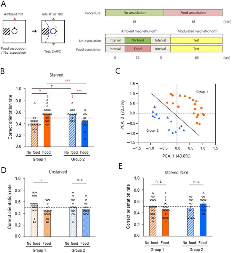
Differential sensitivity in ambient GMF‑responsive orientation. Male subjects were starved or fed
normally, then tested using the rotary chair method combined with a 2-AFC paradigm that required the subjects
to choose one of the two directions on the magnetic north–south axis (Fig. 1A, see “Methods” section). During
the association phase of the experiment, subjects with eyes closed and wearing earmuffs were aligned by experi-
menters so as to face ambient magnetic north while sitting on a rotatable chair and were either conditioned to
associate this direction with food or were not conditioned, depending on the session. During the test phase, in
which the ‘modulated’ magnetic north was randomly set to true magnetic north or true magnetic south, subjects
were asked to indicate the modulated magnetic north direction, without reference to other information includ-
ing visual and auditory cues. The test was initially performed under full-wavelength visible light (350–800 nm)
(Table S1, #1 and Fig. S1A). Subjects that had been starved to produce a significant reduction in blood glucose
levels (Table S2) were divided into two groups. Group 1 (Fig. 1B, left, n = 20, ca. 60%) showed a significant
increase in the ability to orient correctly with food-association compared to subjects who had not been condi-
tioned while Group 2 (Fig. 1B, right, n = 14, ca. 40%) showed a significantly lower rate of food-association (see
Figs. S2 and 1C for clustering of the subjects). In our previous study31, under full wavelength and > 400 nm but
not > 500 nm light conditions, a majority of subjects showed significantly correct magnetic orientations with
food-association, whereas the remaining did not. In an attempt to objectively cluster the subjects in the present
study, principal component analysis36,37 of data recorded for starved subjects with and without food-association
under the full wavelength and > 400 nm light conditions as shown in Fig. 2A,C, produced two subject groups
(Fig. 1C). The results shown in Fig. 1B are consistent with a reanalysis of relevant data from our previous s tudy31
(Fig. S3) and indicate that starved men with food-association were clustered into two groups; one group that
showed significant GMF-orientation toward modulated magnetic north on the magnetic north–south axis, and
one that did not. By contrast, when the subjects were normally fed (Table S2), Group 1 but not Group 2 showed
a significant decrease in correct orientation rate with food-association (Fig. 1D). Subjects were then tested to
identify modulated magnetic north randomly set on the true magnetic east–west axis. Similar to the results in
Fig. 1B, the orientation rate with food-association increased in Group 1 and decreased in Group 2, compared to
no-association with starvation (Fig. S4, Table S2). In our previous study, the ambient GMF was thought to be an
unconditioned stimulus during the association p hase31. Therefore, we examined whether sensing the ambient
GMF during association was necessary for correct magnetic orientation on the magnetic north–south axis, as
shown in Fig. 1B. Under a near-zero GMF condition during the association phase, neither starved group demon-
strated a significant difference in correct orientation rate with and without food association (Fig. 1E), indicating
that sensing the ambient GMF prior to the test was necessary for correct magnetic orientation in both groups.
These results show that there are two groups with different magnetic sensitivity in ambient GMF-orientation,
depending on the food context.
Pivotal role of blue light in wavelength‑dependent distinct magnetic orientation. Based on
our previous study31 which showed that light was necessary for food-associated geomagnetic orientation in
starved men, the effect of wavelength on magnetic orientation was investigated in the two groups. Distinct
magnetic orientation profiles that were highly dependent on food context and Group were obtained under six
light conditions: five different light wavelengths (Table S1, #1–5 and Fig. S1A–E) and ‘dark’ (Fig. 2A–D). The
starved subjects in Group 1 showed significant correct orientation rate with food-association compared to no-
association under full wavelength and > 400 nm light, while the orientation rates were dramatically reversed
under > 500 nm light (Fig. 2A). This result indicates that UV-A–blue (350–500 nm) and green (500–570 nm)
light are either necessary for, or inhibit, correct magnetic orientation and that these effects occur in differ-
ent directions for the two wavelength ranges depending on the association conditions. This finding is reminis-
cent of the antagonistic effect of < 450 nm and > 500 nm light on magnetic orientation in newts and fl ies38. The
unstarved Group 1 subjects showed a low orientation rate under > 400 nm light with no-association, suggest-
ing that UV-A light (350–400 nm) was helpful for correct orientation under the full wavelength whereas blue
light (400–500 nm) decreased the orientation rate (Fig. 2B). By contrast, Group 2 subjects demonstrated no
significant differences in orientation rate for changes of wavelength and association conditions, with or without
starvation (Fig. 2C,D). However, there was a significant change in the rate for > 400 nm light compared to the
full wavelength range, wherein the effect of UV-A light was not negligible and there was a notable rate under red
light (> 600 nm) with food-association (Fig. 2C). There were no significant differences between orientation rates
in the dark condition by association, starvation, or group, except for a slightly higher rate in starved Group 2
with food-association, indicating that light was crucial for correct magnetic orientation. To dissect the interplay
among light wavelengths, blue light alone was sufficient to increase the rate in starved Group 1 subjects (Fig. 2E),
suggesting that together with the data in Fig. 2A red light disrupted an increase in the rate elicited by green light,
but not by blue light, with the food-association. Importantly, this result indicates that blue light plays a pivotal
role in magnetic orientation by overcoming the effect of red light. In addition, UV-A light alone produced a dis-
tinct magnetic orientation profile, depending on the group and food context (Fig. S5). These results demonstrate
that human geomagnetic orientation is highly sensitive to light wavelength, and that blue light plays a critical
role in orientation.
Scientific Reports | (2022) 12:8997 | https://doi.org/10.1038/s41598-022-12460-6 2
Vol:.(1234567890)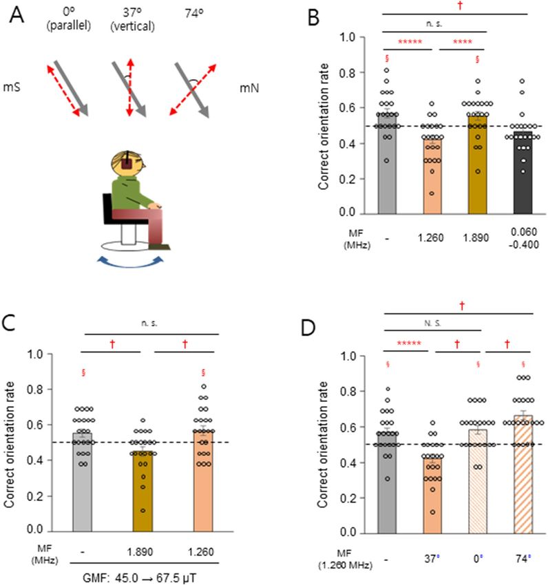
www.nature.com/scientificreports/

                                 Figure 1.  Different sensitivity of geomagnetic orientation in men. (A) Schematic drawing of a 2-AFC behavioral paradigm to test
                                 geomagnetic orientation in men. Left: Top view of the subject facing toward the ambient geomagnetic north during the no-association/
                                 food-association phase, and rotating to search for one of the two modulated magnetic norths randomly provided during the test phase.
                                 Subjects were allowed to rotate clockwise or counterclockwise during the test as shown by arrows within the range indicated by the
                                 dashed blue lines (see “Methods” section). mN, magnetic north; square, the vertical Helmholtz coils; square with hatched lines, stool
                                 to limit the subject’s rotation; circle, the subject; black closed triangle, the facing direction of the subject; red open triangle(s), direction
                                 of the ambient magnetic north (association phase) or modulated magnetic norths (test phase). Right: The procedure and timeline for
                                 the 2-AFC experiment. Food was provided for ‘food-association’ trials only. (B) A significant increase (Group 1) and decrease (Group
                                 2) in correct orientation rate between the different associations under starved conditions. (C) Two subject groups were classified by
                                 principal component analysis. Analyzed parameters; correct orientation rate with the food/no-food association under light wavelength
                                 of 350–800 nm or 400–800 nm (data, n = 136; 34 subjects). Group 1 (orange filled circle), n = 20; Group 2 (blue filled triangle), n = 14.
                                 Solid line, the classifying boundary (PCA 2 =  − 1.1515 PCA 1 − 0.0897) (D) A significant decrease (Group 1) in correct orientation rate
                                 by the change of association under the unstarved condition. (E) No notable change of correct orientation rate with near-zero GMF
                                 during the association phase. NZA, near-zero GMF association; §, P < 0.05 by one-sample t-test; *, P < 0.05; ***, P < 0.005 and n.s., not
                                 significant by paired sample t-test; ###, P < 0.005 by two sample t-test; †, P-value, 0.9999 > 0.9750 (confidence interval, − 0.2170, − 0.0820)
                                 for the data of no-association and food-association in Group 1, and 1.0000 > 0.9750 (confidence interval, − 0.0801, 0.0786) for the
                                 data of no-association in Group 1 and Group 2 by a percentile bootstrap analysis (see Fig. S2); horizontal dashed lines, 0.5 for correct
                                 orientation rate; error bars, standard error of the mean (SEM).

Scientific Reports |   (2022) 12:8997 |                    https://doi.org/10.1038/s41598-022-12460-6                                                                           3

                                                                                                                                                                         Vol.:(0123456789)
www.nature.com/scientificreports/
Figure 2. Blue light plays a pivotal role in wavelength-dependent magnetic orientation. (A–D) Correct
magnetic orientation rate in Group 1 (A, B) or Group 2 (C, D) subjects under starved and unstarved conditions.
The data for > 350 nm light were the same as those in Fig. 1, B or D. Data points connected by straight lines
and dashed lines were obtained from food-association and no-association, respectively. §, P < 0.05 by one-
sample t-test; *, P < 0.05; ***, P < 0.005; *****, P < 0.0005; and n.s., not significant by paired sample t-test between
the corresponding data from food- and no-association; #, P < 0.01 by paired sample t-test between the data
from > 400 nm and > 500 nm in food-association. (E) Correct magnetic orientation rate with the different light
conditions in Group 1 starved. §, P < 0.05 by one-sample t-test; *, P < 0.05; and n.s., not significant by paired
sample t-test; †, significant between the data from > 350 nm and > 400 nm in no-association; N. S., not significant
by a percentile bootstrap analysis (see Appendixes S1 and S2); horizontal dashed lines, 0.5 for correct orientation
rate; error bars, standard error of the mean (SEM). Group 1, n = 20 and Group 2, n = 14.
A magnetic field resonance mechanism‑mediated human magnetic orientation. Blue light is
essential for radical pair-mediated magnetoreception in birds21,39. Therefore, to understand the magnetosensory
Scientific Reports | (2022) 12:8997 | https://doi.org/10.1038/s41598-022-12460-6 4
Vol:.(1234567890)www.nature.com/scientificreports/
mechanism in humans, we tested whether oscillating magnetic fields at particular frequencies35,40,41 disrupt mag-
netic orientation in humans. First, 1.260 MHz, the electron Larmor f requency21 in the ambient GMF (45.0 μT),
or a flanking frequency (1.890 MHz or 60–400 kHz)39,40 was provided vertically to the starved subjects who had
shown a correct orientation rate greater than 0.5 in Fig. 1B (Group 1 with food-association and Group 2 with
no-association, Fig. 3A, middle and Fig. S6). The 1.260 MHz and lower frequency broadband magnetic fields,
but not the 1.890 MHz field, significantly disrupted magnetic orientation compared to the dummy load control
(Fig. 3B). When the strength of the static magnetic field was increased by 50% to 67.5 μT, such that the Larmor
frequency was 1.890 MHz, we found that the 1.890 MHz field, but not the 1.260 MHz field, significantly reduced
the correct orientation rate (Fig. 3C). Given the critical property of the proposed radical pair magnetoreceptor as
a direction sensor20,21, the effect of the angle between the 1.260 MHz field and the ambient GMF was evaluated
(Fig. 3A, left and right). The different orientations of the radiofrequency antenna produced different magnetic
field intensities at the eyes of the subjects, caused by the shadowing e ffects42 of their heads. (Table S3). Compared
to the dummy load control, the rate was not notably changed in the parallel configuration (0°), whereas it was
significantly decreased in the vertical arrangement (37°) and increased for the 74° angle, indicating a differential
effect on an anisotropic receptor (Fig. 3D). These results support the notion that radical pairs probably mediate
the magnetic orientation observed in humans.
A non‑canonical magnetic inclination compass in humans. In principle, radical pair-mediated
magnetoreception confers an inclination compass1,2. The vertical component of the ambient GMF was therefore
cancelled in an attempt to learn more about the properties of the magnetic compass12. The correct magnetic
orientation we had observed for subjects toward true magnetic north (mN) or true magnetic south (mS) when
the modulated magnetic north was set to 0° and 180°, respectively, in the control field inclination (+ 53°), was
almost eliminated when the inclination was ≈ 0°, indicating that the vertical component was essential and that
an inclination compass mediated the observed magnetic orientation (Figs. 4A, S7A,B). Inversion of the vertical
GMF component, another diagnostic tool to discriminate between magnetic compass m echanisms2,12, produced
orientation toward the opposite direction, mS, when the modulated magnetic north was set to 0° (Figs. 4B,
S7C). By contrast, an exact reversal of the GMF, i.e., setting magnetic north to 180° and inverting the vertical
component, failed to produce significant orientation toward mN or mS, deviating from the notion of a canonical
inclination compass (Figs. 4B, S7C). Taken together, the results demonstrate that human magnetic orientation is
mediated by a non-canonical inclination compass.
Discussion
The present study has established the existence of a human magnetic sense to the GMF and for the first time
suggested a magnetic field resonance mechanism as the underlying magnetoreception principle. Consistent with
findings for some species of birds35,39, changes in orientation rates using magnetic fields oscillating at the Larmor
frequency, demonstrate that a magnetic field resonance mechanism mediates the human magnetic sense. The
effect of an external linearly polarized oscillating magnetic field at the Larmor frequency is known to depend
on the angle between the field axis and the ambient G MF21,35,43; therefore not only the absence of an effect in the
parallel arrangement (0°), but also the enhanced correct orientation rate in the 74° condition provides strong
evidence for a probable involvement of radical pair mechanism in humans. Magnetite particles large enough to
rotate in the Earth’s magnetic field are not expected to show such effects because they move too slowly to track the
direction of a field that reverses more than a million times per second21. In agreement with the light-dependent
radical pair model and previous studies of b irds24,39, light effects resulted in distinct orientation profiles with a
strong wavelength dependence. Importantly, blue but not green light seems to play a pivotal role in magnetic
orientation by overcoming the effect of red light, whereas green light is also sufficient for correct magnetic ori-
entation in birds25. The magnetic response to blue light approximately matches the absorption spectrum of fully
oxidised FAD in c ryptochrome21,39 (wavelengths less than 500 nm) and cryptochromes have been reported to
occur in the human retina44, suggesting that cryptochromes might be promising magnetoreceptor in humans. To
understand the complicated food context- and group-dependent orientation profiles, the following is proposed:
radical species, e.g., reactive oxygen species such as O 2·−, occur at different concentrations due to different meta-
bolic rates, depending on starvation, body weight or exposed magnetic fi elds45–48. In turn, O
2·− could form part of
a magnetically sensitive cryptochrome-based radical pair in the eyes35,49–51 (but see Refs52,53). In particular, O2·− in
the optimal range of concentration may be crucial for significant enhancement of the anisotropic magnetic field
effect (direction sensor) through the F ADH·/O2·− radical scavenging s ystem51. Besides the evolutionary adaptive
31
scenario , this might be a plausible speculation explaining the superior magnetic sensitivity of men, compared to
women with lower basal metabolic rate, under fasting rather than normal feeding. Nevertheless, there might be
unknown magnetic sensitivity for women under different contexts, requiring separate investigation. The human
inclination compass identified in the present study deviates from the canonical inclination compass, but still
maintains the compass function: humans are much less mobile than long-distance migratory birds and may not
have evolved to be as sensitive as birds to encountering polarity variation of the GMF. At first sight, the principal
results of this study, i.e., blue light and radical pair mechanism-mediated magnetic orientation, are difficult to
reconcile with the results of Wang et al.32 which suggested a light-independent magnetite-based mechanism
for the human magnetic sense. However, the two studies used very different experimental assays that could be
probing different receptors. This could be compared to birds, which are thought to have radical pairs in the eyes
(for direction sensing) and magnetite elsewhere (for intensity sensing).
The 2-AFC paradigm would be a useful experimental setup particularly for a comparative study on magne-
toreception mechanisms, such as radical pair- or magnetite-involved mechanisms. As shown in Fig. S3, the valid-
ity and feasibility of the paradigm for the magnetic orientation test are supported by the consistency between the
Scientific Reports | (2022) 12:8997 | https://doi.org/10.1038/s41598-022-12460-6 5
Vol.:(0123456789)www.nature.com/scientificreports/
Figure 3. Radical pair mechanism mediates magnetic orientation in humans. (A) Experimental set-up to test
oscillating magnetic field (red dashed arrows) effect on magnetic orientation of the subjects. The selected starved
subjects mentioned in the main text were food/no-food associated and tested under the ambient GMF (45.0
μT, gray arrows) with full wavelength light. (B) Correct orientation rate of the subjects exposed to oscillating
magnetic fields of different frequency. (C) Subjects were tested under an increased intensity of the GMF (67.5
μT), while the ambient GMF was maintained during the association phase. (D) Different directions of the
1.260 MHz magnetic field relative to the ambient GMF; 0° (parallel), 37° (vertical), and 74°. Note that in the
parallel and 74° configurations, the 1.260 MHz magnetic field had the same angle with respect to the subjects.
For 0° and 74° angles, the radiofrequency field was provided only in trials when modulated magnetic north was
set to true magnetic north (mN, 0°), but not true magnetic south (mS, 180°), and the correct orientation rate
was calculated on these trials alone. §, P < 0.05 by one-sample t-test; ****, P < 0.001; *****, P < 0.0005; and n.s.,
not significant by paired sample t-test; †, significant; N. S., not significant by a percentile bootstrap analysis (see
Appendixes S1 and S2); horizontal dashed lines, 0.5 for correct orientation rate; error bars, standard error of the
mean (SEM). Subjects, n = 22.
results of the present and previous s tudy31 that adopted a rotary chair experiment. The 2-AFC permits rotation
within 240° that is larger than the symmetric half of the rotary chair (180°) and subjects can freely rotate clock-
wise or counterclockwise at any position within the angle in both setups. Therefore, in principle, the difference
in rotation extent between the 2-AFC (240°) and the rotary chair (360°) should not necessarily be a difference in
the actual magnetic direction search of subjects. In the 2-AFC paradigm, subjects could be more concentrated on
the correct magnetic discrimination of one of the two directions rather than precise orientation, for modulated
Scientific Reports | (2022) 12:8997 | https://doi.org/10.1038/s41598-022-12460-6 6
Vol:.(1234567890)www.nature.com/scientificreports/
Figure 4. A non-canonical magnetic inclination compass in humans. Selected starved subjects were tested
under different inclinations with full wavelength light. The Y axis represents arbitrary orientation rate toward
true magnetic north (mN) or true magnetic south (mS). (A) Inclination ≈ 0° due to cancellation of the vertical
component of the ambient GMF. (B) Inclination − 53° due to inversion of the vertical component. The data from
the ambient inclination (+ 53°) in (a) and (b) are the same. †, significant; N. S., not significant by a percentile
bootstrap analysis (see Appendixes S1 and S2); error bars, standard error of the mean (SEM). Subjects, n = 22.
magnetic north on, e.g., the magnetic north–south axis, which was available through the direction referring to
the stool that confined the rotation extent. Nonetheless, a significantly correct choice for modulated magnetic
north is still a difficult task that would not be accomplished by chance in the absence of real magnetic responsive-
ness of the subjects as demonstrated in the present study. The 2-AFC paradigm remarkably saved time (≈ 33%)
for the experiment on human subjects as described in the Methods section. Given the realistic or potential time
limit of subjects impeding human magnetoreception studies in the past and the future, time-saving could be an
important advantage. Consequently, researchers may have options to take advantage of a more appropriate test
paradigm for their magnetoreception studies on humans.
To gain further insight into the mechanism, future investigations could be undertaken as follows, using
the experimental setup of the present study as a platform with or without the oscillating magnetic fields: (1) a
magnetic orientation test on subjects with color blindness, including tritanopia, to examine whether function-
ing photoreceptor cells are necessary for magnetic sensing; (2) identification of magnetoreceptive cells and
magnetoreceptors using human eye cultures or eye organoids, combined with spatio-temporal analysis of the
magnetoreception process at the molecular level; and (3) electroencephalography cortical mapping of the mag-
netic sense area to examine whether the radical pair mechanism is critical under different lights or food contexts.
Methods
Subjects. The study comprised 34 men (19–26 years, mean 23 years; body mass index 19–31 kg/m2, mean
24 kg/m ) with no physical disabilities or mental disorders, including color blindness and claustrophobia30,31.
2
All subjects were informed of the aims, the study procedure, and the financial compensation for participa-
tion, and were asked to follow the rules of the study. Prior to each experiment, subjects underwent short-term
starvation31,54 (18–20 h; no food except pure water after lunch (12–1 pm) or dinner (6–7 pm), no later than 1 pm
or 7 pm, respectively, one the day before the test), no medical treatments, and normal sleep (at least 6 h, between
10 pm the day before the test day to 8 am on the test day)31. Prior to starting each experiment, subjects were sta-
bilized on a chair for ~ 10 min in a room next to the testing room. Based on an assessment with a pre-experiment
questionnaire and the first blood glucose level, measured before starting the experiment (see “Geomagnetic
orientation assay” section below), subjects who had not followed these rules were not allowed to take the test on
the day and the test was postponed. The study was approved by the Institutional Review Board of Kyungpook
National University and all the procedures followed the regulations for human subject research. Informed con-
sent was obtained from all subjects.
Modulation of GMF. The ambient GMF in the testing room had a total intensity 45.0 μT, inclination 53°,
and declination − 7° (Daegu city, Republic of Korea); the total intensity of 50.0 μT in our previous study31 was
changed due to a reconstruction of the building; 45.0 μT was maintained throughout the period of this study. To
Scientific Reports | (2022) 12:8997 | https://doi.org/10.1038/s41598-022-12460-6 7
Vol.:(0123456789)www.nature.com/scientificreports/
provide the subjects with various GMF-like magnetic fields (i.e., by modulating of total intensity, inclination, or
direction of magnetic north), the coil system from our previous s tudies6,7,31 was used. It comprised three double-
wrapped, orthogonal, rectangular Helmholtz coils (1.890 × 1.890 m, 1.890 × 1.800 m, and 1.980 × 1.980 m for the
north–south, east–west, and vertical axes, respectively), electrically-grounded with copper mesh shielding. The
subject was seated on a rotatable plastic chair with no metal components, at the center of the three-dimensional
coils with his head positioned in the middle space of the vertical axis of the coils. To modulate the geomag-
netic north, each pair of coils was supplied with direct current from a power supply (MK3003P; MKPOWER,
Republic of Korea). The magnetic field was measured using a 3-axis magnetometer (MGM 3AXIS; ALPHALAB,
USA); the field homogeneity at the position of the subject’s head was found to be 95%. The testing room was
shielded by a six-sided Faraday cage comprising 10 mm thick aluminum plates, and was grounded during the
entire experiment40. Background electromagnetic noise was measured inside the coils at the start and the end of
each experimental day. It was attenuated by the Faraday cage more than 200-fold over the range from 500 Hz to
100 MHz as described in detail in our previous study31. The 60 Hz power frequency magnetic field was no more
than 2 nT (3D NF Analyzer NFA 1000; Gigahertz Solutions, Germany). All electronic devices were placed out-
side the Faraday cage during the experiments, with the exception of the switch button module for GMF modula-
tion and the antenna for generating the oscillating magnetic fields. The temperature experienced by the subjects
was maintained at 25 ± 0.5 °C (Data logger 98,581; MIC Meter Industrial, Taiwan) by air circulation through the
honeycomb on the ceiling of the Faraday cage31.
Geomagnetic orientation assay. Adopting a two-alternative forced choice (2-AFC) paradigm33,34, a
geomagnetic orientation assay was conducted similar to our previous s tudy31. Experiments were performed at
09:30–11:30 am or 1:00–5:00 pm (local time, UTC + 09:00) (each experiment: 50 min–1 h 10 min; mean ≈ 1 h,
which was shorter by approximately 30 min than that in the previous study: 1 h 20 min–1 h 40 min; mean ≈ 1 h
30 min). Depending on the experiment, starved or unstarved subjects were tested individually. Prior to each
experiment, the subjects were asked to remain with their heads facing the front, with eyes closed and earmuffs
on during the experiment. In particular, they were asked to concentrate on sensing, if they could, the ambient
geomagnetic north during the association phase, and to use the sensed information, depending on the experi-
ment, to orient toward one of the two modulated magnetic norths (0°/180° for magnetic north–south axis or
90°/270° for magnetic east–west axis, rotated clockwise with respect to the ambient geomagnetic north) during
the test phase. Subjects were instructed to avoid distracting thoughts and to think immediately “which direc-
tion is modulated magnetic north?” whenever they were distracted during the test phase, or felt they were being
biased by experiences from earlier experiments. While seated on the rotatable chair, the subject’s blood glucose
level was measured shortly before the first session and immediately after each session with eyes open except in
the ‘dark’ experiment (Accu-Chek Guide; Roche, Germany)31. If the determined value before the first session
varied by more than 15% relative to the mean (Table S2)31, the experiment was postponed and repeated at a
later date (approximately 2% of experiments). The subjects were stabilized with eyes closed for 2 min before the
first trial in the absence of visual, auditory, olfactory, and haptic sensory cues. For the ‘dark’ experiment (light
intensity ≈ 0 lx), subjects wore home-made ‘blind’ goggles and were stabilized with eyes closed for 5 m in55,56,
and then asked whether they could see any light. If they could, the goggles were adjusted to prevent leakage of
light, and the subject then had another 5 min of stabilization with eyes closed before starting the experiment.
The subjects were illuminated with light from a filtered/non-filtered diffused light-emitting diode, depending
on the experiment (Table S1). The home-made filter goggles were worn throughout the experiment, including
the association and test phase, when required. The goggles contained glass filters (Tae Young Optics, Republic of
Korea) to provide the eyes with particular wavelengths of light (Spectrometer USB4000-UV-VIS, Ocean Optics,
USA) (Fig. S1). Each experiment consisted of 16 sequential trials for ‘no-association’ and ‘food-association’. For
the food-association, a subject facing toward the ambient geomagnetic north was gently provided with a choco-
late chip31 on his right palm by an experimenter, and given 30 s to eat it, while during no-association trials, food
was not provided during the association phase. After a subsequent 5 s interval, the experimenter gently touched
the subject’s right thenar area using a paper rod, as the cue to start the test. One of the two modulated magnetic
north directions, depending on the experiment, was randomly provided 3 s before the cue for the test. Each of
the modulated magnetic north directions was provided eight times for the no-association and food-association
sessions. Subjects were informed of the nearly equal probability for each of the modulated magnetic north direc-
tions before each experiment. With the touch cue, subjects were asked to rotate freely toward any direction
(clockwise or counterclockwise) by themselves (1–4 cycles of two-thirds rotation) and try to sense the direction
of the modulated magnetic north during a 1 min period. Rotation was allowed within the rotation angle (− 30°
to 210° for the magnetic north–south axis or − 120° to 120° for the east–west axis, depending on experiments,
with respect to the ambient magnetic north), which was confined by the plastic stool (Fig. 1A) touching the
left or right ankle of the subjects. When subjects determined the direction of the magnetic north, they stopped
rotating to face toward the direction and lifted their right hand to indicate the direction to the experimenter.
The direction was measured by the experimenter at 10° intervals using the scale on the walls of the Faraday
cage31. A prerequisite for correct orientation was that the subject indicated the direction within the range of 30°
to the both sides with respect to the magnetic cardinal directions, which was instructed to the subjects before
each experiment. When the direction the subjects indicated was out of the 30° range, the trial was not included
in the data and was repeated (approximately 0.63% of trials). Before the subsequent trial, the subject was gently
rotated to face toward the ambient geomagnetic north and then rested for 5 s. For the ‘dark’ experiment, subjects
were asked whether they could see any leaked light immediately after the last measurement of blood glucose
level at the end of experiment. If the subject could see leaked light, the experiment was nullified and repeated
later on (approximately 3% of experiments; 2/68). All experiments were performed in a double-blind fashion.
Scientific Reports | (2022) 12:8997 | https://doi.org/10.1038/s41598-022-12460-6 8
Vol:.(1234567890)www.nature.com/scientificreports/
The experimenter who conducted the orientation assay knew whether a subject was starved or not, wearing
filter goggles, and food-associated or not, but did not know the random magnetic north sequences that were
controlled by the personal computer (PC) system. Another experimenter responsible for analyzing the data did
not know whether the subject was starved or not, the experimental conditions, including light wavelengths, or
whether an oscillating magnetic field had been provided to the subjects. Thus, none of the experimenters were
aware of all the subject information and data during the experiments and data analysis. The correct orientation
rate was calculated by (the number of correct orientation trials/total number of trials) (raw data, Appendix S3).
All the subjects participated in all the experiments performed in random order with an interval of at least 3 days
between experiments. After each experiment, the subjects were asked to answer a post-experiment question-
naire about whether they closed their eyes when required during the entire period of the experiment. In cases
when a subject did not maintain closed eyes, the experiment was repeated (approximately 1% of experiments).
For each subject, a preliminary experiment on the “magnetic north–south axis” was conducted twice (unstarved
and starved for each) with no goggles for adaption to the experimental procedure. These data were not included
in the results.
Experiments with oscillating magnetic fields. Experiments with oscillating magnetic fields were per-
formed using the standard geomagnetic orientation assay described above. To produce the oscillating magnetic
fields, oscillating currents from a function generator (AFG3021; Tektronix, USA. For each magnetic field, sweep
of 500 ms; interval of 1 ms. See Fig. S6A) were amplified (ENI 2100L RF power amplifier; Bell Electronics, USA)
and fed into a calibrated coil antenna (30 cm diameter, 6509 loop antenna; ETS-LINDGREN, USA) mounted on
a wooden frame, comprised of a single winding of coaxial cable. The oscillating magnetic fields were measured
daily, before the first and after the last experiment of the day, using a spectrum analyzer (SPA-921TG; Com-
Power, USA) with a calibrated loop antenna (48 cm diameter, AL-130R; Com-Power, USA) and a calibrated
magnetometer (Probe HF 3061, NBM-550; Narda, Germany). Magnetic field intensities were measured on the
glabella of the subjects; variations in intensity between subjects due to different seating heights were less than
10% of the average values (Table S3). The function generator and amplifier were placed outside the Faraday cage,
and switched on during the dummy load control experiments with no signal from the PC system. The band
widths of the monochromatic magnetic fields, i.e., 1.260 and 1.890 MHz were 0.020 and 0.019 MHz (“average”,
√3 kHz), respectively, at the bottoms of the peaks. During the test phase, the maximum values of magnetic noise
on the glabella of subjects including the dummy load did not exceed the following values: (1) 5 Hz–9 kHz; 2 nT/√
2 kHz of “average” and 8 nT/√ 9 kHz of “max-hold” (0.05 nT/√ 2 kHz of “average” and 5 nT/√ 9 kHz of “max-
hold” in the dummy load) (3D NF Analyzer NFA 1000; Gigahertz Solutions, Germany); (2) 9 kHz–500 kHz;
5 nT/√ 3 kHz of “average” and 8 nT/√ 3 kHz of “max-hold” (≈ 0 nT/√ 3 kHz of “average” and ≈ 1 nT/√ 3 kHz
of “max-hold” in the dummy load) (the AL-130R antenna) (Fig. S6C); and (3) 500 kHz–30 MHz; 0.006 nT of
3.780 MHz harmonic in the 1.260 MHz, 0.03 nT of 5.670 MHz harmonic in the 1.890 MHz, and ≈ 0 nT in the
dummy load (/√ 10 kHz of “average”) (Fig. S6B), and 0.15 nT/√ 10 kHz of “max-hold” at the same frequencies
above and ≈ 0 nT in the dummy load (the AL-130R antenna).
Statistical analysis. To determine the significance of orientation data from the 2-AFC paradigm, a one-
sample t-test (test mean: 0.5), paired sample t-test, or two-sample t-test was performed using Origin software 11
(Origin, USA). To verify the reasonability of the t-tests, all data sets were checked using the Anderson–Darling
test if the data follow a normal distribution (Appendix S4). To evaluate the difference between the means of two
data sets when at least one of them did not show a normal distribution, the percentile bootstrap m ethod57 was
used (95% confidence interval, see Fig. S2, Appendices S1 and S2 for raw data). To analyze the blood glucose
data, a paired sample t-test was used. Based on the results of previous study31, to describe different response
groups of magnetic orientation in the 2-AFC paradigm, a principal component analysis36,37 was conducted on
correct orientation rates by starved subjects, with no association/food-association under the full wavelength
or > 400 nm light conditions using SPSS 23 (IBM, USA). Following the principal component analysis calculation,
the k-means clustering algorithm—one of the unsupervised learning methods—was used to objectively classify
the groups58. The number of groups was two, and the distance between the center of the cluster and all points was
Euclidean distance. The classification boundary was marked with the perpendicular bisector from the centers
of the two groups. The first two principal components accounted for a significant portion of the total variance
(73.1%; PC1 = 40.8%, PC2 = 32.3%). Statistical values are presented as mean ± SEM.
Received: 29 September 2021; Accepted: 10 May 2022
References
1. Johnsen, S. & Lohmann, K. J. The physics and neurobiology of magnetoreception. Nat. Rev. Neurosci. 6, 703–712 (2005).
2. Wiltschko, R. & Wiltschko, W. Magnetoreception. BioEssays 28, 157–168 (2006).
3. Lohmann, K. J., Putman, N. F. & Lohmann, C. M. The magnetic map of hatchling loggerhead sea turtles. Curr. Opin. Neurobiol.
22, 336–342 (2012).
4. Guerra, P. A., Gegear, R. J. & Reppert, S. M. A magnetic compass aids monarch butterfly migration. Nat. Commun. 5, 4164. https://
doi.org/10.1038/ncomms5164 (2014).
5. Gegear, R., Casselman, A., Waddell, S. & Reppert, S. M. Cryptochrome mediates light-dependent magnetosensitivity in Drosophila.
Nature 454, 1014–1018 (2008).
Scientific Reports | (2022) 12:8997 | https://doi.org/10.1038/s41598-022-12460-6 9
Vol.:(0123456789)www.nature.com/scientificreports/
6. Bae, J. E. et al. Positive geotactic behaviors induced by geomagnetic field in Drosophila. Mol. Brain 9, 55. https://doi.org/10.1186/
s13041-016-0235-1 (2016).
7. Oh, I. T. et al. Behavioral evidence for geomagnetic imprinting and transgenerational inheritance in fruit flies. Proc. Natl. Acad.
Sci. USA 117, 1216–1222 (2020).
8. Begall, S., Cerveny, J., Neef, J., Vojtech, O. & Burda, H. Magnetic alignment in grazing and resting cattle and deer. Proc. Natl. Acad.
Sci. USA 105, 13451–13455 (2008).
9. Hart, V. et al. Dogs are sensitive to small variations of the Earth’s magnetic field. Front. Zool. 10, 80. https://doi.org/10.1186/1742-
9994-10-80 (2013).
10. Bazalova, O. et al. Cryptochrome 2 mediates directional magnetoreception in cockroaches. Proc. Natl. Acad. Sci. USA 113, 1660–
1665 (2016).
11. Maffei, M. E. Magnetic field effects on plant growth, development, and evolution. Front. Plant Sci. 5, 445. https://doi.org/10.3389/
fpls.2014.00445 (2014).
12. Wiltschko, W. & Wiltschko, R. Magnetic compass of European robins. Science 176, 62–64 (1972).
13. Boles, L. C. & Lohmann, K. J. True navigation and magnetic maps in spiny lobsters. Nature 421, 60–63 (2003).
14. Blakemore, R. Magnetotactic bacteria. Science 190, 377–379 (1975).
15. Phillips, J. B. & Borland, S. C. Behavioural evidence for use of a light-dependent magnetoreception mechanism by a vertebrate.
Nature 359, 142–144 (1992).
16. Phillips, J. B. Two magnetoreception pathways in a migratory salamander. Science 233, 765–767 (1986).
17. Marhold, S., Wiltschko, W. & Burda, H. A magnetic polarity compass for direction finding in a subterranean mammal. Naturwis-
senschaften 84, 421–423 (1997).
18. Lohmann, K. et al. Magnetic orientation of spiny lobsters in the ocean: Experiments with undersea coil systems. J. Exp. Biol. 198,
2041–2048 (1995).
19. Schulten, K., Swenberg, C. E. & Weiler, A. A biomagnetic sensory mechanism based on magnetic field modulated coherent electron
spin motion. Z. Phys. Chem. 111, 1–5 (1978).
20. Ritz, T., Adem, S. & Schulten, K. A model for photoreceptor-based magnetoreception in birds. Biophys. J. 78, 707–718 (2000).
21. Hore, P. J. & Mouritsen, H. The radical-pair mechanism of magnetoreception. Annu. Rev. Biophys. 45, 299–344 (2016).
22. Mann, S., Sparks, N. H., Walker, M. M. & Kirschvink, J. L. Ultrastructure, morphology and organization of biogenic magnetite
from sockeye salmon, Oncorhynchus nerka: Implications for magnetoreception. J. Exp. Biol. 140, 35–49 (1988).
23. Xu, J. et al. Magnetic sensitivity of cryptochrome 4 from a migratory songbird. Nature 594, 535–540 (2021).
24. Wiltschko, R., Stapput, K., Thalau, P. & Wiltschko, W. Directional orientation of birds by the magnetic field under different light
conditions. J. R. Soc. Interface 7(Suppl 2), S163-177 (2010).
25. Wiltschko, W., Munro, U., Ford, H. & Wiltschko, R. Red light disrupts magnetic orientation of migratory birds. Nature 364, 525–527
(1993).
26. Maslanyj, M., Lightfoot, T., Schüz, J., Sienkiewicz, Z. & McKinlay, A. A precautionary public health protection strategy for the
possible risk of childhood leukaemia from exposure to power frequency magnetic fields. BMC Public Health 10, 673. https://doi.
org/10.1186/1471-2458-10-673 (2010).
27. Vadalà, M. et al. Mechanisms and therapeutic effectiveness of pulsed electromagnetic field therapy in oncology. Cancer Med. 5,
3128–3139 (2016).
28. Baker, R. R. Goal orientation by blindfolded humans after long-distance displacement: Possible involvement of a magnetic sense.
Science 210, 555–557 (1980).
29. Westby, G. W. & Partridge, K. J. Human homing: Still no evidence despite geomagnetic controls. J. Exp. Biol. 120, 325–331 (1986).
30. Mulligan, B. P. & Persinger, M. A. Experimental simulation of the effects of sudden increases in geomagnetic activity upon quan-
titative measures of human brain activity: Validation of correlational studies. Neurosci. Lett. 516, 54–56 (2012).
31. Chae, K. S., Oh, I. T., Lee, S. H. & Kim, S. C. Blue light-dependent human magnetoreception in geomagnetic food orientation.
PLoS ONE 14, e0211826. https://doi.org/10.1371/journal.pone.0211826 (2019).
32. Wang, C. X. et al. Transduction of the geomagnetic field as evidenced from alpha-band activity in the human brain. eNeuro. 6,
ENEURO.0483-18.2019. https://doi.org/10.1523/ENEURO.0483-18.2019 (2019).
33. Azzopardi, P. & Cowey, A. Is blindsight like normal, near-threshold vision?. Proc. Natl. Acad. Sci. USA 94, 14190–14194 (1997).
34. Tinsley, J. N. et al. Direct detection of a single photon by humans. Nat. Commun. 7, 12172. https://doi.org/10.1038/ncomms12172
(2016).
35. Ritz, T., Thalau, P., Phillips, J. B., Wiltschko, R. & Wiltschko, W. Resonance effects indicate a radical-pair mechanism for avian
magnetic compass. Nature 429, 177–180 (2004).
36. Jolliffe, I. T. & Cadima, J. Principal component analysis: A review and recent developments. Philos. Trans. A. Math. Phys. Eng. Sci.
374, 20150202. https://doi.org/10.1098/rsta.2015.0202 (2016).
37. Mure, L. S., Vinberg, F., Hanneken, A. & Panda, S. Functional diversity of human intrinsically photosensitive retinal ganglion cells.
Science 366, 1251–1255 (2019).
38. Phillips, J. B., Jorge, P. E. & Muheim, R. Light-dependent magnetic compass orientation in amphibians and insects: Candidate
receptors and candidate molecular mechanisms. J. R. Soc. Interface. 7(Suppl 2), S241-256 (2010).
39. Wiltschko, R. & Wiltschko, W. Sensing magnetic directions in birds: Radical pair processes involving cryptochrome. Biosensors
(Basel). 4, 221–242. https://doi.org/10.3390/bios4030221 (2014).
40. Engels, S. et al. Anthropogenic electromagnetic noise disrupts magnetic compass orientation in a migratory bird. Nature 509,
353–356 (2014).
41. Schwarze, S. et al. Weak broadband electromagnetic fields are more disruptive to magnetic compass orientation in a night-migratory
songbird (Erithacus rubecula) than strong narrow-band fields. Front. Behav. Neurosci. 10, 55. https://doi.org/10.3389/fnbeh.2016.
00055 (2016).
42. Januszkiewicz, Ł. Analysis of human body shadowing effect on wireless sensor networks operating in the 2.4 GHz band. Sensors
(Basel) 18, 3412. https://doi.org/10.3390/s18103412 (2018).
43. Thorsten, R. et al. Magnetic compass of birds is based on a molecule with optimal directional sensitivity. Biophys. J. 96, 3451–3457
(2009).
44. Thompson, C. L. et al. Expression of the blue-light receptor cryptochrome in the human retina. Invest. Ophthalmol. Vis. Sci. 44,
4515–4521 (2003).
45. Chen, Y., Azad, M. B. & Gibson, S. B. Superoxide is the major reactive oxygen species regulating autophagy. Cell Death Differ. 16,
1040–1052 (2009).
46. Wohaieb, S. A. & Godin, D. V. Starvation-related alterations in free radical tissue defense mechanisms in rats. Diabetes 36, 169–173
(1987).
47. Arthaut, L.-D. et al. Blue-light induced accumulation of reactive oxygen species is a consequence of the Drosophila cryptochrome
photocycle. PLoS ONE 12, e0171836. https://doi.org/10.1371/journal.pone.0171836 (2017).
48. Sherrard, R. M. et al. Low-intensity electromagnetic fields induce human cryptochrome to modulate intracellular reactive oxygen
species. PLoS Biol. 16, e2006229 (2018).
49. Behndig, A., Svensson, B., Marklund, S. L. & Karlsson, K. Superoxide dismutase isoenzymes in the human eye. Invest. Ophthalmol.
Vis. Sci. 39, 471–475 (1998).
Scientific Reports | (2022) 12:8997 | https://doi.org/10.1038/s41598-022-12460-6 10
Vol:.(1234567890)www.nature.com/scientificreports/
50. Solov’yov, I. A. & Schulten, K. Magnetoreception through cryptochrome may involve superoxide. Biophys. J. 96, 4804–4813 (2009).
51. Kattng, D. R. Radical-pair-based magnetoreception amplified by radical scavenging: Resilience to spin relaxation. J. Phys. Chem.
B. 121, 10215–10227 (2017).
52. Hogben, H. J., Efimova, O., Wagner-Rundell, N., Timmel, C. R. & Hore, P. J. Possible involvement of superoxide and dioxygen
with cryptochrome in avian magnetoreception: Origin of Zeeman resonances observed by in vivo EPR spectroscopy. Chem. Phys.
Lett. 480, 118–122 (2009).
53. Player, T. C. & Hore, P. J. Viability of superoxide-containing radical pairs as magnetoreceptors. J. Chem. Phys. 151, 225101. https://
doi.org/10.1063/1.5129608 (2019).
54. Romijn, J. A., Godfried, M. H., Hommes, M. J., Endert, E. & Sauerwein, H. P. Decreased glucose oxidation during short-term
starvation. Metabolism 39, 525–530 (1990).
55. Ramamurthy, M. & Lakshminarayanan, V. Human Vision and Perception. Handbook of Advanced Lighting Technology (Springer,
2015).
56. Ando, K. & Kripke, D. F. Light attenuation by the human eyelid. Biol. Psychiatry 39, 22–25 (1996).
57. Efron, B. The jackknife, the bootstrap and other resampling plans. In CBMS-NSF Regional Conference Series in Applied Mathemat-
ics, Monograph, vol. 38 (SIAM, 1982).
58. Lloyd, S. P. Least squares quantization in PCM. IEEE Trans. Inf. Theory. 28, 129–137 (1982).
Acknowledgements
We thank the undergraduate students for technical assistance in calculation of the raw data of orientation. We
are grateful to Dr. Peter Hore (University of Oxford, UK) for helpful comments on the manuscript. This research
was supported by the Basic Science Research Program (2021R1A2C1094765) to KSC through the NRF funded
by the Korea government (MSIT).
Author contributions
K.S.C. initiated the study and designed the experiments; K.S.C. and H.J.K. performed experiments; K.S.C., S.C.K.,
Y.K. and H.J.K. analyzed the data; S.C.K. and K.S.C. constructed the experimental apparatuses; K.S.C. wrote the
paper. All the authors discussed and commented on the manuscript.
Competing interests
The authors declare no competing interests.
Additional information
Supplementary Information The online version contains supplementary material available at https://doi.org/
10.1038/s41598-022-12460-6.
Correspondence and requests for materials should be addressed to K.-S.C.
Reprints and permissions information is available at www.nature.com/reprints.
Publisher’s note Springer Nature remains neutral with regard to jurisdictional claims in published maps and
institutional affiliations.
Open Access This article is licensed under a Creative Commons Attribution 4.0 International
License, which permits use, sharing, adaptation, distribution and reproduction in any medium or
format, as long as you give appropriate credit to the original author(s) and the source, provide a link to the
Creative Commons licence, and indicate if changes were made. The images or other third party material in this
article are included in the article’s Creative Commons licence, unless indicated otherwise in a credit line to the
material. If material is not included in the article’s Creative Commons licence and your intended use is not
permitted by statutory regulation or exceeds the permitted use, you will need to obtain permission directly from
the copyright holder. To view a copy of this licence, visit http://creativecommons.org/licenses/by/4.0/.
© The Author(s) 2022
Scientific Reports | (2022) 12:8997 | https://doi.org/10.1038/s41598-022-12460-6 11
Vol.:(0123456789)You can also read