Endosomal trafficking defects alter neural progenitor proliferation and cause microcephaly - Nature
←
→
Page content transcription
If your browser does not render page correctly, please read the page content below

ARTICLE
                  https://doi.org/10.1038/s41467-021-27705-7                 OPEN

                  Endosomal trafficking defects alter neural
                  progenitor proliferation and cause microcephaly
                  Jacopo A. Carpentieri1, Amandine Di Cicco1, Marusa Lampic1, David Andreau1, Laurence Del Maestro2,
                  Fatima El Marjou1, Laure Coquand1, Nadia Bahi-Buisson3,4, Jean-Baptiste Brault1 & Alexandre D. Baffet                                                1,5 ✉
1234567890():,;

                  Primary microcephaly and megalencephaly are severe brain malformations defined by
                  reduced and increased brain size, respectively. Whether these two pathologies arise from
                  related alterations at the molecular level is unclear. Microcephaly has been largely associated
                  with centrosomal defects, leading to cell death. Here, we investigate the consequences of
                  WDR81 loss of function, which causes severe microcephaly in patients. We show that WDR81
                  regulates endosomal trafficking of EGFR and that loss of function leads to reduced MAP
                  kinase pathway activation. Mouse radial glial progenitor cells knocked-out for WDR81 exhibit
                  reduced proliferation rate, subsequently leading to reduced brain size. These proliferation
                  defects are rescued in vivo by expressing a megalencephaly-causing mutant form of Cyclin
                  D2. Our results identify the endosomal machinery as an important regulator of proliferation
                  rates and brain growth, demonstrating that microcephaly and megalencephaly can be caused
                  by opposite effects on the proliferation rate of radial glial progenitors.

                  1 Institut Curie, PSL Research University, CNRS UMR144, 75005 Paris, France. 2 Epigenetics and Cell Fate (EDC) department, UMR7216, Centre National de

                  la Recherche Scientifique (CNRS), Université de Paris, F-75013 Paris, France. 3 INSERM U1163, Institut Imagine, Necker Hospital, 75015 Paris, France.
                  4 Pediatric Neurology, Necker Enfants Malades Hospital, Université de Paris, 75015 Paris, France. 5 Institut national de la santé et de la recherche médicale

                  (Inserm), Paris, France. ✉email: alexandre.baffet@curie.fr

                  NATURE COMMUNICATIONS | (2022)13:16 | https://doi.org/10.1038/s41467-021-27705-7 | www.nature.com/naturecommunications                                       1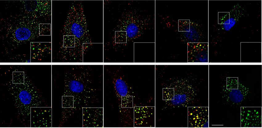
ARTICLE                                                                  NATURE COMMUNICATIONS | https://doi.org/10.1038/s41467-021-27705-7

D
         evelopment of the neocortex relies on neural stem cells              proliferation defects can be rescued by expressing a
         called radial glial (RG) cells, that generate the majority of        megalencephaly-causing mutated cyclin D2, indicating that
         cortical neurons1. Neuronal production is restricted to a            microcephaly and megalencephaly can be due to opposite effects
short period during which all excitatory neurons are produced2.               of the proliferation rates of radial glial cells.
This leaves little room for compensatory mechanisms to occur,
and alterations during this critical period lead to brain
                                                                              Results
malformations3. Indeed, the developing neocortex is highly sen-
                                                                              WDR81 KO mice display reduced brain size and altered neu-
sitive to perturbations, and a large number of mutations have
                                                                              ronal positioning. In mice, two WDR81 isoforms have been
been described to specifically alter its growth, but not that of
                                                                              identified. A long isoform (Isoform 1, 210 kDa) encompassing an
other organs4.
                                                                              N-terminal BEACH domain, a central transmembrane region,
   Primary microcephaly is a severe neurodevelopmental disorder
                                                                              and a C-terminal WD40 repeat domain; and a shorter isoform
characterized by a head circumference that is more than 3 stan-
                                                                              (Isoform 2, 81 kDa) lacking the BEACH domain (Fig. 1a). Mea-
dard deviations (SD) below the mean5. The major molecular
                                                                              surements of mRNA isolated from embryonic E14.5 cortex
cause of microcephaly lies in defects in centrosome number6,7,
                                                                              extracts indicated that WDR81 isoform 1 was highly dominant,
maturation8 and mitotic spindle regulation9–11, leading to
                                                                              with only trace levels of the short isoform (Fig. 1b). Isoform 1
apoptotic cell death. In fact, apoptosis appears to be the leading
                                                                              expression gradually increased from E12.5 to E16.5 (Supple-
cause of microcephaly in animal models, irrespective of the
                                                                              mentary Fig. 1a). We generated two WDR81 KO mice using
upstream affected molecular pathway12–14. Reduced proliferation
                                                                              gRNAs targeting the beginning of exon 1 (KO-1, affecting iso-
rates of progenitors, while proposed to be a putative cause of
                                                                              form 1), and the end of exon 1 (KO-2, affecting both isoforms)
microcephaly15, has received much less experimental support.
                                                                              (Fig. 1a). Both lines displayed frameshifts leading to the appear-
One notable example is the gene encoding IGFR1, which is
                                                                              ance of a premature STOP codon (Supplementary Fig. 1b). QPCR
mutated in syndromic forms of microcephaly, and when deleted
                                                                              measurements in KO1 did not reveal any upregulation of isoform
in mouse leads to reduced proliferation and small brain size16,17.
                                                                              2, indicating an absence of compensation (Fig. 1b). Moreover, a
   On the opposite end of the spectrum, megalencephaly (MEG)
                                                                              strong reduction of isoform 1 mRNA levels was observed, likely
is a neuronal disorder characterized by brain overgrowth (3 SD
                                                                              due to non-sense mRNA decay (Fig. 1b). WDR81 homozygote
over the mean)18. The causes of megalencephaly are diverse, but
                                                                              mutant embryos and pups were detected at sub-mendelian rates,
activating mutations in the Pi3K-AKT-mTOR and the Ras-
                                                                              and did not live for more than 21 days (Supplementary Fig. 1c).
MAPK pathways have been identified as important underlying
                                                                                 We then analyzed brain size and organization in WDR81−/−
events19–21. Mouse and cerebral organoid models for these
                                                                              pups. Both KO lines were severely microcephalic, with a reduced
activated pathways demonstrated increased proliferation of
                                                                              hemisphere area at P7 (Fig. 1c, d). Cortical thickness was also
radial glial cells leading to tissue overgrowth22–24. Stabilizing
                                                                              greatly reduced (by ~54%), suggesting defects both in tangential
mutations in the downstream target Cyclin D2 were also
                                                                              and radial expansion of the brain (Fig. 1e, f). As in patients,
reported, and its ectopic expression in mouse brain stimulated
                                                                              microcephaly was present at birth and progressed, with reduced
progenitor proliferation25.
                                                                              cortical thickness and reduced number of NEUN+ neurons at P0
   The EGF receptor (EGFR) and its ligands are major regulators
                                                                              (Fig. 1f, g, h). We next analyzed neuronal positioning in
of tissue growth26. Accordingly, knock-out of EGFR leads to a
                                                                              WDR81−/− P7 cortices. The localization of upper layer late-
dramatic atrophy of the cerebral cortex27. Progenitor cells appear
                                                                              born neurons was severely affected, with a large number of
to become responsive to EGF at mid-neurogenesis, while at ear-
                                                                              CUX1-positive neurons dispersed throughout the cortex (Fig. 1i).
lier stages they rather exhibit FGR2 dependence28. Endosomal
                                                                              Deeper neurons, which are born earlier during cortical develop-
trafficking of EGFR plays a major role in the regulation of its
                                                                              ment were however correctly positioned, as indicated by the
activity: while most EGFR signaling is believed to occur at the
                                                                              localization of CTIP2-positive neurons (Fig. 1j). Overall, mice
plasma membrane, internalization of EGFR is critical for signal
                                                                              knocked out for WDR81 have reduced brain size and altered
termination29. Following endocytosis, internalized cargos follow
                                                                              neuronal positioning, largely recapitulating the microcephaly and
different trafficking routes including recycling towards the plasma
                                                                              lissencephaly phenotypes reported in humans. These phenotypes
membrane or delivery to lysosomes for degradation30. Phospha-
                                                                              were observed for both WDR81 KO lines and we therefore next
tidylinositols (PtdIns) are major regulators of this process,
                                                                              focused our analysis on KO-1 (referred to as WDR81−/− from
defining endosomal compartment identity. Early endosomes are
                                                                              here on).
characterized by the presence of the small GTPase RAB5 and
PtdIns3P, and late endosomes by RAB7 and PtdIns(3,5)P231.
Recently, WDR81 and its partner WDR91 were shown to act as                    WDR81 KO alters radial glial progenitor proliferation. To
negative regulators of class III phosphatidylinositol 3-kinase                identify the causes of reduced brain size in WDR81−/− pups, we
(PI3K)-dependent PtdIns3P generation, therefore promoting                     tested for alterations of neocortex development at embryonic
early to late endosomal conversion32. In WDR81 knock-out (KO)                 stages. To test for proliferation defects, we first measured the
HeLa cells, endosomal maturation defects led to delayed EGFR                  mitotic index of WDR81−/− radial glial progenitors, as defined by
degradation32.                                                                the percentage of phospho-Histone H3 (PH3)-positive cells out of
   We recently reported compound heterozygous mutations in                    total PAX6-positive cells. While at E12.5, proliferation appeared
the human WDR81 gene, that result in severe microcephaly at                   normal, mitotic index of WDR81−/− radial glial progenitors was
birth that progresses in the first years of life, associated with              severely reduced at E14.5 and E16.5 (Fig. 2a). Strikingly, TBR2-
reduced gyrification of the neocortex33. Here, we generated a                  positive intermediate progenitors appeared to cycle normally
knock-out mouse model that largely recapitulates the human                    throughout development (Fig. 2b). Therefore, WDR81 mutation
phenotype. Mutant brains are not only smaller but also display                specifically alters proliferation of radial glial progenitors at mid
altered neuronal layering. We demonstrate that microcephaly is                and late neurogenic stages. We next analyzed further these pro-
the result of reduced proliferation rates of radial glial progeni-            liferation defects and measured the percentage of cells in S phase.
tors, but not of cell death. Mechanistically, we show that WDR81              A 30-minute BrdU pulse revealed an increased amount of
mutation delays EGFR endosomal trafficking and leads to                        WDR81−/− radial glial progenitors in S-phase (Fig. 2c, d). To test
reduced activation of the MAPK signaling pathway. These                       whether this was due to a longer duration of S-phase, we

2                 NATURE COMMUNICATIONS | (2022)13:16 | https://doi.org/10.1038/s41467-021-27705-7 | www.nature.com/naturecommunications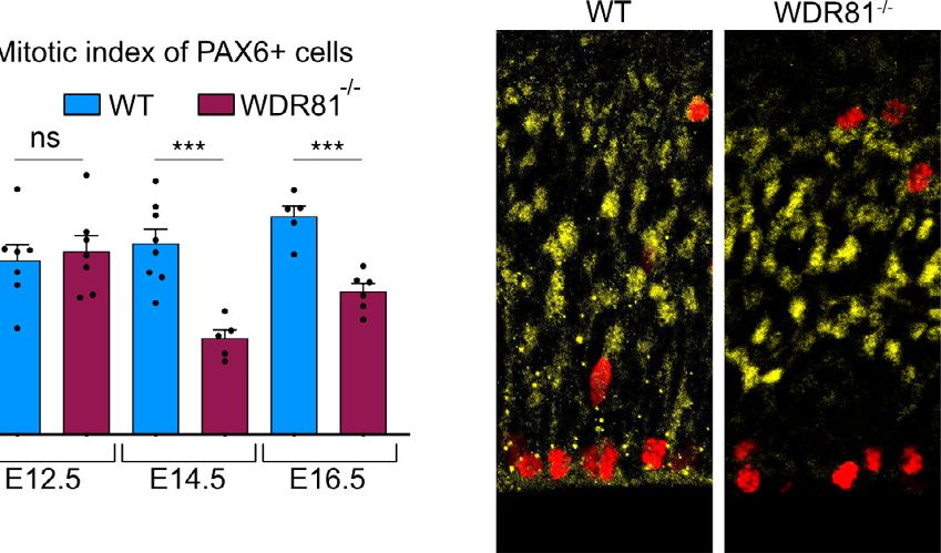
NATURE COMMUNICATIONS | https://doi.org/10.1038/s41467-021-27705-7                                                                               ARTICLE

Fig. 1 WDR81 KO mice display reduced brain size and altered neuronal positioning. a Schematic representation of mouse WDR81 isoforms and predicted
structure. b Quantification of WDR81 isoforms 1 and 2 mRNA levels in WT (p < 0.0001) and WDR81−/− (p = 0.0002) E14.5 cortices (n = 3 independent brains
for each genotype). Isoform 1 (ISO1) is the dominant isoform and its levels are strongly reduced in WDR81−/− cortices. c WDR81−/− postnatal day 7 brains are
microcephalic and display reduced cortical surface area as compared to WT brains. d Quantification of hemisphere area at P7 in WT and WDR81 KO1 brains
(p < 0.0001) (n = 4 independent brains for each genotype). e DAPI staining of P7 WT and WDR81−/− cross sections reveals reduced cortical thickness in
mutants. f Quantification of cortical thickness in WT, KO1 and KO2 brains at P0 and P7 (At P0, WT vs KO1 p = 0.0112; WT vs KO2 p = 0.0158. At P7, WT vs KO1
p < 0.0001; WT vs KO1 p < 0.0001) (n = 4 independent brains for each genotype and stage). g NeuN staining of WT and WDR81−/− cortical plates (CP) at P0.
h Quantification of NEUN+ cells in WT and WDR81 KO1 cortical plates at P0 in 600 × 300 μm crops reveals reduced number of neurons at birth (p = 0.0002)
(n = 3 independent brains for each genotype). i CUX1 staining in P7 WT and WDR81−/− cortices. Quantification of CUX1+ neuronal positioning reveals dispersion
throughout the thickness of the neocortex (Bin 5, p = 0.0003) (n = 5 independent brains for each genotype). j CTIP2 staining in P7 WT and WDR81−/− cortices.
Quantification does not neuronal positioning defects, with CTIP2+ neurons still concentrated in the third bin (n = 5 independent brains for each genotype). i, j WT
and mutant cortices were divided into 5 bins of equal size to measure neuronal relative positioning, independently of cortical thickness. All data are expressed as
mean ± standard deviation (SD). *p < 0.05; **p < 0.01; ***p < 0.001; ****p < 0.0001 by two-tailed unpaired t tests.

NATURE COMMUNICATIONS | (2022)13:16 | https://doi.org/10.1038/s41467-021-27705-7 | www.nature.com/naturecommunications                                           3
ARTICLE                                                                 NATURE COMMUNICATIONS | https://doi.org/10.1038/s41467-021-27705-7

performed a double BrdU-EdU pulse, in order to measure the rate              not observe any decrease in the proportion of progenitors out of the
of S-phase exit. Mice were first injected with BrdU, followed by a            total cell population throughout development indicating they did
second injection with EdU 4 h later. This assay revealed a                   not prematurely differentiate (Fig. 2g). In fact, we even detected a
decreased proportion of cells that exited S phase (BrdU+/EdU−)               reduction of the proportion of neurons at mid and late neurogenesis
in WDR81−/− brains, indicating a longer S phase in mutant                    (Fig. 2g). Finally, we analyzed apoptotic cell death in WDR81−/−
radial glial progenitors (Fig. 2e, f).                                       cortices. Staining for cleaved caspase-3 (CC3) did not reveal any
   An alternative potential cause of reduced brain size is premature         increased apoptosis, which remained almost undetectable both in
differentiation of progenitor cells. To test this, embryonic cortices        WT and mutant embryos (Fig. 2h). Therefore, reduced brain size in
were stained for PAX6 (radial glial progenitors), TBR2 (intermediate         WDR81−/− mice is not the result of premature progenitor
progenitors) and NEUN (neurons) at different developmental stages            differentiation or increased apoptotic cell death, but appears to be
and the proportion of each cell population was measured. We did              a consequence of reduced radial glial progenitor proliferation rates.

4                NATURE COMMUNICATIONS | (2022)13:16 | https://doi.org/10.1038/s41467-021-27705-7 | www.nature.com/naturecommunications

NATURE COMMUNICATIONS | https://doi.org/10.1038/s41467-021-27705-7 ARTICLE
Fig. 2 WDR81 KO alters radial glial progenitor proliferation. a PAX6 and PH3 double staining in E14.5 WT and WDR81−/− brains. Quantification of the
mitotic index of PAX6+ cells reveals decreased proliferation of WDR81−/− radial glial progenitors at E14.5 (p = 0.0005) and E16.5 (p = 0.0003) (n = 5–8
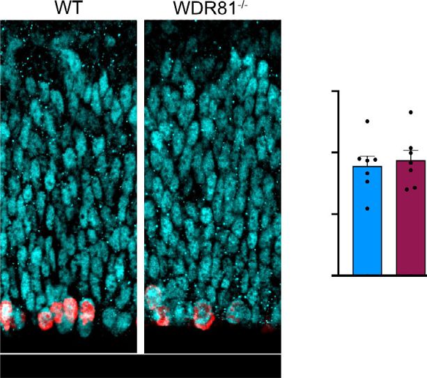
independent brains for each genotype and stage). b TBR2 and PH3 double staining in E14.5 WT and WDR81−/− brains. Quantification of the mitotic index
of TBR2+ cells indicates that proliferation of WDR81−/− intermediate progenitors in not affected (n = 3 independent brains for each genotype and stage).
c Schematic representation of the BrdU labeling experimental approach and Pax6 and BrdU staining in E14.5 WT and WDR81−/− brains. d Quantification of
the percentage of BrdU+ PAX6+ out of total PAX6 cells reveals increased number of cells in S phase in WDR81−/− radial glial progenitors at E14.5
(p = 0.0007) (n = 3 independent brains for each genotype). e Schematic representation of the BrdU/EdU double labeling experimental approach and
PAX6, BrdU and EdU staining in E14.5 WT and WDR81−/− brains. Arrowheads indicate EdU+ BrdU+ cells. f Quantification of the percentage of of BrdU+
EdU− PAX6+ out of the total BrdU+ PAX6+ cells reveals a decreased proportion of cells that exited S phase following BrdU injection in WDR81−/− radial
glial progenitors at E14.5 (p = 0.0174) (n = 3 independent brains for each genotype). g Staining for the cell fate markers PAX6 (radial glial progenitors),
TBR2 (Intermediate progenitors) NEUN (Neurons) in E14.5 WT and WDR81−/− brains, and quantification of cell fate distribution at E12.5, E14.5 (p = 0.071
for NeuN) and E16.5 (p = 0.0005 for NeuN). (n = 3–5 independent brains for each genotype, staining and stage) h Staining for Cleaved Caspase-3 (CC3)
and DAPI in E14.5 WT and WDR81−/− brains, showing an absence of apoptosis induction. All data are expressed as mean ± standard deviation (SD).
*p < 0.05; **p < 0.01; ***p < 0.001 by two-tailed unpaired t tests.
Reduced proliferation and EGFR signaling in WDR81 patient Importantly, these two functions are independent and act via the
cells. We next tested whether similar proliferation defects could WDR81 specific binding partners WDR91 and p62, respectively.
be observed in patient cells mutated for WDR81. Two mutant We therefore tested whether one of these factors affected neo-
primary fibroblast lines, derived from skin biopsies, were cortex development similarly to WDR81. To perform this, we in
analyzed and compared to two control fibroblast lines. Both utero electroporated shRNA-expressing constructs for WDR81,
patient lines (Patient1: 1882C-T/3713C-G; Patient2: 1582C-T/ WDR91 and p62 in E13.5 developing brains and analyzed cell
Del4036_4041) display compound heterozygous mutations, distribution at E17.5 (Supplementary Fig. 3a). Consistent with the
with one severe mutation (premature STOP) and one point KO data, WDR81 knock-down (KD) strongly affected neurode-
mutation leading to a single amino acid change. The mitotic velopment (Fig. 4a, b). In particular, a large fraction of KD cells
index of both patient cells was strongly decreased, mimicking accumulated in the intermediate zone (IZ), at the expense of the
the mouse radial glial progenitor phenotype (Fig. 3a, b). As an germinal zones and cortical plate. This phenotype was phe-
alternative measurement of proliferation, cells were stained for nocopied by WDR91 KD, but not by p62 KD which did not
Ki67, which also revealed a substantial decrease for both patient appear to affect cell distribution (Fig. 4a, b). These results support
cell lines (Fig. 3c, d). the endosomal function of WDR81 as a critical player for proper
In radial glial progenitors, we detected proliferation defects neocortex development.
from mid-neurogenesis (Fig. 2a), which fits with the time when We next tested whether endosomal defects could be observed
these cells start responding to EGF28. This observation suggested in WDR81 KO radial glial progenitors. Staining for various
a potential alteration in the EGFR signaling pathway. In order to endolysosomal compartments revealed a specific alteration of
test this, we monitored the activity of this signaling pathway in EEA1+ early endosomes, which appeared strongly enlarged
control and patient fibroblasts. Strikingly, we observed that the (Fig. 4c). Quantification of their size confirmed this observation,
protein levels of EGFR itself were drastically reduced in both revealing a 63% average increase (Fig. 4e). To test whether this is
patient cell lines (Fig. 3e, f). We next measured the activation of a conserved feature of WDR81 patient cells, we measured early
the mitogen-activated protein kinase (MAPK) signaling pathway endosome size in mutant fibroblasts. Again, EEA1+ endosomes
in response to EGF stimulation, perfomed after a short (2H) EGF were found to be swollen, with an increased proportion of large
starvation. Consistent with the decreased levels of EGFR, the endosomes (>0.5 µm) (Fig. 4d, f). These results are consistent
phosphorylation of ERK was reduced in both patient fibroblasts with previous observations made in KO HeLa cells and
following EGF pulse (Fig. 3g, h, i and Supplementary Fig. 2a, b). demonstrate a role for WDR81 in negative regulation of Class
No effect on the levels of phospho-AKT was detected, suggesting III Pi3K32.
no effect on the PI3K pathway (Supplementary Fig. 2c). There- Because these endosomal defects are a potential cause of altered
fore, WDR81 patient cells display reduced EGFR levels, leading to EGFR signaling, we next tested whether EGFR endosomal
a reduced activation of the MAPK signaling pathway upon EGF trafficking was affected in WDR81 mutant cells. Cells were first
stimulation. starved for 24 h to restore EGFR to the levels of control cells (See
The levels of EGFR are known to be tightly regulated, through Fig. 3j), and subsequently pulsed with fluorescent EGF555, to
complex feedback loops and the balance between recycling and monitor internalization and clearance of EGF-bound EGFR. In
degradation of the internalized receptor34. We therefore asked both patient cells, EGF555 was shown to accumulate longer within
whether reduced EGFR levels were a consequence of defects EEA1+ early endosomes (Fig. 4g). Quantification of the
within the EGFR pathway itself. To test this, we performed a colocalization between EGF555 and EEA1 revealed that this delay
long-term EGF starvation (24H) in order to abolish EGFR was particularly important 120 minutes after EGF internalization
signaling, and subsequently measured the levels of EGFR. In (Fig. 4h). We confirmed these defects in vivo, where EGFR was
patient cells, starvation rescued EGFR levels to the ones of control observed to accumulate in larger and more numerous intracel-
cells (Fig. 3j, k). These results indicate that reduced EGFR levels lular foci within KO radial glial progenitors (Supplementary
are only seen when the pathway is activated. Given that EGFR Fig. 3b, c, d). Finally, we tested if endosomal maturation defects
internalization upon EGF binding is a major regulator of the also led to impaired recycling, using a Transferrin uptake assay.
pathway, this data points towards intracellular processing defects Transferrin546 was observed to be strongly delayed into EEA1+
of EGFR. early endosomes of patient cells after 1 h, indicating defective
processing of the Transferrin receptor (Supplementary Fig. 3e, f).
WDR81 is required for endosomal trafficking of EGFR. Overall, these results show that WDR81 is critical for endosomal
WDR81 is known to regulate endosomal maturation as well as homeostasis and trafficking of internalized EGFR following EGF
autophagic clearance of aggregated proteins (aggrephagy)32,35. binding.
NATURE COMMUNICATIONS | (2022)13:16 | https://doi.org/10.1038/s41467-021-27705-7 | www.nature.com/naturecommunications 5
ARTICLE                                                                   NATURE COMMUNICATIONS | https://doi.org/10.1038/s41467-021-27705-7

Fig. 3 Reduced proliferation and EFGR signaling in WDR81 patient cells. a PH3 and DAPI staining in control and WDR81 patient fibroblasts.
b Quantification of the percentage of PH3+ cells reveals decreased mitotic index in patient cells (control1-patient1 p = 0.0051; control1-patient2
p = 0.0043; control2-patient1 p = 0.0015; control2-patient2 p = 0.0016) (n = 3 independent experiments). c Ki67 and DAPI staining in control and
WDR81 patient fibroblasts. d Quantification of the percentage of Ki67+ cells shows decreased proliferation in patient cells (control1-patient1 p = 0.0207;
control1-patient2 p = 0.0476; control2-patient1 p = 0.0245; control2-patient2 p = 0.0473) (n = 3 independent experiments). e Western Blot for EGFR in
control and WDR81 patient fibroblasts (n = 3 independent experiments). f Quantification reveals a strong reduction of EGFR levels in patient cells
(control1-patient1 p = 0.0017; control1-patient2 p = 0.0004) (n = 5 independent experiments). g Time course of EGFR and P-ERK levels in control1 and
WDR81 patient1 fibroblasts following an EGF pulse. h Quantification of EGFR levels, normalized to control levels at T0 in control1 and patient1 (T0
p = p < 0.0001; T5 p = p < 0.0001; T15 p = 0.0005; T30 p < 0.0001; T60 p = 0.0075) (n = 5 independent experiments). i Quantification of P-ERK levels,
normalized to control levels at T5 in control1 and patient1 (T5 p < 0.0001; T30 p < 0.0001) (n = 5 independent experiments). j Western Blot for EGFR in
control and WDR81 patient fibroblasts at steady state (+EGF) and cultivated for 24H in the absence of EGF. k Quantification reveals a restoration of EGFR
levels following starvation (at steady state, control1-patient1 p = 0.0161; control1-patient2 p = 0.0368; control2-patient1 p = 0.0075; control2-patient2
p = 0.0176) (n = 4 independent experiments). All data are expressed as mean ± standard deviation (SD). *p < 0.05; **p < 0.01; ***p < 0.001; ****p < 0.0001
by two-tailed unpaired t tests.

Megalencephaly-causing mutation rescues progenitor pro-                        during development18. Major causes include gain-of-function
liferation in WDR81 mutant brains. Our results indicate that                   mutations in AKT3 and its downstream target Cyclin D225,36.
trafficking defects of EGFR can arise from mutations in WDR81,                  Together, these data suggest that microcephaly and megalencephaly
and lead to reduced activation of the MAPK signaling pathway.                  can be the consequence of opposite effects on the proliferation rates
They further show that reduced radial glial progenitor proliferation           of radial glial progenitors. To further test this, we analyzed the effect
is a cause of primary microcephaly. Megalencephaly is characterized            of a megalencephaly-causing Cyclin D2Thr280Ala mutant on the
by brain overgrowth and can be due to increased cell proliferation             proliferation of WDR81 KO radial glial progenitors. Degradation-

6                  NATURE COMMUNICATIONS | (2022)13:16 | https://doi.org/10.1038/s41467-021-27705-7 | www.nature.com/naturecommunications
NATURE COMMUNICATIONS | https://doi.org/10.1038/s41467-021-27705-7                                                                    ARTICLE

resistant Cyclin D2Thr280Ala, WT Cyclin D2 or a control vector were             progenitor proliferation rates (Fig. 5a, b). Strikingly, expression of
expressed using in utero electroporation in WT and WDR81-                       degradation-resistant Cyclin D2Thr280Ala in WDR81−/− brains res-
mutant mice brains at E14.5, and the mitotic index of PAX6 cells                cued the mitotic index reduction (Fig. 5a, b). WT cyclin D2 was also
was measured at E16.5. Following expression of the control empty                able to rescue proliferation, although to a lesser extent, indicating
vector, we confirmed the reduced mitotic index in WDR81−/−                       that large amounts of this protein, either due to overexpression or
brains (Fig. 5a, b). Moreover, expression of Cyclin D2Thr280Ala in              impaired degradation is able to restore proliferation (Fig. 5a, b).
WT brain increased mitotic index, indicating that this                          Together, these results indicate that a megalencephaly-causing
megalencephaly-causing mutation indeed stimulates radial glial                  mutation can overcome the effect of a microcephaly-causing

NATURE COMMUNICATIONS | (2022)13:16 | https://doi.org/10.1038/s41467-021-27705-7 | www.nature.com/naturecommunications                              7
ARTICLE NATURE COMMUNICATIONS | https://doi.org/10.1038/s41467-021-27705-7
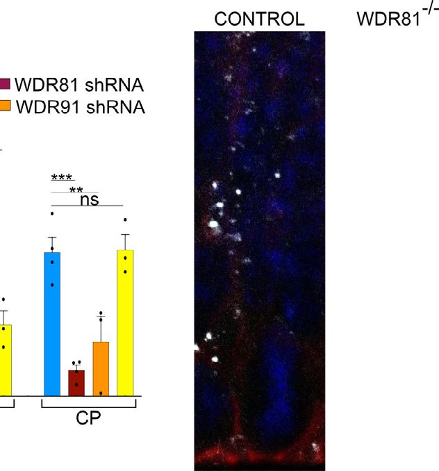
Fig. 4 WDR81 is required for endosomal trafficking of EGFR. a Expression of control shRNA and shRNA-mediated knockdown constructs for WDR81,
WDR91 and p62, together with pCAG-GFP. Plasmids were delivered by in utero electroporation at E13.5 and analysis was performed at E16.5. Ventricular
Zone and Sub-Ventricular Zone (VZ + SVZ), Intermediate Zone (IZ) and Cortical Plate (CP) were identified based on DAPI staining. b Quantification of
electroporated cell distribution reveals major accumulation in the IZ following WDR81 and WDR91 knockdown (VZ + SVZ, WDR91 shRNA p = 0.0025; IZ,
WDR81 shRNA p = 0.0001, WDR91 shRNA p = 0.067; CP, WDR81 shRNA p = 0.0003, WDR91 shRNA p = 0.0234) (n = 3–4 independent brains per
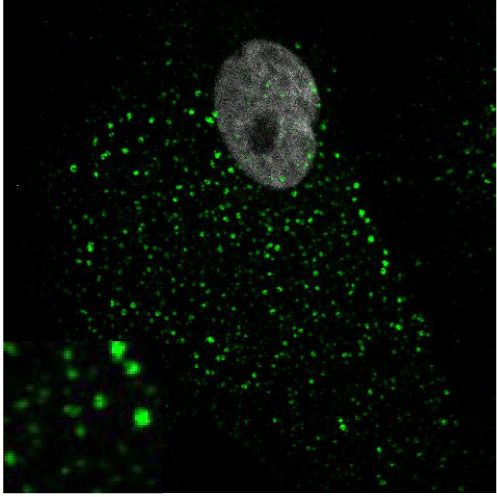
condition). c Ventricular zone of E14.5 WT and WDR81−/− mice cortices stained for EEA1 and Actin. d Control and WDR81 patient fibroblasts stained for
EEA1. e Quantification of individual EEA1+ early endosomes in WT and WDR81−/− VZ in 200 × 100 μm crops reveals increased size in mutant brains
(p < 0.0001) (n = 3 independent experiments). f Quantification of individual EEA1+ early endosomes in control and WDR81 patient fibroblasts reveals
increased size in mutant cells (For all control-patient couples p < 0.0001) (n = 3 independent experiments). g EGF555 uptake assay in control and WDR81
patient fibroblasts, and stained for EEA1. h Quantification of EGF555 and EEA1 colocalization during EGF555 uptake reveals prolonged colocalization between
EGF and early endosomes in WDR81 patient cells (At T15, C1 vs P2 p = 0.0158; C2 vs P1 p < 0.0001; C2 vs P2 p < 0.0001. At T30, C2 vs P1 p = 0.0149, C2
vs P2 p = 0.0033. At T60, C1 vs P1 p < 0.0001; C1 vs P2 p = 0.001; C2 vs P1 p = 0.0028. At T120, C1 vs P1 p = 0.0008; C1 vs P2 p = 0.0011; C2 vs P1
p = 0.0072; C2 vs P2 p = 0.0148. At T360, C1 vs P1 p = 0.0349; C2 vs P1 p < 0.0001. C2 vs P2 p = 0.0001) (n = 7 independent experiments). All
experiments were performed at least three independent times. All data are expressed as mean ± standard deviation (SD). *p < 0.05; **p < 0.01;
***p < 0.001; ****p < 0.0001 by two-tailed unpaired t test (A & H) and Mann–Whitney tests (E & F).
mutation on the proliferation of radial glial progenitors. These two described, reduced proliferations rates have received little experi-
pathologies can therefore arise from a highly related cause: an mental support16. In mouse, RG cells produce eight to nine
imbalance in cell cycle regulation leading either to reduced brain neurons during a short neurogenic period, before differentiating2.
growth or to brain overgrowth (Fig. 5c). We show here that WDR81 mutation does not affect the modes of
division of RG cells nor cell survival, but act solely through a
reduction of their proliferation rate, leading to reduced brain size.
Discussion This highlights the absence of compensatory mechanisms in the
In this study, we investigated the mechanisms by which mutation developing neocortex, where all neurons must be produced in a
in the WDR81 gene leads to severe microcephaly in patients. We short temporal window. During corticogenesis, G1 lengthening is
show that KO mouse recapitulates many features of the pheno- associated with increased neurogenic divisions at the expense of
type previously observed in patients and that the endosomal symmetric amplifying divisions41,42. We did not detect such cell
maturation function of WDR81 is critical for neocortex devel- fate changes in WDR81−/− brains. This is likely due to the fact
opment. WDR81 is required for endosomal clearance of inter- that the proliferation rate of mutant RG cells is only affected at a
nalized EGFR and normal activation of the mitogenic MAPK stage where the vast majority of cells already perform neurogenic
signaling pathway. In the absence of WDR81, the proliferation divisions1. At the macroscopic level, microcephaly and mega-
rate of radial glial cells is affected, leading to reduced brain size. lencephaly appear as opposite phenotypes. We show here that they
Importantly, cell death does not appear to contribute to this can be due to opposite effects on the proliferation rates of RG cells,
phenotype. Proliferation defects can be rescued by the expression and can therefore be viewed as two sides of the same coin.
of a megalencephaly-causing mutated cyclin D2, highlighting a
tight functional link between these two pathologies. Methods
Membrane trafficking has been poorly investigated in radial Animals. All experiments involving mice were carried out according to the
glial cells, albeit its predicted implication in many important recommendations of the European Community (2010/63/UE). The animals were
processes including cargo polarized transport, secretion of bred and cared for in the Specific Pathogen Free (SPF) Animal Facility of Institut
Curie (agreement C 75-05-18). All animal procedures were approved by the ethics
extracellular matrix components, or endocytic processing of committee of the Institut Curie CEEA-IC #118 and by French Ministry of Research
surface receptors for lysosomal degradation or recycling. We (2016-002). Animals were housed at a temperature of 22 °C, 50% humidity and a
show here that the endosomal maturation machinery plays a 12/12 hour light/dark cycle.
critical role in the processing of internalized EGFR in RG cells,
and is required for their proliferation. Neurogenesis depends on Guide RNA selection and preparation. gRNA sequences targeting exon 1 of
EGFR activity, with radial glial cells becoming responsive to EGF WDR81 have been identified and selected using the online software CRIPSOR
(crispor.tefor.net). Forward and reverse oligonucleotides were annealed and cloned
from mid-neurogenesis27,28. We find that WDR81 expression into px330 plasmid. To generate Cas9 mRNA and gRNA, in vitro transcriptions
rises at from E14.5 and that KO RG cells are specifically affected were performed on the Cas9 pCR2.1-XL plasmid and gRNA plasmids, using the
at E14.5 and E16.5 stages of development. Why the proliferation mMESSAGE mMACHINE T7 ULTRA kit and the MEGAshortscript T7 kit (Life
rate of IPs was not affected is unclear but EGF is secreted into the Technologies), respectively. Cas9 mRNA and sgRNAs were then purified using the
MEGAclear Kit (Thermo Fisher Scientific) and eluted in RNAse-free water. The
cerebrospinal fluid from the choroid plexus and apical contact gRNA and Cas9mRNA quality were evaluated on agarose gel.
may be critical for responsiveness37,38. EGFR was previously
reported to be asymmetrically inherited during radial glial cell Generation of WDR81 knock-out mice. Eight-week-old B6D2F1 (C57BL/6 J ×
division, generating a daughter cell with higher proliferative DBA2) females from Charles River France, were superovulated by intraperitoneal
potential37. Later in development, EGFR also acts as an important (i.p.) administration of 5 IU of Pregnant Mare Serum Gonadotropin followed by an
regulator of astrocyte differentiation39. Our data point to the additional i.p. injection of 5 IU Human Chorion Gonadotropin 48 h later. Females
intracellular processing of EGFR as an important level of control were mate to a stud male of the same genetic background. Cytoplasmic micro-
injection was performed into mouse fertilized oocytes using Cas9 mRNA and
for the regulation of proliferation in radial glial cells. WDR81 is sgRNA at 100 ng/μl and 50 ng/μl, respectively in Brinster buffer (10 mM Tris-HCl
likely to affect the trafficking of other cargos40, which may also pH 7.5; 0.25 mM EDTA). Microinjected zygotes were cultured in Cleave medium
impact radial glial cell proliferation. Moreover, the trafficking of (Cook, K-RVCL-50) at 37 °C under 5% CO2 and then implanted at one cell stage
neuronal cargos, such as adhesion molecules, is likely to lead to into infundibulum of E0.5 NMRI pseudo-pregnant females (25–30 injected zygotes
per female). According to the genotyping strategy, 3 mice showed modified allele
the altered neuronal positioning observed in KO mice, and to the out of a total of 22 pups. The founders were then backcrossed to C57BL6/J.
lissencephaly phenotype in human.
In principle, microcephaly can be the consequence of premature Genotyping WDR81−/− animals. Mice DNA was extracted from a piece of ear
progenitor differentiation, reduced proliferation rates, or cell (adult) or tail (dissected embryos), put at 96 degrees in lysis tampon overnight. The
death. While centrosomal defects leading to apoptosis have been DNA was then amplified via PCR using WDR81 specific primers: for KO1
8 NATURE COMMUNICATIONS | (2022)13:16 | https://doi.org/10.1038/s41467-021-27705-7 | www.nature.com/naturecommunications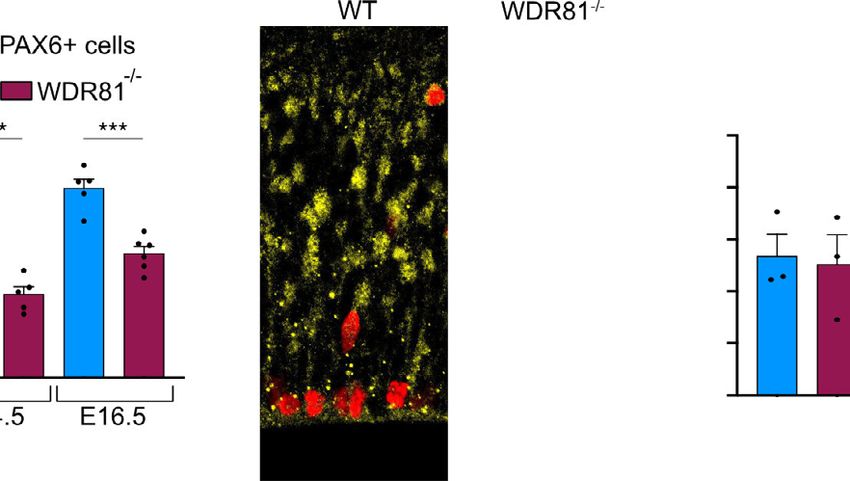
NATURE COMMUNICATIONS | https://doi.org/10.1038/s41467-021-27705-7                                                                                ARTICLE

Forward: GGCGGAAAGTGGTTCTTACA, Reverse: AGCCACCTCCTGCATG                           Real-time reverse-transcription PCR. Wild type and WDR81−/− cortices were
AACC; for KO2 Forward: GGCTTGTAGTGGTTCTGTAC, Reverse: GATCC                        dissected at E14.5 in 1 ml of TRIZOL (Thermo Fisher 15596026). The mRNA was
TTCTGCATTCCAA. For KO1 the amplicon was purified using the nucleospin               isolated as follows: TRIZOL+ sample solution was exposed to chlorophorm for
purification kit (Machenery and Nagel) and then exposed to the restriction enzyme   7 min at room temperature and centrifuged at 15.000 g for 30 min at 4 degrees. The
AfeI (New England Bioscience). The restriction enzyme only cuts the mutant DNA     translucent solution formed was then transferred in 1 ml of isopropanol, incubated
giving rise to two DNA pieces of 200 bp. For KO2, the amplicon was sanger-         7 min at room temperature and centrifuged at 4 degrees 10.000 g. The pellet of
sequenced using the GATC-Eurofins platform.                                         nucleic acid formed was then washed in ethanol 70% and centrifuged 5 min at

NATURE COMMUNICATIONS | (2022)13:16 | https://doi.org/10.1038/s41467-021-27705-7 | www.nature.com/naturecommunications                                             9
ARTICLE                                                                            NATURE COMMUNICATIONS | https://doi.org/10.1038/s41467-021-27705-7

Fig. 5 Undegradable Cyclin D2Thr280Ala rescues WDR81−/− proliferation index. a Expression of Cyclin D2, Cyclin D2Thr280Ala and empty vector in WT
and WDR81−/− brains. Constructs were in utero electroporated at E14.5, and brains were fixed at 16.5 and stained for PAX6 and PH3. b Quantification of
the percentage of mitotic (PH3+) electroporated radial glial cells (PAX6+) out of total electroporated radial glial cells reveals rescue of mitotic index in
WDR81−/− cells expressing Cyclin D2Thr280Ala (WT-empty vector vs WT-Cyclin D2Thr280Ala p = 0.0382; WT-empty vector vs WDR81−/− empty vector
p = 0.0247; WDR81−/− empty vector vs WDR81−/− Cyclin D2 p = 0.031; WDR81−/− empty vector vs WDR81−/− Cyclin D2Thr280Ala p = 0.0023) (n = 3
independent brains per genotype and condition). c Model. WDR81 loss of function leads to reduced activation of the MAPK signaling pathway downstream
of EGFR, to reduced radial glial progenitor proliferation, and to microcephaly. Gain of function in the Pi3K-AKT pathway or stabilizing mutations in Cyclin
D2 lead to increased radial glial progenitor proliferation, and to megalencephaly. Cyclin D2 mutants can rescue proliferation defects in WDR81−/− brains,
indicating that these two pathologies can arise from opposite effects on the proliferation rates of radial glial progenitor. All data are expressed as
mean ± standard deviation (SD). *p < 0.05; **p < 0.01 by two-tailed unpaired t tests.

10.000 g at 4 degrees. The pellet was then resuspended in water. The nucleic acids      45 min at room temperature in blocking solution with Alexa Fluor coupled sec-
solution was purified from DNA using TURBO DNA-free Kit (Thermofisher). The               ondary antibodies. Cells were then washed and mounted.
mRNA obtained was then retrotranscribed using the RT reverse transcription Kit
(Thermofisher). Real time RT-PCR was performed using the qPCR Master Mix kit
                                                                                        Antibodies. Primary antibodies used: mouse anti Ctip2 (Abcam ab18465, 1/300),
(Thermofisher) and the WDR81, WDR91 and p62 Forward/Reverse primers;
                                                                                        rabbit anti Pax6 (Biolegend 901301 1/500), Sheep anti TBR2/EOMES (R&D system
GAPDH gene was used for internal control and for normalization. Primers used for
                                                                                        AF6166 1/500), rabbit anti NEUN (Abcam ab177487 1/1000), goat anti Phospho
WDR81 isoform 1 are forward: AGTGGATCCTTCAGACAGCC, Reverse: GAAG
                                                                                        Histone3 (Santa Cruz SC-12927 1/1000), rabbit anti CUX1 (Santa Cruz, dis-
CCAGCCACAACACTC. Primers used for WDR81 isoform 2 are Forward: AGTG
                                                                                        continued 1/100), rabbit anti BRDU (Abcam AB152095 1/500), rabbit cleaved
GATCCTTCAGACAGCC, Reverse: CTGACTTGTAGTGGTGCGTG. Primers
                                                                                        caspase-3 (CST 9661, 1/2000), rabbit anti Ki67 (abcam ab15580, 1/500), rabbit anti
used for WDR91 are forward: AGTGCTGAGCCAAGAAGAGT, Reverse: CTAG                         EGFR (CST 4267, 1/100), mouse anti p-ERK (CST 9106, 1/200), rabbit anti GAPDH
GGAGAGCAGTGGTGAC. Primers used for p62 are forward: TATCTTCTGGG
                                                                                        (Sigma–AldrichG9545, 1/5000), anti p-AKT (CST 4060, 1/200) and mouse anti
CAAGGAGGA, Reverse: TGTCAGCTCCTCATCACTGG.
                                                                                        EEA-1 (BD biosciences 610457, 1/500). Secondary antibodies used: donkey Alexa
                                                                                        Fluor 488 anti-mouse (1/250), anti-rabbit, anti-goat (Jackson laboratories 715-545-
                                                                                        150, 711-165-152, 715-605-152), donkey Alexa Fluor 555 (1/250) anti-mouse, anti-
In utero electroporation of mouse embryonic cortex. Pregnant mice at
                                                                                        rabbit, anti-goat (Jackson laboratories 715-545-150, 711-165-152, 715-605-152),
embryonic day 13.5 or 14.5 were anesthetized with isoflurane gas, and injected
                                                                                        donkey Alexa Fluor 647 (1/250) anti-mouse, anti-rabbit, anti-goat (Jackson
subcutaneously first with buprenorphine (0.075 mg/kg) and a local analgesic,
                                                                                        laboratories 715-545-150, 711-165-152, 715-605-152).
bupivacaine (2 mg/kg), at the site of the incision. Lacrinorm gel was applied to the
eyes to prevent dryness/irritation during the surgery. The abdomen was shaved and
disinfected with ethanol and antibiotic swabs, then opened, and the uterine horns       Expression constructs and shRNAs. For WDR81 knockdown experiments,
exposed. Plasmid DNA mixtures were used at a final concentration of 1 µg/µl per          WDR81 shRNA was provided by GenecopoeiaTM. The small interfering RNA
plasmid, dyed with Fast Green and injected into the left lateral ventricle of several   sequence was ggagataagcaattggacttc and was cloned in psi-mU6.1 vector coex-
embryos. The embryos were then electroporated through the uterine walls with a          pressing mcherryFP. For WDR91 knockdown experiments shRNA was provided
NEPA21 Electroporator (Nepagene) and a platinum plated electrode (5 pulses of           by Tebu-bio (217MSH024100-mU6). For p62 knockdown experiments shRNA was
50 V for 50 ms at 1 second intervals). The uterus was replaced and the abdomen          provided by Tebu-bio (217CS-MSH079315-mU6-01). Constructs were co-injected
sutured. The mother was allowed to recover from surgery and supplied with               with GFP-pCagIG (Addgene 11159) at a concentration of 1 ug/ul. Plasmids were
painkillers in drinking water post-surgery. Electroporated brains were harvested at     introduced in the in vivo developing cortex by intraventricular injection and
E16.5 and E17.5.                                                                        electroporation. For validation of shRNA efficiency, Neuro2A cells (ATCC, CCL-
                                                                                        131) were transfected with WDR81, WDR91, p62 and scramble control plasmids
                                                                                        using Lipofectamin-3000 (Thermo Fisher Scientific) and lyzed in TRIZOL after
Immunostaining of brain slices. Mouse embryonic brains were dissected out of            3 days for QPCR. For WDR81 rescue experiments, Cyclin D2 and Cyclin D2
the skull, fixed in 4% Pfa for 2 h, and 80 µm-thick slices were prepared with a Leica    Thr280Ala were synthetized in vitro (Genescript). They were then cloned into
VT1200S vibratome in PBS. Slices were boiled in citrate sodium buffer (10 mM,           GFP-pCagIG (Addgene 11159) after digestion by restriction enzymes EcoRI
pH6) for 20 min and cooled down at room temperature (antigen retrieval). Slices         and EcoRV.
were then blocked in PBS-Triton X100 0.3%-Donkey serum 2% at room tem-
perature for 2 h, incubated with primary antibody overnight at 4 °C in blocking
                                                                                        EGF pulse assay, EGF555 uptake assay and Transferrin546 uptake assay. For
solution, washed in PBS-Tween 0.05%, and incubated with secondary antibody
                                                                                        EGF pulse assay, fibroblast cultures were EGF starved for two hours before the
overnight at 4 °C in blocking solution before final wash and mounting in aqua-
                                                                                        assay. EGF was added directly to the culture medium at 0.1 mg/ml. Cells were
polymount. Imaging was performed on a fully motorized spinning disk wide
                                                                                        then harvested at 0, 5, 15, 30, 60, 120 min and processed for protein extraction.
microscope driven by Metamorph software (Molecular Devices) and equipped with
                                                                                        Proteins were then mixed with 4x Leammli (Biorad) and BME solution and used
a Yokogawa CSU-W1 scanner unit to increase the field of view and improve the
                                                                                        for Western Blot analysis. For EGF555 pulse assay, fibroblast cells were cultivated
resolution deep in the sample. Image analysis, modifications of brightness and
                                                                                        on glass coverslips; cells were starved for 24 h and then exposed to 0,1 mg/ml
contrast were carried out with Fiji. Statistical analysis was carried out with Prism.
                                                                                        EGF555 (Thermo Fisher). Cells were fixed in paraformaldehyde 4% at 15, 30, 60,
Figures were assembled in Affinity Designer.
                                                                                        120, 360 min and used for immunostaining. For Transferrin546 uptake assay,
                                                                                        fibroblast cells were cultivated on glass coverslips; cells were serum-starved for
                                                                                        1 h and then exposed to at 0.1 mg/ml Transferrin546 (Thermo Fisher). Cells were
Brdu/Edu labeling. For BrDU labeling experiments, BrDU (Invitrogen B23151)
                                                                                        fixed in paraformaldehyde 4% at 15, 30, 60, 120, 360 min and used for
was injected at 50 mg/kg intraperitoneally 30 min prior to harvesting embryos. For
                                                                                        immunostaining.
BrDU/EDU labeling experiments, BrDU was injected at 50 mg/kg intraperitoneally
4 h prior to harvesting embryos, and EdU (Thermofisher Click-iT EdU Alexa Fluor
555) was injected at 50 mg/kg 30 min prior to harvesting embryos. After fixation,        Statistical analysis. Quantitative data are described as mean ± standard deviation
brain slices were incubated in 2 N HCL for 30 min at 37 degrees and then washed 3       (SD) for n ≥ 3. No data were excluded from the analyses and the experiments were
times with PBS prior to immunostaining.                                                 not randomized. Statistical analysis was performed using two-tailed unpaired
                                                                                        Student’s t test using GraphPad Prism 9 software (GraphPad Software, San Diego,
                                                                                        CA, USA). P values lower than 0.05 were considered statistically significant.
WDR81 patient cells and immunostaining. Control and WDR81 mutant primary
fibroblasts were provided by Institut Imagine, Paris. The genotype of patient 1 cells
                                                                                        Reporting summary. Further information on research design is available in the Nature
was compound heterozygote 1882C > T/3713 C > G and the genotype of patient 2
                                                                                        Research Reporting Summary linked to this article.
cells was compound heterozygote 1582 C > T/4036_4041dup. Cells were grown in
OPTIMEM + 10%FBS at 37 degrees in humid air containing 5% CO2. Fibroblasts
were fixed in 4% paraformaldehyde for 20 min, treated with 50 mM NH4Cl for               Data availability
10 min, washed three times with PBS and left in a blocking solution (PBS 1%             The original pictures of WB membranes are provided in the file “Supplementary
donkey serum 0.1% Triton X) for 30 min. Cells were then incubated 1 h at room           information_WB membranes.pdf” and all quantifications are available in the
temperature with primary antibodies, washed three times in PBS and incubated for        “Quantifications_Carpentieri et al.xlsx” file, within the Source Data File. The

10                   NATURE COMMUNICATIONS | (2022)13:16 | https://doi.org/10.1038/s41467-021-27705-7 | www.nature.com/naturecommunications
NATURE COMMUNICATIONS | https://doi.org/10.1038/s41467-021-27705-7 ARTICLE
immunofluorescence data that support the findings of this study, due to their large size, 28. Tropepe, V. et al. Distinct neural stem cells proliferate in response to EGF and
are available from the corresponding author (alexandre.baffet@curie.fr) or from the first FGF in the developing mouse telencephalon. Dev. Biol. 208, 166–188 (1999).
author (jacopocarpentieri@gmail.com) upon request. WDR81 KO mice strains will be 29. Tomas, A., Futter, C. E. & Eden, E. R. EGF receptor trafficking: consequences
shared upon request. Source data are provided with this paper. for signaling and cancer. Trends Cell Biol. 24, 26–34 (2014).
30. Cullen, P. J. & Steinberg, F. To degrade or not to degrade: mechanisms and
Received: 18 August 2020; Accepted: 30 November 2021; significance of endocytic recycling. Nat. Rev. Mol. Cell Biol. 19, 679–696
(2018).
31. Stenmark, H. Rab GTPases as coordinators of vesicle traffic. Nat. Rev. Mol.
Cell Biol. 10, 513–525 (2009).
32. Liu, K. et al. Negative regulation of phosphatidylinositol 3-phosphate levels in
early-to-late endosome conversion. J. Cell Biol. 212, 181–198 (2016).
33. Cavallin, M. et al. WDR81 mutations cause extreme microcephaly and impair
References mitotic progression in human fibroblasts and Drosophila neural stem cells.
1. Uzquiano, A. et al. Cortical progenitor biology: key features mediating Brain 140, 2597–2609 (2017).
proliferation versus differentiation. J. Neurochem. 146, 500–525 (2018). 34. Avraham, R. & Yarden, Y. Feedback regulation of EGFR signalling: decision
2. Gao, P. et al. Deterministic progenitor behavior and unitary production of making by early and delayed loops. Nat. Rev. Mol. Cell Biol. 12, 104–117
neurons in the neocortex. Cell 159, 775–788 (2014). (2011).
3. Homem, C. C. F., Repic, M. & Knoblich, J. A. Proliferation control in neural 35. Liu, X. et al. The BEACH-containing protein WDR81 coordinates p62 and
stem and progenitor cells. Nat. Rev. Neurosci. 16, 647–659 (2015). LC3C to promote aggrephagy. J. Cell Biol. 216, 1301–1320 (2017).
4. Pirozzi, F., Nelson, B. & Mirzaa, G. From microcephaly to megalencephaly: 36. Rivière, J.-B. et al. De novo germline and postzygotic mutations in AKT3,
determinants of brain size. Dialogues Clin. Neurosci. 20, 267–282 (2018). PIK3R2 and PIK3CA cause a spectrum of related megalencephaly syndromes.
5. Jayaraman, D., Bae, B.-I. & Walsh, C. A. The genetics of primary Nat. Genet. 44, 934–940 (2012).
microcephaly. Annu Rev. Genomics Hum. Genet. 19, 177–200 (2018). 37. Sun, Y., Goderie, S. K. & Temple, S. Asymmetric distribution of EGFR
6. Insolera, R., Bazzi, H., Shao, W., Anderson, K. V. & Shi, S.-H. Cortical receptor during mitosis generates diverse CNS progenitor cells. Neuron 45,
neurogenesis in the absence of centrioles. Nat. Publ. Group 17, 1528–1535 (2014). 873–886 (2005).
7. Marthiens, V. et al. Centrosome amplification causes microcephaly. Nat. Cell 38. Lehtinen, M. K. & Walsh, C. A. Neurogenesis at the brain–cerebrospinal fluid
Biol. 15, 731–740 (2013). interface. Annu Rev. Cell Dev. Biol. 27, 653–679 (2011).
8. Lizarraga, S. B. et al. Cdk5rap2 regulates centrosome function and 39. Beattie, R. et al. Mosaic analysis with double markers reveals distinct
chromosome segregation in neuronal progenitors. Development 137, sequential functions of Lgl1 in neural stem cells. Neuron 94, 517–533 (2017).
1907–1917 (2010). 40. Wang, M. et al. WDR81 regulates adult hippocampal neurogenesis through
9. Johnson, M. B. et al. Aspm knockout ferret reveals an evolutionary mechanism endosomal SARA-TGFβ signaling. Mol. Psychiatry 17, 385–16 (2018).
governing cerebral cortical size. Nature 556, 370–375 (2018). 41. Pilaz, L.-J. et al. Forced G1-phase reduction alters mode of division, neuron
10. Chen, J.-F. et al. Microcephaly disease gene Wdr62 regulates mitotic number, and laminar phenotype in the cerebral cortex. Proc. Natl Acad. Sci.
progression of embryonic neural stem cells and brain size. Nat. Commun. 5, USA 106, 21924–21929 (2009).
3885–13 (2014). 42. Lange, C., Huttner, W. B. & Calegari, F. Cdk4/cyclinD1 overexpression in
11. Hu, W. F. et al. Katanin p80 regulates human cortical development by limiting neural stem cells shortens G1, delays neurogenesis, and promotes the
centriole and cilia number. Neuron 84, 1240–1257 (2014). generation and expansion of basal progenitors. Cell Stem Cell 5, 320–331
12. Kim, S. et al. The apical complex couples cell fate and cell survival to cerebral (2009).
cortical development. Neuron 66, 69–84 (2010).
13. Mao, H., McMahon, J. J., Tsai, Y.-H., Wang, Z. & Silver, D. L.
Haploinsufficiency for core exon junction complex components disrupts Acknowledgements
embryonic neurogenesis and causes p53-mediated microcephaly. PLoS Genet We acknowledge Institut Curie, member of the French National Research Infrastructure
12, e1006282 (2016). France-BioImaging (ANR10-INBS-04) and the Nikon BioImaging Center (Institut Curie,
14. Gruber, R. et al. MCPH1 regulates the neuroprogenitor division mode by France). We thank Renata Basto, Veronique Marthiens, Iva Simeonova, Cedric Delevoye
coupling the centrosomal cycle with mitotic entry through the Chk1-Cdc25 (I. Curie) and Fiona Francis (IFM), for helpful discussions and critical reading of the
pathway. Nat. Cell Biol. 13, 1325–1334 (2011). manuscript. J.A.C. was funded by the IC3i Institut Curie doctoral program founded by
15. Reynolds, J. J. et al. Mutations in DONSON disrupt replication fork stability the European Union’s Horizon 2020 research and innovation program under the Marie
and cause microcephalic dwarfism. Nat. Genet. 49, 537–549 (2017). Sklodowska-Curie actions grant agreement and from Fondation de la Recherche Medi-
16. Lehtinen, M. K. et al. The cerebrospinal fluid provides a proliferative niche for cale (FRM). A.D.B. is an Inserm researcher. This work was supported by the CNRS,
neural progenitor cells. Neuron 69, 893–905 (2011). Institut Curie, the Ville de Paris “Emergences” program, Labex CelTisPhyBio (11-LBX-
17. Juanes, M. et al. Three novel IGF1Rmutations in microcephalic patients with 0038) and PSL university.
prenatal and postnatal growth impairment. Clin. Endocrinol. 82, 704–711
(2014).
18. Dobyns, W. B. & Mirzaa, G. M. Megalencephaly syndromes associated with Author contributions
mutations of core components of the PI3K‐AKT–MTOR pathway: PIK3CA, J.A.C. and A.D.B. conceived the project. J.A.C. & A.D.B. analyzed the data. J.A.C., J.B.B.
PIK3R2, AKT3, and MTOR. Am. J. Med. Genet. C. Semin Med. Genet. 181, & A.D.B. wrote the manuscript. J.A.C., A.D.C. and M.L. did most of the experimental
582–590 (2019). procedures. J.A.C., L.D.M. and F.E.M. created the mutant mouse lines. D.A. supervised
19. Mirzaa, G. M. & Poduri, A. Megalencephaly and hemimegalencephaly: the mouse mutant colonies and performed all the crossings. L.C. and J.A.C. set up in
breakthroughs in molecular etiology. Am. J. Med. Genet. C. Semin Med. Genet. utero electroporation experiments. N.B.B. provided biological samples of the affected
166C, 156–172 (2014). patients A.D.B. supervised the project.
20. Kang, M. & Lee, Y.-S. The impact of RASopathy-associated mutations on CNS
development in mice and humans. Mol. Brain 12, 96–17 (2019). Competing interests
21. Rauen, K. A. The RASopathies. Annu Rev. Genomics Hum. Genet. 14, 355–369 The authors declare no competing interests.
(2013).
22. Li, Y. et al. Induction of expansion and folding in human cerebral organoids.
Cell Stem Cell 20, 385–396 (2017). Additional information
23. Groszer, M. et al. Negative regulation of neural stem/progenitor cell Supplementary information The online version contains supplementary material
proliferation by the Pten tumor suppressor gene in vivo. Science 294, available at https://doi.org/10.1038/s41467-021-27705-7.
2186–2189 (2001).
24. Hegedus, B. et al. Neurofibromatosis-1 regulates neuronal and glial cell Correspondence and requests for materials should be addressed to Alexandre D. Baffet.
differentiation from neuroglial progenitors in vivo by both cAMP- and Ras-
dependent mechanisms. Cell Stem Cell 1, 443–457 (2007). Peer review information Nature Communications thanks Christian Gonzalez-Billault
25. Mirzaa, G. M. et al. De novo CCND2 mutations leading to stabilization of and the other, anonymous, reviewer(s) for their contribution to the peer review of this
cyclin D2 cause megalencephaly-polymicrogyria-polydactyly-hydrocephalus work. Peer reviewer reports are available.
syndrome. Nat. Genet. 46, 510–515 (2014).
26. Sigismund, S., Avanzato, D. & Lanzetti, L. Emerging functions of the EGFR in Reprints and permission information is available at http://www.nature.com/reprints
cancer. Mol. Oncol. 12, 3–20 (2018).
Publisher’s note Springer Nature remains neutral with regard to jurisdictional claims in
27. Threadgill, D. W. et al. Targeted disruption of mouse EGF receptor: effect of
genetic background on mutant phenotype. Science 269, 230–234 (1995). published maps and institutional affiliations.
NATURE COMMUNICATIONS | (2022)13:16 | https://doi.org/10.1038/s41467-021-27705-7 | www.nature.com/naturecommunications 11ARTICLE NATURE COMMUNICATIONS | https://doi.org/10.1038/s41467-021-27705-7
Open Access This article is licensed under a Creative Commons
Attribution 4.0 International License, which permits use, sharing,
adaptation, distribution and reproduction in any medium or format, as long as you give
appropriate credit to the original author(s) and the source, provide a link to the Creative
Commons license, and indicate if changes were made. The images or other third party
material in this article are included in the article’s Creative Commons license, unless
indicated otherwise in a credit line to the material. If material is not included in the
article’s Creative Commons license and your intended use is not permitted by statutory
regulation or exceeds the permitted use, you will need to obtain permission directly from
the copyright holder. To view a copy of this license, visit http://creativecommons.org/
licenses/by/4.0/.
© The Author(s) 2022
12 NATURE COMMUNICATIONS | (2022)13:16 | https://doi.org/10.1038/s41467-021-27705-7 | www.nature.com/naturecommunicationsYou can also read