Thyroid Hormone Receptor Beta as Tumor Suppressor: Untapped Potential in Treatment and Diagnostics in Solid Tumors
←
→
Page content transcription
If your browser does not render page correctly, please read the page content below
Preprints (www.preprints.org) | NOT PEER-REVIEWED | Posted: 23 July 2021                    doi:10.20944/preprints202107.0549.v1

                   Review

                   Thyroid Hormone Receptor Beta as Tumor
                   Suppressor: Untapped Potential in Treatment and
                   Diagnostics in Solid Tumors
                   Cole D. Davidson 1,2, Noelle E. Gillis 1,2, and Frances E. Carr 1,2*
                     1   Department of Pharmacology, Larner College of Medicine, University of Vermont Burlington VT
                         05405 USA
                     2   University of Vermont Cancer Center, Burlington VT 05401 USA
                         cole.d.davidson@uvm.edu, negillis@uvm.edu

                     * Correspondence: frances.carr@med.uvm.edu Tel.: 802-656-1318

                   Simple Summary: Dysregulation of the thyroid hormone receptor beta (TRβ) is characteristic of
                   many solid and endocrine-related tumors. Despite a recognized role as a tumor suppressor, the
                   mechanisms by which TRβ regulates tumor growth are not yet clear. As a transcription factor that
                   responds to changes in thyroid hormone levels, TRβ plays a key role in regulating many cell
                   signalling nodes that are important for maintenance of normal cell identity and tumor progression.
                   This review will address the need for a deeper understanding of TRβ tumor suppressor mechanisms
                   to inform the development of more effective thyroid cancer diagnostics and therapies.

                   Abstract: There is compelling evidence that the nuclear receptor TRβ, a member of the thyroid
                   hormone receptor (TR) family, is a tumor suppressor in thyroid, breast and other solid tumors. Cell-
                   based and animal studies reveal that the liganded TRβ induces apoptosis, reduces an aggressive
                   phenotype, decreases stem cell populations, and slows tumor growth through modulation of a
                   complex interplay of transcriptional networks. TRβ-driven tumor suppressive transcriptomic
                   signatures include repression of known drivers of proliferation such as PI3K/Akt pathway and
                   activation of novel signaling (JAK1/STAT1) and metabolic reprogramming in both thyroid and breast
                   cancers. The presence of TRβ is also correlated with a positive prognosis and response to therapeutics
                   in BRCA+ and triple-negative breast cancers respectively. Ligand activation of TRβ enhances
                   sensitivity to chemotherapeutics. TRβ co-regulators and bromodomain-containing chromatin
                   remodeling proteins are emergent therapeutic targets. This review considers TRβ as a potential
                   biomolecular diagnostic and therapeutic target.

                     Keywords: TRβ, tumor suppression, co-regulators, therapeutics

                   1. Introduction
                        Altered gene expression programming in cancer cells is often a consequence of a loss of function
                   of cell-type specific transcriptional control mechanisms. Genetic mutations and epigenetic silencing
                   of thyroid hormone receptor beta (TRβ) is characteristic of a number of solid tumors, and can be a
                   marker for dedifferentiation [1-5]. TRβ is a ligand-dependent transcription factor that responds
                   primarily to triiodothyronine (T3). TRβ is recognized as a tumor suppressor and a positive prognostic
                   indicator, however the mechanisms by which it regulates tumor growth remain unclear[6-9]. Recent
                   studies indicate that TRβ tumor suppressive effects are mediated in part through intracellular
                   signaling pathways including PI3K/Akt, Ras/MAPK, and JAK-STAT pathways, and induction of the
                   mesenchymal-to-epithelial transition. Mutations in TRβ that lead to thyroid hormone resistance have
                   also been shown to be oncogenic [10]. Transcriptional regulation by TRβ is critical for its function as
                   a tumor suppressor because it acts as both a signal transducer and facilitator of long-term epigentic

            © 2021 by the author(s). Distributed under a Creative Commons CC BY license.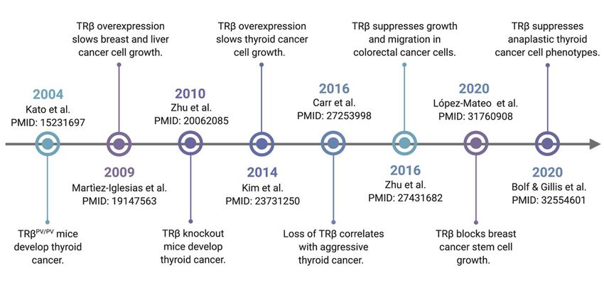
Preprints (www.preprints.org) | NOT PEER-REVIEWED | Posted: 23 July 2021 doi:10.20944/preprints202107.0549.v1
2 of 14
programming for maintenance of cell identity. The purpose of this review is to discuss recent
advances in our understanding of TRβ tumor suppression, and highlight its potential utility as a
diagnostic indicator and therapeutic target.
2.1 TRβ Satisfies the Criteria for a Tumor Suppressor
There is an abundance of evidence both in vivo and at the molecular level that highlight TRβ
tumor suppressor function. Loss of the transcription factor TRβ, a member of the thyroid hormone
receptor (TR) family, through mutation or epigenetic silencing is characteristic of thyroid and other
endocrine-related cancers [1, 2, 6-9]. Restoration of TRβ function in malignant cells decreases tumor
growth in xenograft studies, supporting a tumor suppressor role for TRβ [7, 11, 12]. In order to
designate a particular factor as a tumor suppressor there are criteria that need to be met: 1) loss of the
factor must result in cancer growth, and 2) restoration of the factor must reduce cancer growth [13,
14]. Over the course of the last two decades, it has been demonstrated through multiple studies that
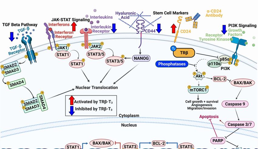
TRβ does indeed meet these criteria (Figure 1).
One of the first reports of potential TRβ tumor suppressor activity was from a study of resistance
to thyroid hormone [10]. Mice with a point mutation in the ligand binding domain of TRβ that renders
TRβ unable to bind ligand (TRβPV) unexpectedly exhibited enlarged thyroid glands, in addition to
the symptoms of resistance to thyroid hormone syndrome. Further examination of the enlarged
thyroid glands in TRβPV mutant mice by histology revealed that the mice had developed thyroid
cancer. Further studies from the same group demonstrated that thyroid-specific knockout mice also
spontaneously develop thyroid cancer [15]. Histological sections from tissues of TRβ knockout mice
showed evidence of anaplasia, capsular invasion, vascular invasion, and metastatic lesions in the
lung. In human thyroid cancer tissues, loss of TRβ expression is correlated with dedifferentiation [16].
Normal thyroid epithelial cells have the highest TRβ expression, while TRβ expression is lowest in
anaplastic thyroid cancer cells, the most aggressive form of thyroid cancer. Combined, these results
establish that loss of TRβ results in cancer growth.
Restoration of TRβ signaling in TRβ-low or TRβ-null cell lines has been shown to slow tumor
growth in vivo. This was first shown in MDA-MB-468 triple negative breast cancer cells with TRβ
restored, in a nude mouse xenograft model [6]. In the same study, SK-hep1 hepatocarcinoma cells
with TRβ restored showed reduced tumor growth [6]. Cells with restored TRβ expression were also
shown to have less metastatic potential than control cells. A separate study showed that FTC-133
follicular thyroid cancer cells with TRβ restored show reduced growth in a xenograft study [9]. These
TRβ-expressing tumors showed evidence of reduced PI3K-Akt signaling and less blood vessel
formation compared to tumors without TRβ expression. Most recently, TRβ restoration has been
shown to suppress growth and migration in colorectal cancer cells [17], and block cancer stem cell
out growth in luminal A breast cancer cell lines [18]. Our lab demonstrated that restoration of TRβ in
anaplastic thyroid cancer cells re-programs the transcriptome, promotes apoptosis, and suppresses
many of their aggressive phenotypic traits [19]. Taken together, these results establish that restoration
of TRβ slows cancer growth.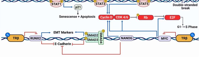
Preprints (www.preprints.org) | NOT PEER-REVIEWED | Posted: 23 July 2021                      doi:10.20944/preprints202107.0549.v1

                                                                                                                      3 of 14

                       Figure 1. Timeline of seminal studies of TRβ tumor suppression. TRβ function was first linked to
                       cancer growth when it was shown that a expression of ligand-binding domain mutant (TRBPV) in mice
                       led to spontaneous development of thyroid tumors. Increasing numbers of studies have demonstrated
                       over the following two decades that TRβ is a classically-defined tumor suppressor.

                   2.2 TRβ Attenuates the PI3K-Akt Signaling Pathway via Genomic and Nongenomic Mechanisms
                        Phosphoinositide 3-kinase (PI3K) signaling is a potent tumor activating pathway that is
                   implicated in many solid and hematologic tumors [20-24]. PI3K is recruited to ligand-bound,
                   phosphorylated receptor tyrosine kinases (RTKs) and phosphorylates the membrane lipid
                   phosphatidylinositol 4,5-bisphosphate (PIP2) to phosphatidylinositol (3,4,5)-trisphosphate (PIP3).
                   PIP3 is a docking lipid that anchors protein kinase B (Akt) for phosphorylation and activation by
                   phosphoinositide-dependent kinase-1 (PDK1) on thr308 and mammalian target of rapamycin
                   complex 2 (mTORC2) on ser473. Akt is a multi-substrate kinase that phosphorylates targets involved
                   in apoptosis regulation, cell cycle progression, angiogenesis, and metabolism. An important function
                   of Akt is the indirect activation of mTORC1, which phosphorylates a myriad of targets such as
                   p70S6K and eukaryotic translation initiation factor 4B (eIF4B) to increase cell metabolism and protein
                   translation. PI3K-Akt pathway activation is frequently overactive due to gain-of-function mutations
                   in PI3K, loss of expression of phosphatase and tensin homolog (PTEN), or amplifications in RTKs
                   and Akt [21, 23, 25].
                        TRβ has long been understood to directly alter the PI3K pathway through nongenomic
                   mechanisms. Simoncini et al. first reported on the potential for hormone receptors to increase PIP3
                   content in endothelial cells [26]. Estrogen, dexamethasone, and thyroid hormone receptors all
                   increased PIP3 levels following respective hormone treatment, suggesting a shared yet noncanonical
                   role for hormone receptors in PI3K activation. In other normal cells such as human fibroblasts,
                   treatment with 15 min of 10 nM T3 induced Akt phosphorylation, and phosphorylation of mTORC2
                   and p70S6K was observed after 30 min T3 exposure[27]. This effect was abrogated with PI3K
                   inhibitors LY294002 and wortmannin. This rapid impact of T3 was unlikely due to TRβ-meditated
                   transcription; indeed TRβ was shown to directly bind to the RTK localization subunit of PI3K, p85⍺,
                   independently of ligand (Figure 2) [27]. Interestingly, 100 nM of T3 did result in reduced TRβ-p85⍺
                   binding and increase in PI3K activity in fibroblasts [27]. These findings were confirmed in GH4C1
                   and CHO cells treated with 100 nM T3 for only five minutes [28], suggesting a rapid and conserved
                   nongenomic function of TRβ across diverse mammalian cell types.
Preprints (www.preprints.org) | NOT PEER-REVIEWED | Posted: 23 July 2021                           doi:10.20944/preprints202107.0549.v1

                                                                                                                             4 of 14

                        Figure 2. TRβ regulates various oncogenic signaling pathways in cancer models that govern cell
                        proliferation, migration, and apoptosis. TRβ has shown to be a significant regulator of various
                        oncogenic cell signaling pathways in diverse models of normal and cancer cells. TRβ decreases TGF-
                        β signaling resulting in decreased SMAD phosphorylation and transcription of EMT markers. TRβ
                        differentially regulates JAK-STAT signaling pathways which results in decreased STAT3 signaling
                        and enhanced STAT1 response to induce apoptosis. TRβ activation can reduce the cancer stem cell
                        population as evident by decreased levels of CD44 and increased CD24 in breast and thyroid cancer
                        models, leading to attenuated NANOG levels. TRβ also has potent inhibitory effects on PI3K signaling
                        whether via direct binding to the p85⍺ subunit or through genomic mechanisms to increase
                        transcripts of phosphoinositol phosphatases and decrease receptor tyrosine kinases. TRβ also
                        modulates cell cycle genes in various cancer models to enhance expression and phosphorylation of
                        Rb to stall the cell cycle. These potent effects on cancer cell transcriptional reprogramming allow for
                        enhanced efficacy of targeted inhibitors on the PI3K pathway and cell cycle.

                         In cancer models however, the role of TRβ on modulating PI3K appears to be more nuanced. TRβ
                   first appeared to have a role in regulating PI3K with the TRβPV/PV mouse model [29], in which Akt was
                   hyperphosphorylated. It was later revealed that TRβPV/PV bound to p85⍺ with a higher affinity
                   compared to wildtype TRβ [30]. This is likely due to the C terminal frameshift in the ligand-binding
                   domain resulting in higher p85⍺ binding affinity. Zhu et al. reported that absence of thyroid hormone
                   receptors correlated with higher levels of phosphorylated Akt, mTORC1, and p70S6K in thyroid cancer
                   [15]. Moriggi et al. showed that TRβ could complex with p85⍺ in four of the six cancer cell lines
                   investigated which resulted in either an increase or decrease in Akt phosphorylation [31]. This may
                   reflect the levels of endogenous TRβ in cell lines as well as individual genetic backgrounds of the cells.
Preprints (www.preprints.org) | NOT PEER-REVIEWED | Posted: 23 July 2021 doi:10.20944/preprints202107.0549.v1
5 of 14
Notably, the experiments were conducted at 24 and 48 hours of T 3 exposure, indicating that gene
expression could have been at play in conjunction with p85⍺ binding. In MDA-MB-468 and SK-Hep1
cells transduced with TRβ, pAkt was blunted in the presence of Insulin-like growth factor 1 (IGF-1)
compared to control cells, suggesting a protective effect of TRβ on the PI3K-Akt pathway [6]. Indeed,
qPCR revealed a reduction in the RTKs epidermal growth factor receptor 1 (EGFR1), HER3 (ERBB3),
and IGFR1 in both cell lines [6].
In vivo studies using the follicular thyroid cancer cell lines FTC-133 and FTC-236 transfected with
TRβ revealed a decrease in pAkt, mTORC1, p70S6K, and eIF4B [9]. There was also a decrease in vascular
endothelial growth factor (VEGF) levels which stimulates endothelial cells to promote angiogenesis
by way of the PI3K pathway, further implicating a broad-spectrum tumor suppressive role of TRβ.
Additionally, TRβ transduction resulted in reduced autocrine signaling in an MCF-7 model [18]. TRβ-
T3 decreased expression of vascular endothelial growth factor receptor 9 (FGFR9) and cognate ligands
FGF3 and FGF4, while blunting estrogen-mediated induction of FGF9. While PI3K activity itself was not
measured, this study confirmed an additional mechanism of TRβ modulation of players in the PI3K-
Akt pathway.
There were similar findings in colorectal cancer cells in which long-term T 3 exposure resulted in
reduced ser473 Akt phosphorylation by an unknown mechanism [32]. However, as we have recently
described, TRβ may play a critical genomic role in PI3K regulation in anaplastic thyroid cancer (ATC)
[33]. While short term (30 min) T3 exposure failed to reduce pAkt levels, long term exposure (24 hours)
reduced pAkt and pmTORC1 levels concordantly with changes in gene expression. T 3-TRβ in ATC cells
increased phosphatase levels such as phosphatidylinositol 4,5-bisphosphate 5-phosphatase A
(INPP5J), inositol polyphosphate 4-phosphatase type II ( INPP4B), and PH domain and leucine rich
repeat protein phosphatase 1 (PHLPP1). Importantly, INPP4B is a potent tumor suppressor of thyroid
cancer in vivo [34]. Conversely, TRβ-T3 decreased levels of receptor tyrosine kinases ERBB3 (HER3),
FGFR3, and FGFR4. This impact on PI3K pathway attenuation resulted in increased sensitivity to the
PI3K inhibitors LY294002 and buparlisib, providing a provocative implication on the relationship
between TRβ status in cancer patients and response to therapies.
A major consequence of PI3K-Akt signaling in cancer cells is the increase in cell metabolism.
While the impact of TRβ on normal metabolism is well studied [35, 36], there is little known on the
tumor suppressor’s role in cancer cell metabolism. We were able to determine that TRβ and T3
potently modulate key metabolic pathways in triple negative breast cancer and ATC cells. Stearic acid
is known to exhibit tumor suppressor functions in breast cancer by inducing apoptosis[37]. In our
MDA-MB-468 cells transduced with TRβ, T3 induced expression of enzymes in the stearic acid
synthesis pathway including Acetyl-CoA synthetase short chain family member 2 (ACSS2),
glutaminase, and 6-phosphofructo-2-kinase/fructose-2,6-biphosphatase 3 (PFKFB1). In ATC cells, we
noticed the potential for TRβ and T3 to regulate glycogen metabolism, an oncogenic metabolic
pathway that’s been observed in many cancer models such as breast, colorectal, and pancreatic
cancer[38-41]. Glycogen is a storage form of glucose for cancer cells to advantageously breakdown
via glycogen phosphorylase in times of low cellular energy or oxidative stress (Figure 3)[42]. TRβ and
T3 decreased expression of the brain isoform of glycogen phosphorylase (PYGB) and differentially
regulated expression of key signaling proteins that activate PYGB such as cell migration inducing
hyaluronidase (CEMIP), and the beta inhibitory subunit of phosphorylase kinase (PHK)[43]. Since these
studies highlight two specific metabolic pathways regulated by TRβ in cancer cells, further exploration
will be required to ascertain the extent of how TRβ induces global metabolic changes in cancer cells.Preprints (www.preprints.org) | NOT PEER-REVIEWED | Posted: 23 July 2021 doi:10.20944/preprints202107.0549.v1
6 of 14
Figure 3. TRβ may regulate glycogen metabolism in anaplastic thyroid cancer cells. RNA-
sequencing[19] revealed a novel function of liganded TRβ on metabolism in a cancer cell model. TRβ
and T3 induced significant changes in gene expression in the glycogen pathway to decrease levels of
PYGB and the glycogen signaling protein CEMIP. TRβ also enhanced the inhibitory beta subunit of
PHK which phosphorylates and activates PYGB to enhance glycogen breakdown and potentially cell
survival.
2.3 TRβ Differentially Influences JAK-STAT Signaling
Another important signaling cascade in cancer is the JAK-STAT pathway. Immune modulators
such as interferons and interleukins activate cognate receptors to recruit Janus kinases (JAKs) to the
membrane to phosphorylate specific signal transducer and activator of transcription proteins
(STATs)[44]. Although the JAK-STAT pathways are best studied in immune cells to promote cell growth
and the immune response, cancer cells can take advantage of the signaling pathway to contribute to
malignant phenotypes[45-47]. STAT3 and STAT5 typically demonstrate tumor promoting activity by
inhibiting apoptosis through repression of BAX and BAK transcription along with induction of BCL2
transcription (Figure 2)[48-50]. Conversely, JAK1 and STAT1 have shown to induce apoptosis in a
variety of cell models, likely through BCL2 repression and CDKN1A (p21) transcription[51-54]. TRβ has
been shown to specifically regulate activity of certain JAK-STAT pairs. For example, Guignon et al.
showed that TRβPV/PV in a mouse model of breast cancer resulted in enhanced prolactin expression
and sustained phosphorylation of STAT5[55], which is known to inhibit apoptosis and encourage cell
cycle progression by promoting BCL2 and CCND1 (cyclin D) transcription, respectively[50, 56].
Introduction of wildtype TRβ attenuated prolactin-induced p-STAT5 levels which were further reduced
with addition of T3. Park et al. showed that TRβ decreased JAK2, STAT3, and STAT5 phosphorylation
and activation in MCF-7-TRβ cells[8]. This led to a decrease in tumor size and proliferation with an
increase in apoptosis. In an ATC model with stably-transduced TRβ, we observed differential
regulation in opposing JAK-STAT pathways[19]. STAT3 signaling was downregulated in the ATC cells
while STAT1 signaling was induced compared to control cells. STAT1 activation led to induction of
apoptosis as evident from Caspase 3 and poly (ADP-ribose) polymerase (PARP) cleavage. Importantly,
we were able to activate STAT1 independently of TRβ with 2-(1,8-Naphthyridin-2-ly)phenol. This study
revealed a novel target in ATC, highlighting the importance of better understanding the tumor
suppressor program of TRβ.
2.4 TRβ Regulation of Cell Cycle Progression
The cell cycle is a crucial target for aggressive cancers (Figure 2). Perez-Juste et al. first observed
that overexpression of TRβ with T3 had profounds effects on neuron development in murine neural
crest-derived cells[57]: TRβ activation led to a decrease in MYC and cyclin D1 expression with a
concomitant increase in the cell cycle regulator p27. Since p27 can directly inhibit cyclin-dependent
kinases (CDKs), the investigators unsurprisingly observed a decrease in phosphorylatedPreprints (www.preprints.org) | NOT PEER-REVIEWED | Posted: 23 July 2021 doi:10.20944/preprints202107.0549.v1
7 of 14
retinoblastoma protein (Rb), resulting in cell cycle arrest. The authors later confirmed the direct
relationship of TRβ to the cell cycle promoter MYC by elucidating a negative thyroid response element
(TRE) on the MYC promoter[58]. Porlan et al. expanded on these observations by showing that TRβ in
3T3 cells inhibited proliferation and the cell cycle via decreasing pRb, cyclin D 1, 2, and 3, and cyclin
E[59]. Yen et al. showed similar findings in HepG2 cells using 10 nM T 3 for multiple days; there was an
increase in p21 and decrease in cyclin E and pRb[60]. In pancreatic adenocarcinoma cell lines, TRβ
decreased cyclins D1 and E but increased p21, which led to an increase in p27 [61]. This excitingly
allowed for an enhanced response to the antiproliferative agents gemcitabine and cisplatin. The
impact of TRβ on cell cycle has also been observed in vivo; Martinez et al. observed a decrease in
cyclin E in hypothyroid patients with hepatocellular carcinoma or breast cancer[62]. These authors
also noted increased p27 expression in their SK-TRβ mouse model.
Lin et al. expanded on the mechanism by showing that TRβ with T3 increases endoglin expression
to stabilize p21 protein in HCC cells[63]. Cell cycle arrest was also measured in MCF-7-TRβ cells with
long term T3 exposure; transcriptomic analysis revealed a decrease in cell cycle related gene transcripts
including MYC and members of the E2F family[18]. Finally, we recently performed RNA-sequencing
to capture the full cell cycle signaling pathway with TRβ and 24 hours of T3[19]. We observed a
decrease in CCND1 and MYC expression and an increase in CDKN1A, highlighting the importance of
TRβ on cell cycle regulation. Importantly, we showed that TRβ enhanced the efficacy of palbociclib, a
CDK4/6 inhibitor. Palbociclib is frequently used to treat ER + and HER2+ breast cancers and is in clinical
trials for several solid tumors[64-67]. However, resistance to CDK inhibition often develops,
highlighting a potentially useful diagnostic and role for TRβ in predicting response to CDK inhibitors.
2.5 Impact of TRβ on TGF-β Signaling
Another important signaling pathway in cancer is the transforming growth factor beta (TGF-β)
signaling cascade. TGF-β stimulates its cognate receptor to induce dimerization and
autophosphorylation to phosphorylate mothers against decapentaplegic homolog (SMADs) [68-70].
The SMAD complex is translocated to the nucleus to regulate gene transcription (Figure 2). In normal
cells, TGF-β signaling regulates the cell cycle and can promote apoptosis. In cancer cells however,
mutations in TGF-β, SMADs, or SMAD binding partners can induce a tumor promoting gene
expression program[68-70]. While TR⍺ and T3 positively regulated TGF-β signaling in liver cancer
cells[60], TRβ and T3 appear to negatively regulate TGF-β. This was first observed in GH4C1 cells as
well as an in vivo model[71]. TRβ and T3 downregulated TGF-β signaling induction, partly by directly
competing for SMAD binding sites, resulting in decreased fibrosis. In transduced MCF-7 cells, López-
Mateo et al. noted that TRβ-T3 could blunt TGF-β-induced SMAD2 and SMAD3 phosphorylation in
MCF-7 cells, leading to a decrease in both SMAD2 and SMAD3 transcriptional activity [18]. TGF-β
signaling attenuation is a notable and conserved feature of liganded TRβ that further highlights its
function as a broad-spectrum, potent tumor suppressor.
2.6 TRβ Inhibits Epithelial–Mesenchymal Transition
Aggressive cancers display hallmarks of epithelial–mesenchymal transition (EMT) as the cell
becomes more metastatic. TRβ has been shown to reduce EMT in several cancer models (Figure 2).
First, Martinez-Iglesias et al. demonstrated that TRβ reduced expression of vimentin, beta catenin,
and matrix metallopeptidase 1 and 9 (MMP1 and MMP9) in breast and liver cancer models, leading
to a decrease in malignancy in vivo[6]. Dentice et al. further showed an increase in E cadherin with
T3 in colon cancer cells [72]. We also showed that there is a direct and positive relationship between
TRβ expression/activation and mesenchymal—epithelial transition (MET) in thyroid cancer[73]. TRβ
directly regulates the expression of the RUNX family transcription factor 2 (RUNX2), a master EMT
transcription factor. There was an inverse relationship observed between TRβ and RUNX2 expression
in thyroid cancer from normal cells to the highly dedifferentiated anaplastic thyroid cancer. T 3
reduced RUNX2 expression in normal and ATC cells, and TRβ knockdown increased RUNX2,
increasing the expression of MMP2, MMP13, cyclin D1, osteopontin (OPN), and cadherin 6.
Transfected TRβ also repressed RUNX2 and EMT markers in MDA-MB-231 cells; conversely, TRβ-Preprints (www.preprints.org) | NOT PEER-REVIEWED | Posted: 23 July 2021 doi:10.20944/preprints202107.0549.v1
8 of 14
knockdown in breast epithelial-like MCF10A cells caused an increase in RUNX2 and EMT markers
[74]. López-Mateo et al. confirmed a decrease in EMT genes such as vimentin and snail family
transcriptional repressor 2 (SLUG) in estrogen receptor alpha positive (ER⍺+) breast cancer cells[18].
We recently observed more evidence of MET in a transduced ATC model in which TRβ and T 3
increased transcript levels of E cadherin and decreased vimentin mRNA [19].
2.2 TRβ Promotes Cancer Cell Re-Differentiation
EMT is closely correlated with dedifferentiation, a phenotype that does not resemble the original
tissue of origin but more closely resembles a stem cell [75]. Dedifferentiation is a crucial process in
the cancer cell to evade the immune system, enhance proliferation, promote angiogenesis, and
develop drug resistance [76, 77]. Modulation in expression of specific tissue markers is often
correlated with dedifferentiation. For example, breast cancer cells increaseexpression of cytokeratins,
proteins that are secreted into the extracellular matrix (ECM) or attached at the cell surface [78, 79].
These keratins are advantageous modulators of the ECM that allow for enhanced cancer cell
migration and invasion [80]. TRβ decreased the expression of keratins 8 and 18 in MCF-7 cells [6, 62].
These two keratins, amongst other keratin isoforms, were also reduced in stem cell models of ER⍺+
breast cancer [18]. Furthermore, we observed a decrease in mRNA and protein levels of keratins 5
and 14 in a triple negative breast cancer model. It may be possible that TRβ canonically regulates
keratin expression, as this has been observed in organisms such as Xenopus laevis [81].
TRβ appears to induce re-differentiation in cells to encourage expression of normal cell markers,
agnostic of cell type. Perra et al. noted a striking difference in rats with preneoplastic liver lesions
treated with the selective TRβ agonist sobetirome (GC-1): TRβ activation resulted in loss of
dedifferentiation markers and reacquisition of differentiated liver proteins [82]. This phenomenon
was also detected in colon cancer cells, in which T3 induced robust expression of normal colon
markers sucrase isomaltase and intestinal alkaline phosphatase and slowed the proliferation of the
cancer cells[72].
In addition to breast and colon cancer models, TRβ has shown to induce re-differentiation in
thyroid cancer cells. We recently demonstrated that TRβ and T 3 induced the re-expression of several
key thyroid specific genes that are lost in dedifferentiated thyroid cancer[19]. These included
iodothyronine deiodinase 2 (DIO2), dual oxidase 1 (DUOX1), thyroid peroxidase (TPO), and
thyroglobulin (TG). Excitingly, we were also able to induce expression of these genes plus six other
thyroid specific markers by using the potent TRβ-specific analog GC-1 to activate the low level of
TRβ expressed in ATC [83]. The sodium iodide symporter (NIS) transcript and protein level were
increased using GC-1, which allowed for a significantly higher intake of iodide in cell culture models.
These studies not only demonstrate the role of TRβ in re-differentiation programming but could have
functional significance as induction of NIS could be exploited for radioactive iodide treatment in
thyroid and breast cancers.
Finally, in the course of dedifferentiation, aggressive cancers become more stemlike. This allows
for unlimited replicative potential and evasion from the immune system [75]. TRβ appears to reduce
the stem cell population in both breast and thyroid cancers. López-Mateo et al. showed that TRβ
activation in MCF-7 cells reduced the mammosphere population[18]. This was associated with a
decrease in the breast stem cell markers aldehyde dehydrogenase (ALDH1) and CD44, with an
increase in the monolayer marker CD24. An increase in CD24 expression is not only notable for
demonstrating a decrease in the stem cell population, but it can also be used as a drug target for
promoting tumor cell clearing by macrophages in the tumor microenvironment[84, 85]. They also
observed a decrease in SRY-box transcription factor 2 (SOX2) and NANOG, which are downstream
of CD44 and TGF-β signaling. We also observed a decline in the stem cell population in both ATC
and MDA-MB-468 breast cancer cells by manipulating TRβ levels[19]. We noted that ALDH, POU5F1
(encodes OCT3/4), CD44, FUT4 (encodes SSEA-1), and PROM1 expression were significantly
downregulated in cancer cells transduced with TRβ. We have also recently been able to see this
change in ATC cells with GC-1 that resulted in stem cell death, increase in CD24 expression, and
decrease in CD44 expression [83]. We also observed an enhanced efficacy to the inhibitors buparlisibPreprints (www.preprints.org) | NOT PEER-REVIEWED | Posted: 23 July 2021 doi:10.20944/preprints202107.0549.v1
9 of 14
(PI3K), sorafenib (MAPK), and palbociclib (cell cycle) in the stem cell population. The use of TRβ
screening and specific activation may help reduce the stem cell population and increase the efficacy
of clinically relevant therapeutics in ATC patients.
2.8 TRβ interactions with Epigenetic Modulators are Key to Tumor Suppression
In its capacity as a transcription factor, TR is a hub for incoming molecular signals that include
fluctuations in hormone levels, input from cellular signaling pathways, and post-translational
modifications. It must integrate all of these signals to recruit the necessary coregulators and excute a
transcriptional response. TR has a diverse repertoire of potential binding partners in normal cells,
as evidenced by immunoprecipitation to mass spectrometry studies [86, 87], and by our own
proximity ligation assays [88]. By complexing with a variety of co-regualtors TR acts to coordinate
complex gene regulatory events that have variety of implications in maintenance of cellular
homeostasis and tumor suppression. Disruption of these crucial interactions in cancer cells may lead
to either a loss of response to T3 or to abberant transcription.
Many coactivators have been implicated in T3-dependent gene activation by TR, including the
steroid receptor coactivator (SRC), p300/CBP histone acetyl transferases, and the mediator-like TR
associated proteins (TRAP)/DRIP) [89]. SRC interacts directly with liganded TR and serves as an
adapter molecule to facilitate recruitment of p300/CBP to acetylate histones and interact with
components of basal transcriptional machinery. The TRAP complex is a mutlisubunit coactivator
complex that interacts with liganded TRs and recruits RNA polymerase II to promoters. Chromatin
immunoprecipitation experiments have demonstrated that upon T3 binding, TRβ first recruits SRC
proteins and p300, resulting in histone acetylation, followed by the TRAP complex [90]. Together
these coactivators facilitate transcriptional activation through a stepwise process of acetylation of
histones to decompact the local chromatin and subsequent recruitement of the basal transcriptional
machinery. Compounds that target the specific interactions between nuclear hormone receptors and
SRC have been developed, and have shown early signs of therapeutic benefit in vivo [91].
TR also complexes with a variety of nuclear co-repressors. Specifically, evidence suggests that
TR represses gene expression via the recruitment of either nuclear co-repressor 1 (NCoR1) or
silencing mediator for retinoid or thyroid-hormone receptors (SMRT, NCoR2) [92-94]. NCoR1 and
SMRT are highly homologous and contain three similar nuclear receptor interaction domains.
Furthermore, both NCoR1 and SMRT bind to TR, as well as other nuclear hormone receptors via
similar mechanisms at specific residues [95]. The seminal article that first identified NCoR1 as a
crucial regulatory protein found that it bound TR at amino acid residues 203-230 but that amino
acid residues 230-260 act to stabilize this interaction [96]. NCoR1 and SMRT directly recruit and
interact with Class II histone deacetylases (HDAC), and recruit Class I HDAC’s via linker proteins
such as Sin3a or Sin3b [97-100]. NCoR1 itself has been demonstrated to be critical for suppression of
breast cancer growth in coordination with TR [101]. HDAC inhibitors may be useful in combinatiton
with hormone therapy, particularly in the context of anti-estrogen or anti-androgen [102, 103]
resistance. The future use of compounds that can promote interactions between TR and its
coregulators, or block interactions with negative regulators, may be an attractive way to enhance the
beneficial effects of thyroid hormone or thyromimetics.
3. Conclusions
Despite a recognized role as a tumor suppressor, the potential for TRβ as a therapeutic target
and diagnostic indicator remains untapped. This is in part because, until recently, the mechanisms
by which TRβ regulates tumor growth were unclear. TRβ is a ligand-dependent nuclear receptor that
mediates the effects of T3 on many biological processes, and therefore has potent effects throughout
the cell as disussed in this review. At the genomic level, TRβ mediates the effects of T3 via the
regulation of gene expression through the recruitment of co-regulators and chromatin remodeling
complexes to genomic regulatory elements to alter target gene transcription. Disruption of TRβ is
therefore expected to alter assembly of co-regulator complexes needed for initiation of gene
transcription. TRβ-selective thyromimetics have been developed [104] and have been shown to elicitPreprints (www.preprints.org) | NOT PEER-REVIEWED | Posted: 23 July 2021 doi:10.20944/preprints202107.0549.v1
10 of 14
the same transcriptional response as T3 [105]. Current work in our group is focused on stimulation of
endogenous TRβ, even when it is expressed at the low levels in cancer cells, to elicit an anti-tumor
response [83]. Our work suggests TRβ agonists can be combined with modulators of other pathways
to enhance their efficacy, and opens many new avenues for exploration of TRβ tumor suppressive
action.
Author Contributions: Conceptualization, CDD, NEG, and FEC; writing—original draft preparation, CDD and
NEG.; visualization, CDD and NEG; writing—review and editing, CDD, NEG, FEC. All authors have read and
agreed to the published version of the manuscript.
Funding: This work was supported by grants from National Institutes of Health U54 GM115516 for the Northern
New England Clinical and Translational Research Network; National Cancer Institute 1F99CA245796-01; UVM
Cancer Center-Lake Champlain Cancer Research Organization (C3) 12577-21; and UVM Larner College of
Medicine.
Acknowledgements: Biorender vector graphics were used to construct the figures.
Conflicts of Interest: The authors declare no conflict of interest.
References
1 Kim WG, Cheng SY. Thyroid hormone receptors and cancer. Biochimica et biophysica acta 2013; 1830:
3928-3936.
2 Aranda A, Martinez-Iglesias O, Ruiz-Llorente L, Garcia-Carpizo V, Zambrano A. Thyroid receptor:
roles in cancer. Trends in endocrinology and metabolism: TEM 2009; 20: 318-324.
3 Landa I, Ibrahimpasic T, Boucai L, Sinha R, Knauf JA, Shah RH et al. Genomic and transcriptomic
hallmarks of poorly differentiated and anaplastic thyroid cancers. J Clin Invest 2016; 126: 1052-1066.
4 Joseph B, Ji M, Liu D, Hou P, Xing M. Lack of mutations in the thyroid hormone receptor (TR) alpha
and beta genes but frequent hypermethylation of the TRbeta gene in differentiated thyroid tumors. The
Journal of clinical endocrinology and metabolism 2007; 92: 4766-4770.
5 Puzianowska-Kuznicka M, Krystyniak A, Madej A, Cheng SY, Nauman J. Functionally impaired TR
mutants are present in thyroid papillary cancer. The Journal of clinical endocrinology and metabolism 2002;
87: 1120-1128.
6 Martínez-Iglesias O, Garcia-Silva S, Tenbaum SP, Regadera J, Larcher F, Paramio JM et al. Thyroid
hormone receptor beta1 acts as a potent suppressor of tumor invasiveness and metastasis. Cancer Res
2009; 69: 501-509.
7 Kim W, Zhu X, Kim D, Zhang L, Kebebew E, Cheng S. Reactivation of the silenced thyroid hormone
receptor B gene expression delays thyroid tumor progression., vol. 154: Endocrinology, 2013, pp 25-35.
8 Park JW, Zhao L, Cheng SY. Inhibition of estrogen-dependent tumorigenesis by the thyroid hormone
receptor beta in xenograft models. Am J Cancer Res 2013; 3: 302-311.
9 Kim WG, Zhao L, Kim DW, Willingham MC, Cheng SY. Inhibition of tumorigenesis by the thyroid
hormone receptor beta in xenograft models. Thyroid 2014; 24: 260-269.
10 Kato Y, Ying H, Willingham MC, Cheng SY. A tumor suppressor role for thyroid hormone beta receptor
in a mouse model of thyroid carcinogenesis. Endocrinology 2004; 145: 4430-4438.
11 Martinez-Iglesias O, Garcia-Silva S, Tenbaum S, Regadera J, Larcher F, Paramio J et al. Thyroid hormone
receptor β1 acts as a potent suppressor of tumor invasiveness and metastasis., vol. 69: Cancer Res, 2009,
pp 501-509.
12 Park J, Zhao L, Cheng S. Inhibition of estrogen-dependent tumorigenesis by the thyroid hormone
receptor B in xenograft models., vol. 3: Am J Cancer Res, 2013, pp 302-311.
13 Hanahan D, Weinberg RA. Hallmarks of cancer: the next generation. Cell 2011; 144: 646-674.
14 Weinberg RA. Oncogenes and tumor suppressor genes. CA Cancer J Clin 1994; 44: 160-170.
15 Zhu XG, Zhao L, Willingham MC, Cheng SY. Thyroid hormone receptors are tumor suppressors in a
mouse model of metastatic follicular thyroid carcinoma. Oncogene 2010; 29: 1909-1919.
16 Carr FE, Tai PW, Barnum MS, Gillis NE, Evans KG, Taber TH et al. Thyroid Hormone Receptor-β (TRβ)
Mediates Runt-Related Transcription Factor 2 (Runx2) Expression in Thyroid Cancer Cells: A Novel
Signaling Pathway in Thyroid Cancer. Endocrinology 2016; 157: 3278-3292.
17 Zhu L, Tian G, Yang Q, De G, Zhang Z, Wang Y et al. Thyroid hormone receptor β1 suppresses
proliferation and migration by inhibiting PI3K/Akt signaling in human colorectal cancer cells. Oncol
Rep 2016; 36: 1419-1426.Preprints (www.preprints.org) | NOT PEER-REVIEWED | Posted: 23 July 2021 doi:10.20944/preprints202107.0549.v1
11 of 14
18 López-Mateo I, Alonso-Merino E, Suarez-Cabrera C, Park JW, Cheng SY, Alemany S et al. Thyroid
Hormone Receptor β Inhibits Self-Renewal Capacity of Breast Cancer Stem Cells. Thyroid 2020; 30: 116-
132.
19 Bolf EL, Gillis NE, Davidson CD, Rodriguez PD, Cozzens L, Tomczak JA et al. Thyroid Hormone
Receptor Beta Induces a Tumor-Suppressive Program in Anaplastic Thyroid Cancer. Molecular Cancer
Research 2020; 18: 1443-1452.
20 Carnero A, Blanco-Aparicio C, Renner O, Link W, Leal JF. The PTEN/PI3K/AKT signalling pathway in
cancer, therapeutic implications. Curr Cancer Drug Targets 2008; 8: 187-198.
21 Martini M, De Santis MC, Braccini L, Gulluni F, Hirsch E. PI3K/AKT signaling pathway and cancer: an
updated review. Annals of medicine 2014; 46: 372-383.
22 Revathidevi S, Munirajan AK. Akt in cancer: mediator and more. Seminars in cancer biology 2019.
23 Jiang N, Dai Q, Su X, Fu J, Feng X, Peng J. Role of PI3K/AKT pathway in cancer: the framework of
malignant behavior. Molecular biology reports 2020; 47: 4587-4629.
24 Hoxhaj G, Manning BD. The PI3K-AKT network at the interface of oncogenic signalling and cancer
metabolism. Nature reviews Cancer 2020; 20: 74-88.
25 Wang Y, Hou P, Yu H, Wang W, Ji M, Zhao S et al. High prevalence and mutual exclusivity of genetic
alterations in the phosphatidylinositol-3-kinase/akt pathway in thyroid tumors. The Journal of clinical
endocrinology and metabolism 2007; 92: 2387-2390.
26 Simoncini T, Hafezi-Moghadam A, Brazil DP, Ley K, Chin WW, Liao JK. Interaction of oestrogen
receptor with the regulatory subunit of phosphatidylinositol-3-OH kinase. Nature 2000; 407: 538-541.
27 Cao X, Kambe F, Moeller LC, Refetoff S, Seo H. Thyroid hormone induces rapid activation of
Akt/protein kinase B-mammalian target of rapamycin-p70S6K cascade through phosphatidylinositol 3-
kinase in human fibroblasts. Molecular endocrinology (Baltimore, Md) 2005; 19: 102-112.
28 Storey NM, Gentile S, Ullah H, Russo A, Muessel M, Erxleben C et al. Rapid signaling at the plasma
membrane by a nuclear receptor for thyroid hormone. Proc Natl Acad Sci U S A 2006; 103: 5197-5201.
29 Kim CS, Vasko VV, Kato Y, Kruhlak M, Saji M, Cheng SY et al. AKT activation promotes metastasis in
a mouse model of follicular thyroid carcinoma. Endocrinology 2005; 146: 4456-4463.
30 Furuya F, Lu C, Willingham MC, Cheng SY. Inhibition of phosphatidylinositol 3-kinase delays tumor
progression and blocks metastatic spread in a mouse model of thyroid cancer. Carcinogenesis 2007; 28:
2451-2458.
31 Moriggi G, Verga Falzacappa C, Mangialardo C, Michienzi S, Stigliano A, Brunetti E et al. Thyroid
hormones (T3 and T4): dual effect on human cancer cell proliferation. Anticancer Res 2011; 31: 89-96.
32 Zhu L, Tian G, Yang Q, De G, Zhang Z, Wang Y et al. Thyroid hormone receptor β1 suppresses
proliferation and migration by inhibiting PI3K/Akt signaling in human colorectal cancer cells. Oncology
reports 2016; 36: 1419-1426.
33 Davidson CD, Bolf EL, Gillis NE, Cozzens LM, Tomczak JA, Carr FE. Thyroid Hormone Receptor Beta
Inhibits PI3K-Akt-mTOR Signaling Axis in Anaplastic Thyroid Cancer via Genomic Mechanisms.
Journal of the Endocrine Society 2021; 5.
34 Li Chew C, Lunardi A, Gulluni F, Ruan DT, Chen M, Salmena L et al. In Vivo Role of INPP4B in Tumor
and Metastasis Suppression through Regulation of PI3K-AKT Signaling at Endosomes. Cancer Discov
2015; 5: 740-751.
35 Pramfalk C, Pedrelli M, Parini P. Role of thyroid receptor β in lipid metabolism. Biochimica et biophysica
acta 2011; 1812: 929-937.
36 Master AN, A. THRB (Thyroid Hormone Receptor, Beta). Atlas Genet Cytogenet Oncol Haematol 2014; 18:
400-433.
37 Evans LM, Cowey SL, Siegal GP, Hardy RW. Stearate preferentially induces apoptosis in human breast
cancer cells. Nutr Cancer 2009; 61: 746-753.
38 Favaro E, Bensaad K, Chong MG, Tennant DA, Ferguson DJ, Snell C et al. Glucose utilization via
glycogen phosphorylase sustains proliferation and prevents premature senescence in cancer cells. Cell
metabolism 2012; 16: 751-764.
39 Pelletier J, Bellot G, Gounon P, Lacas-Gervais S, Pouyssegur J, Mazure NM. Glycogen Synthesis is
Induced in Hypoxia by the Hypoxia-Inducible Factor and Promotes Cancer Cell Survival. Frontiers in
oncology 2012; 2: 18.
40 Lee WN, Guo P, Lim S, Bassilian S, Lee ST, Boren J et al. Metabolic sensitivity of pancreatic tumour cell
apoptosis to glycogen phosphorylase inhibitor treatment. British journal of cancer 2004; 91: 2094-2100.
41 Davidson CD CF. Review of pharmacological inhibition of thyroid cancer metabolism. . J Cancer
Metastasis Treat 2021; 7:[Accept]. http://dx.doi.org/10.20517/2394-4722.2021.77.
42 Dauer P, Lengyel E. New Roles for Glycogen in Tumor Progression. Trends Cancer 2019; 5: 396-399.Preprints (www.preprints.org) | NOT PEER-REVIEWED | Posted: 23 July 2021 doi:10.20944/preprints202107.0549.v1
12 of 14
43 Terashima M, Fujita Y, Togashi Y, Sakai K, De Velasco MA, Tomida S et al. KIAA1199 interacts with
glycogen phosphorylase kinase beta-subunit (PHKB) to promote glycogen breakdown and cancer cell
survival. Oncotarget 2014; 5: 7040-7050.
44 Harrison DA. The Jak/STAT pathway. Cold Spring Harb Perspect Biol 2012; 4.
45 Seif F, Khoshmirsafa M, Aazami H, Mohsenzadegan M, Sedighi G, Bahar M. The role of JAK-STAT
signaling pathway and its regulators in the fate of T helper cells. Cell Commun Signal 2017; 15: 23.
46 Owen KL, Brockwell NK, Parker BS. JAK-STAT Signaling: A Double-Edged Sword of Immune
Regulation and Cancer Progression. Cancers (Basel) 2019; 11.
47 Brooks AJ, Putoczki T. JAK-STAT Signalling Pathway in Cancer. Cancers (Basel) 2020; 12.
48 Kamran MZ, Patil P, Gude RP. Role of STAT3 in cancer metastasis and translational advances. BioMed
research international 2013; 2013: 421821.
49 Huynh J, Chand A, Gough D, Ernst M. Therapeutically exploiting STAT3 activity in cancer - using
tissue repair as a road map. Nature reviews Cancer 2019; 19: 82-96.
50 Halim CE, Deng S, Ong MS, Yap CT. Involvement of STAT5 in Oncogenesis. Biomedicines 2020; 8.
51 Chin YE, Kitagawa M, Kuida K, Flavell RA, Fu XY. Activation of the STAT signaling pathway can cause
expression of caspase 1 and apoptosis. Mol Cell Biol 1997; 17: 5328-5337.
52 Stephanou A, Latchman DS. STAT-1: a novel regulator of apoptosis. Int J Exp Pathol 2003; 84: 239-244.
53 Sironi JJ, Ouchi T. STAT1-induced apoptosis is mediated by caspases 2, 3, and 7. The Journal of biological
chemistry 2004; 279: 4066-4074.
54 Su Q, Wang F, Dong Z, Chen M, Cao R. IFN-γ induces apoptosis in human melanocytes by activating
the JAK1/STAT1 signaling pathway. Molecular medicine reports 2020; 22: 3111-3116.
55 Guigon CJ, Kim DW, Willingham MC, Cheng SY. Mutation of thyroid hormone receptor-β in mice
predisposes to the development of mammary tumors. Oncogene 2011; 30: 3381-3390.
56 Debierre-Grockiego F. Anti-apoptotic role of STAT5 in haematopoietic cells and in the pathogenesis of
malignancies. Apoptosis 2004; 9: 717-728.
57 Perez-Juste G, Aranda A. The cyclin-dependent kinase inhibitor p27(Kip1) is involved in thyroid
hormone-mediated neuronal differentiation. The Journal of biological chemistry 1999; 274: 5026-5031.
58 Pérez-Juste G, García-Silva S, Aranda A. An element in the region responsible for premature
termination of transcription mediates repression of c-myc gene expression by thyroid hormone in
neuroblastoma cells. The Journal of biological chemistry 2000; 275: 1307-1314.
59 Porlan E, Vega S, Iglesias T, Rodríguez-Peña A. Unliganded thyroid hormone receptor beta1 inhibits
proliferation of murine fibroblasts by delaying the onset of the G1 cell-cycle signals. Oncogene 2004; 23:
8756-8765.
60 Yen CC, Huang YH, Liao CY, Liao CJ, Cheng WL, Chen WJ et al. Mediation of the inhibitory effect of
thyroid hormone on proliferation of hepatoma cells by transforming growth factor-beta. Journal of
molecular endocrinology 2006; 36: 9-21.
61 Michienzi S, Bucci B, Verga Falzacappa C, Patriarca V, Stigliano A, Panacchia L et al. 3,3',5-Triiodo-L-
thyronine inhibits ductal pancreatic adenocarcinoma proliferation improving the cytotoxic effect of
chemotherapy. The Journal of endocrinology 2007; 193: 209-223.
62 Martinez-Iglesias O, Garcia-Silva S, Regadera J, Aranda A. Hypothyroidism enhances tumor
invasiveness and metastasis development. PLoS One 2009; 4: e6428.
63 Lin YH, Huang YH, Wu MH, Wu SM, Chi HC, Liao CJ et al. Thyroid hormone suppresses cell
proliferation through endoglin-mediated promotion of p21 stability. Oncogene 2013; 32: 3904-3914.
64 Turner NC, Ro J, André F, Loi S, Verma S, Iwata H et al. Palbociclib in Hormone-Receptor-Positive
Advanced Breast Cancer. The New England journal of medicine 2015; 373: 209-219.
65 Karasic TB, O'Hara MH, Teitelbaum UR, Damjanov N, Giantonio BJ, d'Entremont TS et al. Phase II Trial
of Palbociclib in Patients with Advanced Esophageal or Gastric Cancer. Oncologist 2020; 25: e1864-e1868.
66 Sepúlveda-Sánchez JM, Gil-Gil M, Alonso-García M, Vaz Salgado M, Vicente E, Mesía Barroso C et al.
Phase II Trial of Palbociclib in Recurrent Retinoblastoma-Positive Anaplastic Oligodendroglioma: A
Study from the Spanish Group for Research in Neuro-Oncology (GEINO). Target Oncol 2020; 15: 613-
622.
67 Serra F, Lapidari P, Quaquarini E, Tagliaferri B, Sottotetti F, Palumbo R. Palbociclib in metastatic breast
cancer: current evidence and real-life data. Drugs Context 2019; 8: 212579.
68 Hata A, Chen YG. TGF-β Signaling from Receptors to Smads. Cold Spring Harb Perspect Biol 2016; 8.
69 Morikawa M, Derynck R, Miyazono K. TGF-β and the TGF-β Family: Context-Dependent Roles in Cell
and Tissue Physiology. Cold Spring Harb Perspect Biol 2016; 8.
70 Vander Ark A, Cao J, Li X. TGF-β receptors: In and beyond TGF-β signaling. Cellular signalling 2018; 52:
112-120.Preprints (www.preprints.org) | NOT PEER-REVIEWED | Posted: 23 July 2021 doi:10.20944/preprints202107.0549.v1
13 of 14
71 Alonso-Merino E, Martín Orozco R, Ruíz-Llorente L, Martínez-Iglesias OA, Velasco-Martín JP,
Montero-Pedrazuela A et al. Thyroid hormones inhibit TGF-β signaling and attenuate fibrotic
responses. Proc Natl Acad Sci U S A 2016; 113: E3451-3460.
72 Dentice M, Luongo C, Ambrosio R, Sibilio A, Casillo A, Iaccarino A et al. β-Catenin regulates deiodinase
levels and thyroid hormone signaling in colon cancer cells. Gastroenterology 2012; 143: 1037-1047.
73 Carr FE, Tai PW, Barnum MS, Gillis NE, Evans KG, Taber TH et al. Thyroid Hormone Receptor-beta
(TRbeta) Mediates Runt-Related Transcription Factor 2 (Runx2) Expression in Thyroid Cancer Cells: A
Novel Signaling Pathway in Thyroid Cancer. Endocrinology 2016; 157: 3278-3292.
74 Bolf EL, Gillis NE, Barnum MS, Beaudet CM, Yu GY, Tomczak JA et al. The Thyroid Hormone Receptor-
RUNX2 Axis: A Novel Tumor Suppressive Pathway in Breast Cancer. Hormones & cancer 2020; 11: 34-
41.
75 Nassar D, Blanpain C. Cancer Stem Cells: Basic Concepts and Therapeutic Implications. Annu Rev Pathol
2016; 11: 47-76.
76 Liu J. The dualistic origin of human tumors. Seminars in cancer biology 2018; 53: 1-16.
77 Wang H, Unternaehrer JJ. Epithelial-mesenchymal Transition and Cancer Stem Cells: At the Crossroads
of Differentiation and Dedifferentiation. Dev Dyn 2019; 248: 10-20.
78 Vora HH, Patel NA, Rajvik KN, Mehta SV, Brahmbhatt BV, Shah MJ et al. Cytokeratin and vimentin
expression in breast cancer. Int J Biol Markers 2009; 24: 38-46.
79 Alshareeda AT, Soria D, Garibaldi JM, Rakha E, Nolan C, Ellis IO et al. Characteristics of basal
cytokeratin expression in breast cancer. Breast Cancer Res Treat 2013; 139: 23-37.
80 Karantza V. Keratins in health and cancer: more than mere epithelial cell markers. Oncogene 2011; 30:
127-138.
81 Mathisen PM, Miller L. Thyroid hormone induces constitutive keratin gene expression during Xenopus
laevis development. Mol Cell Biol 1989; 9: 1823-1831.
82 Perra A, Kowalik MA, Pibiri M, Ledda-Columbano GM, Columbano A. Thyroid hormone receptor
ligands induce regression of rat preneoplastic liver lesions causing their reversion to a differentiated
phenotype. Hepatology 2009; 49: 1287-1296.
83 Gillis NE, Davidson CD, Cozzens LM, Wilson E, Bolf EL, Tomczak JA et al. A Thyroid Hormone
Receptor Beta Specific Agonist Suppresses Anaplastic Thyroid Cancer Cell Phenotype and Increases
Efficacy of Therapeutic Agents. bioRxiv 2021: 2021.2006.2009.447689.
84 Salnikov AV, Bretz NP, Perne C, Hazin J, Keller S, Fogel M et al. Antibody targeting of CD24 efficiently
retards growth and influences cytokine milieu in experimental carcinomas. British journal of cancer 2013;
108: 1449-1459.
85 Barkal AA, Brewer RE, Markovic M, Kowarsky M, Barkal SA, Zaro BW et al. CD24 signalling through
macrophage Siglec-10 is a target for cancer immunotherapy. Nature 2019; 572: 392-396.
86 Hahm JB, Schroeder AC, Privalsky ML. The two major isoforms of thyroid hormone receptor, TRα1
and TRβ1, preferentially partner with distinct panels of auxiliary proteins. Mol Cell Endocrinol 2014; 383:
80-95.
87 Fozzatti L, Lu C, Kim D-W, Cheng S-y. Differential Recruitment of Nuclear Coregulators Directs the
Isoform-Dependent Action of Mutant Thyroid Hormone Receptors. Molecular Endocrinology 2011; 25:
908-921.
88 Gillis NE, Boyd JR, Tomczak JA, Frietze S, Carr FE. Thyroid Hormone Dependent Transcriptional
Programming by TRβ Requires SWI/SNF Chromatin Remodelers. bioRxiv 2021: 2021.2003.2022.436429.
89 Lee KC, Li J, Cole PA, Wong J, Kraus WL. Transcriptional Activation by Thyroid Hormone Receptor-β
Involves Chromatin Remodeling, Histone Acetylation, and Synergistic Stimulation by p300 and Steroid
Receptor Coactivators. Molecular Endocrinology 2003; 17: 908-922.
90 Sharma D, Fondell JD. Ordered recruitment of histone acetyltransferases and the TRAP/Mediator
complex to thyroid hormone-responsive promoters in vivo. Proceedings of the National
Academy of Sciences 2002; 99: 7934-7939.
91 Skowron KJ, Booker K, Cheng C, Creed S, David BP, Lazzara PR et al. Steroid receptor/coactivator
binding inhibitors: An update. Mol Cell Endocrinol 2019; 493: 110471.
92 Astapova I, Hollenberg AN. The in vivo role of nuclear receptor corepressors in thyroid hormone
action. Biochim Biophys Acta 2013; 1830: 3876-3881.
93 Yen PM. Physiological and molecular basis of thyroid hormone action. Physiol Rev 2001; 81: 1097-1142.
94 Astapova I. Role of co-regulators in metabolic and transcriptional actions of thyroid hormone. J Mol
Endocrinol 2016; 56: 73-97.
95 Hu X, Lazar MA. The CoRNR motif controls the recruitment of corepressors by nuclear hormone
receptors. Nature 1999; 402: 93-96.Preprints (www.preprints.org) | NOT PEER-REVIEWED | Posted: 23 July 2021 doi:10.20944/preprints202107.0549.v1
14 of 14
96 Horlein AJ, Naar AM, Heinzel T, Torchia J, Gloss B, Kurokawa R et al. Ligand-independent repression
by the thyroid hormone receptor mediated by a nuclear receptor co-repressor. Nature 1995; 377: 397-
404.
97 Aranda A, Pascual A. Nuclear hormone receptors and gene expression. Physiol Rev 2001; 81: 1269-1304.
98 Zhang Y, Dufau ML. Dual mechanisms of regulation of transcription of luteinizing hormone receptor
gene by nuclear orphan receptors and histone deacetylase complexes. The Journal of steroid biochemistry
and molecular biology 2003; 85: 401-414.
99 Yao YL, Yang WM. The metastasis-associated proteins 1 and 2 form distinct protein complexes with
histone deacetylase activity. J Biol Chem 2003; 278: 42560-42568.
100 Fleischer TC, Yun UJ, Ayer DE. Identification and characterization of three new components of the
mSin3A corepressor complex. Mol Cell Biol 2003; 23: 3456-3467.
101 Martínez-Iglesias O, Olmeda D, Alonso-Merino E, Gómez-Rey S, González-López AM, Luengo E et al.
The nuclear corepressor 1 and the thyroid hormone receptor β suppress breast tumor
lymphangiogenesis. Oncotarget 2016; 7: 78971-78984.
102 Kaushik D, Vashistha V, Isharwal S, Sediqe SA, Lin MF. Histone deacetylase inhibitors in castration-
resistant prostate cancer: molecular mechanism of action and recent clinical trials. Ther Adv Urol 2015;
7: 388-395.
103 Hodges-Gallagher L, Valentine CD, Bader SE, Kushner PJ. Inhibition of histone deacetylase enhances
the anti-proliferative action of antiestrogens on breast cancer cells and blocks tamoxifen-induced
proliferation of uterine cells. Breast Cancer Res Treat 2007; 105: 297-309.
104 Elbers LP, Kastelein JJ, Sjouke B. Thyroid Hormone Mimetics: the Past, Current Status and Future
Challenges. Curr Atheroscler Rep 2016; 18: 14.
105 Yuan C, Lin JZ, Sieglaff DH, Ayers SD, Denoto-Reynolds F, Baxter JD et al. Identical gene regulation
patterns of T3 and selective thyroid hormone receptor modulator GC-1. Endocrinology 2012; 153: 501-
511.You can also read