Identification of a Glycerophosphocholine Phosphodiesterase, GDE5, in chicken
←
→
Page content transcription
If your browser does not render page correctly, please read the page content below
E3S Web of Conferences 245, 03054 (2021) https://doi.org/10.1051/e3sconf/202124503054
AEECS 2021
Identification of a Glycerophosphocholine Phosphodiesterase,
GDE5, in chicken
Hao Gao1, Pingan Chang1*
(1Chongqing Key Laboratory of Big Data for Bio-intelligence, College of Bio-information, Chongqing University of Posts and
Telecommunications, Chongqing 400065, P. R. China)
Abstract: Glycerophosphodiester phosphodiesterase (GDPD/GDE) catalyzes the hydrolysis of
glycerophosphodiesters to glycerol 3-phosphate and alcohol. It was discovered that the
glycerophosphodiesterase family plays a role in lipid metabolism and signal pathway in recent years, but
little has been known about the characteristics of chicken GDEs. Here, chicken GDE5 (cGDE5) was
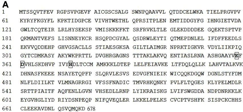
identified and characterized for the first time. The full length coding cDNA sequence of cGDE5 was cloned,
which encoded a polypeptide with 678 amino acids containing a carbohydrate-binding module 20 (CBM20)
and a GDPD domain. Tissue expression profiles showed that cGDE5 mRNA was high in various tissues.
such as heart, brain, skeletal muscle and testis. Moreover, cGDE5 was demonstrated to exhibit
glycerophosphocholine phosphodiesterase activity. These results together suggested that cGDE5, as a
unique member of GDE family, may play multiple roles as a cytoplasmic glycerophosphocholine
phosphodiesterase.
1 Introduction regulates skeletal muscle development[9]. Moreover,
adipose tissue weight and blood triglyceride levels were
Two important factors considered by the poultry industry increased in the transgenic mice specifically expressing
are meat yield and quality. At present, chickens are GDE5DeltaC471 in skeletal muscle[10]. These results
marketed in about half the time and at about twice the indicated GDE5 played a key role in muscle and adipose
body weight compared to 50 years ago, which is closely tissue development.
related to a significant increase in the muscle The chicken is not only an important animal for
proportion[1]. As a complicated life process, the growth providing meat, but it is also serves as a valuable avian
and development of livestock is controlled by a model. In contrast to mammals, there are only six chicken
significant amount of genetic factors. A lot of metabolites, GDE members to be predicted until now. Little has been
including deacylated glycerophospholipids, the known about chicken GDEs, except that GDE2 was
glycerophosphodiesters (GPs), have been investigated in involved in neuron differentiation[9,10]. In our study,
adult skeletal muscles. Although the amounts of skeletal chicken cGDE5 was first cloned and built a unique 3-D
muscle GPs in human and mouse skeletal muscle tissues molecular model. Then cGDE5 mRNA was found to
are reportedly altered in response to physical activity, the express in various tissues. Finally, cGDE5 tagged with
environment, and pathological conditions[2-4], the green fluorescence protein (GFP) was observed to be a
biological function of skeletal muscle GP has not been cytological protein and identified to have
studied to date. GPC-hydrolyzing activity.
Glycerophosphodiester phosphodiesterase (GDPD/
GDE, EC3.1.4.46) catalyzes the hydrolysis of GPs to
glycerol 3-phosphate (G3P) and alcohol[5]. The GDE 2 Results
family proteins have attracted attention recently for their
emerging physiological roles. GDEs are highly conserved 2.1 Molecular cloning and analysis of cGDE5
proteins in prokaryotes and eukaryotes[6]. Previous gene coding sequence
studies were mainly focused on mammalian GDE1-3 and
showed they were involved in diverse physiological The full length coding cDNA sequence of cGDE5 is
functions. GDE5 not only has a GDE domain, but also 2037 bp (GenBank accession No. KC967655.1), and 678
contains a carbohydrate-binding domain[7]. Mouse GDE5 amino acid polypeptide is encoded(Fig. 1A). There is
was initially identified to be a GPC phosphodiesterase 99% identity between the deduced proteins from the
and controls skeletal muscle development[8]. cloned and the predicted cGDE5 gene (data not shown).
GDE5DeltaC471 that contains GDE sequence but lacks The theoretical molecular weight and isoelectric point of
GPC phosphodiesterase activity also innegatively cGDE5 are 76.9 kDa and 5.85 respectively, which were
* Corresponding author: * E-mail address: changpingan@aliyun.com.
© The Authors, published by EDP Sciences. This is an open access article distributed under the terms of the Creative Commons Attribution License 4.0
(http://creativecommons.org/licenses/by/4.0/).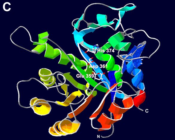
E3S Web of Conferences 245, 03054 (2021)                                                              https://doi.org/10.1051/e3sconf/202124503054
AEECS 2021

       determined by the Compute pI/Mw tool on the ExPASy                     mammalian identified GDEs, the three amino acid sites
       server. Protein domain analysis showed that cGDE5 had                  necessary for GDE activity are E359, D361 and
       a carbohydrate-binding module 20 (CBM20) (residues                     H374(Fig. 1A). However, unlike other GDEs, there is not
       3–105) in the N-terminal region and a GDPD domain                      any transmembrane region in cGDE5 as predicted by the
       (residues 324-612) in the C-terminal region (Fig. 1B). By              TMHMM2 program.
       the most conserved sequence comparison of cGDE5 with

                                           Fig. 1 Analysis of the deduced cGDE5 amino acid sequence.

                                                                              high similarity between cGDE5 and other chicken GDEs
       2.2 cGDE5 is a unique member of chicken GDE                            (data not shown), a high homology exists in the GDPD
       family                                                                 domain of chicken GDE protein family (Fig. 2A). Based
                                                                              on phylogenetic analysis of the GDE domain, we further
       Because there is a 56-residue conserved region in the
                                                                              classified the family into two distinct groups: one group
       GDPD domain among all identified mammalian GDE
                                                                              contains GDE4 and GDE5, the other includes GDE1-3
       proteins and the bacterial GDEs sequence[5], a search of
                                                                              and GDE6 (Fig. 2B). Domain–structure analysis showed
       chicken expressed sequence tags (EST) database based
                                                                              that GDE1-4 and GDE6 appear to be membrane proteins
       on their amino acid homology is expected to find out all
                                                                              and contain multiple putative transmembrane regions
       chicken GDEs cDNA sequences. However, there are only
                                                                              (data not shown). In contrast, GDE5 has not any
       six chicken GDE members. By the comparison of the
                                                                              transmembrane region but contains an N-terminal
       most conserved sequence in all chicken GDEs, it was
                                                                              CBM20 (Fig. 1B). The diverse domain structures of these
       found that the three amino acid positions E, D and H
                                                                              GDEs indicate that cGDE5 is a unique member in
       were the most conserved(Fig. 2A). The multiple
                                                                              chicken GDE family.
       sequence alignments indicate that although there are not

       Fig. 2 The GDE domain sequence alignment and phylogram of the chicken GDE family. A, Identical amino acids in all six proteins
       were marked with an asterisk (*), conservative substitutions with a colon (:), and semi-conservative substitutions with a period (.). B,
                    The numbers indicate the bootstrap confidence values obtained for each nodes after 1000 replications.

                                                                              structure with two starch binding sites. By sequence
       2.3 Molecular 3D model of cGDE5 protein                                alignment, binding site 1 of cGDE5 CBM20 consists of
       domains                                                                residue 32, 63, 86-87, 92. The indole rings of Trp32 and
                                                                              Trp86 form the central part of a carbohydrate binding
       To further study the relationship between structure and
                                                                              platform (Fig 3A). By contrast, binding site 2, defined by
       function, we generated molecular 3D models of cGDE5
                                                                              residue 15,16-19,46,48, is more extended and has higher
       CBM20 and GDPD domains respectively using
                                                                              structural plasticity than binding site 1(Fig 3B).
       SWISS-MODEL(Fig. 3). It showed a β-sandwich fold
                                                                                  As shown in Fig. 3C, The model of the cGDE5
       with eight β-strands distributed in two β-sheets and an
                                                                              GDPD domain contains a 302 residue region (residues
       immunoglobulin-like topology. This fold made an
       open-sided distorted β-barrel with six loops of significant            316 – 617) by using Silicibacter pomeroyi
       length. CBM20 folded as an antiparallel beta-barrel                    glycerophosphoryldiester phosphodiesterase (Protein
                                                                              Data Bank code: 3l12.1.A) as the template. The model

                                                                          2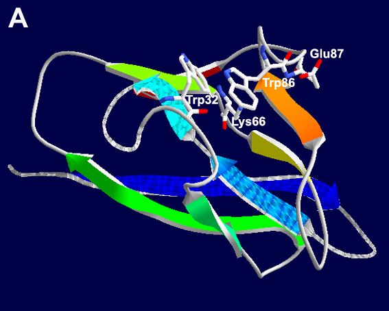
E3S Web of Conferences 245, 03054 (2021) https://doi.org/10.1051/e3sconf/202124503054
AEECS 2021
consists of a central β sheet made up of eight strands and domain. We further located the most probable three sites
10 surrounding α-helices. The predicted α/β structure for catalytic activity, Glu359, Asp361, and His374, in the
resembles a triose-phosphate-isomerase (TIM) barrel 3-D structure (Fig. 3C).
Fig. 3 Molecular 3-D model of the CBM20 (A and B) and GDPD (C) domains of cGDE5. A and B were the 3-D model of
carbohydrate-binding site1 and 2 respectively. Side chains of the conserved carbohydrate-binding sites and the GDPD catalytic active
sites were rendered in stick format.
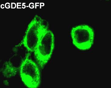
showed that cGDE5 was a cytosolically-located protein.
2.4 Expression profiles of cGDE5 in adult
chicken tissues
We used real-time quantitative PCR to detect the
expression of GDE5 mRNA in chicken tissues to
evaluate the physiological role of cGDE5. As shown in
Fig 4, the highest mRNA level was observed in heart,
and the transcript was also highly expressed in chicken
brain, skeletal muscle and testis. In contrast, there were
relative low levels in liver, kidney and adipose tissue. Fig. 5 Distribution of GFP-tagged cGDE5 protein in
18
mammalian cells.
16
Relative mRNA expression
14
(normalized to actin)
12
2.6 GPC phosphodiesterase activity assay of
10
8
cGDE5
6
4
Previous studies identified that mouse and human GDE5
2 were GPC phosphodiesterase, so cGDE5 may have the
0 same catalytic activity. First, the expression of
brain liver kidney heart skeletal
muscle
adipose testis
cGDE5-GFP was detected using the GFP antibody (Fig.
6A). Then an enzymatic assay using HEK293T cells
Fig. 4 Expression profiles of GDE5 gene in chicken various overexpressing cGDE5-GFP showed that GPC
tissues. phosphodiesterase activity was significantly increased in
cGDE5-GFP expressing cells compared with
2.5 Subcellular localization of cGDE5 GFP-expressing cells (Fig. 6B). In addition, there was no
difference in protein stability between GFP and
We transfected cGDE5 labeled with GFP at the cGDE5-GFP (data not shown). These results indicated
C-terminal and GPF into HEK293T cells to study the that GPC can be hydrolyzed by cGDE5.
subcellular distribution of cGDE5. In HEK293T cells
expressing GFP itself, intense fluorescence was
distributed throughout the cytoplasm (Fig. 5). The result
700
B *
600
GDE activity(% of control)
500
400
300
200
100
0
GFP cGDE5-GFP
Fig. 6 GPC phosphodiesterase activity of cGDE5 in mammalian cells. A, The expression of cGDE5-GFP in HEK293T cells. B,
Assay of GPC phosphodiesterase activity.
3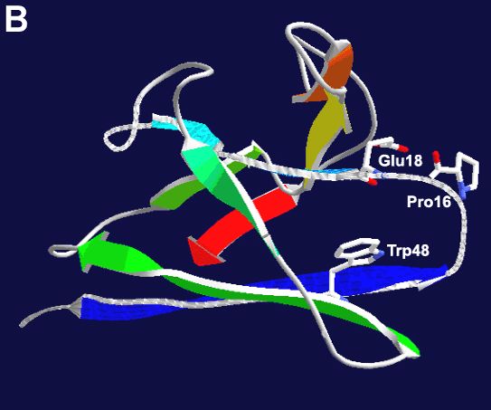
E3S Web of Conferences 245, 03054 (2021)                            https://doi.org/10.1051/e3sconf/202124503054
AEECS 2021

       Reference
       1.  Barbut S, Sosnicki AA, Lonergan SM, Knapp T,
           Ciobanu DC, Gatcliffe LJ, Huff-Lonergan E, Wilson
           EW. Progress in reducing the pale, soft and
           exudative (PSE) problem in pork and poultry meat.
           Meat Sci. 2008;79: 46–63.
       2. Burt CT, Glonek T, Bárány M. Phosphorus-31
           nuclear magnetic resonance detection of unexpected
           phosphodiesters in muscle. Biochemistry 1976;
           15:4850–4853.
       3. Younkin DP, Berman P, Sladky J Chee C, Bank W,
           Chance B. 31P NMR studies in Duchenne muscular
           dystrophy:     age-related    metabolic   changes.
           Neurology 1987;37:165–169.
       4. Sprott H, Rzanny R, Reichenbach JR, Kaiser WA,
           Hein G, Stein G. 31P magnetic resonance
           spectroscopy in fibromyalgic muscle. Rheumatology
           2000;39:1121–1125.
       5. Yanaka N. Mammalian glycerophosphodiester
           phosphodiesterases. Biosci. Biotechnol. Biochem.
           2007;71:1811-1818.
       6. Corda D, Mosca MG, Ohshima N, Grauso L, Yanaka
           N, Mariggiò S. The emerging physiological roles of
           the glycerophosphodiesterase family. FEBS J. 2014;
           281: 998-1016.
       7. Okazaki Y, Ohshima N, Yoshizawa I, Kamei Y,
           Mariggiò S, Okamoto K, Maeda M, Nogusa Y,
           Fujioka Y, Izumi T, Ogawa Y, Shiro Y, Wada M,
           Kato N, Corda D, Yanaka N. A novel
           glycerophosphodiester phosphodiesterase, GDE5,
           controls skeletal muscle development via a
           non-enzymatic mechanism. J. Biol. Chem. 2010;
           285:27652-27663.
       8. Hashimoto T, Yang B, Okazaki Y, Yoshizawa I,
           Kajihara K, Kato N, Wada M, Yanaka N.Time
           Course analysis of skeletal muscle pathology of
           GDE5       transgenic      mouse.    PLoS     One.
           2016;11(9):e0163299.
       9. Rao M, Sockanathan S. Transmembrane protein
           GDE2 induces motor neuron differentiation in vivo.
           Science 2005;309: 2212-2215.
       10. Yan Y, Sabharwal P, Rao M. The antioxidant
           enzyme Prdx1 controls neuronal differentiation by
           thiol-redox-dependent activation of GDE2. Cell
           2009; 138:1209-1221.

                                                                4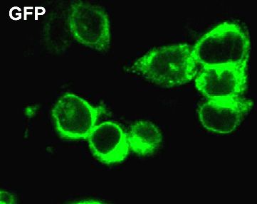
You can also read