Identification of GAPDH as a protein target of the saframycin antiproliferative agents
←
→
Page content transcription
If your browser does not render page correctly, please read the page content below
Identification of GAPDH as a protein target of the
saframycin antiproliferative agents
Chengguo Xing, Jacob R. LaPorte, Joseph K. Barbay, and Andrew G. Myers*
Department of Chemistry and Chemical Biology, Harvard University, 12 Oxford Street, Cambridge, MA 02128
Edited by Douglas C. Rees, California Institute of Technology, Pasadena, CA, and approved March 3, 2004 (received for review November 12, 2003)
Saframycin A (SafA) is a member of a class of natural products with
potent antiproliferative effects in leukemia- and tumor-derived
cells. This activity is frequently conjectured to derive from the
ability of saframycins to covalently modify duplex DNA. We used
a DNA-linked affinity purification technique to identify GAPDH as
a protein target of DNA–small molecule adducts of several mem-
bers of the saframycin class. Nuclear translocation of GAPDH occurs
upon treatment of cancer cells with saframycins, and depletion of
cellular GAPDH levels by small interfering RNA transfection confers
drug resistance. Roeder and coworkers have recently suggested
that GAPDH is a key transcriptional coactivator necessary for entry
into S phase. Our data suggest that GAPDH is also capable of
forming a ternary complex with saframycin-related compounds
and DNA that induces a toxic response in cells. These studies
implicate a previously unknown molecular mechanism of antipro-
liferative activity and, given that one member of the saframycin
class has shown efficacy in cancer treatment, suggest that GAPDH
may be a potential target for chemotherapeutic intervention.
M any small-molecule agents for cancer therapy are known to
covalently modify duplex DNA, but the pathway(s) that
may link DNA alkylation with the inhibition of cancer cell
growth are frequently not well understood. Examples are the
structurally related natural products saframycin A (SafA) (Fig.
1), a bacterial fermentation product active against various tumor
cell lines (1), and ecteinascidin (Et743) (2), a marine alkaloid
currently in advanced clinical trials for the treatment of sarcomas Fig. 1. Molecular structures of SafA, Et743, the quinaldic acid derivatives
of soft tissue and bone (3). Both natural products have been (QAD) of SafA (QADCN, X ⫽ CN; QADOH, X ⫽ OH), and phthalascidin (Pt650),
shown to bind reversibly in the minor groove of duplex DNA by an analog of Et743.
covalent bond formation between a drug-derived iminium ion
intermediate and the exocyclic amino group of guanosine resi-
tography technique. We also provide several independent lines
dues (4–10). The bis-quinone alkaloid SafA requires prior
of experimental evidence to support the hypothesis that the
reductive activation for iminium ion formation and DNA alky-
interaction of these molecules with duplex DNA and GAPDH is
lation to occur (4), whereas the nonquinoid molecule Et743 does
an integral and common component of their antiproliferative
not (10). The development of efficient and versatile synthetic
effects.
routes to each compound series has led to the discovery of
synthetic molecules with enhanced antiproliferative activities Materials and Methods
and兾or improved properties, such as the Et743 analog phthalas- Cells and Reagents. HeLa-S3 and A549 cells (American Type
cidin (Pt650) (11), and the nonquinoid, quinaldic acid derivatives Culture Collection) were cultured as monolayers in DMEM
(QAD) of SafA (Fig. 1) (12). Chemical and biological data supplemented with 10% FBS at 37°C in a 5% CO2 atmosphere.
suggest that the saframycin and Et743 compound series are Resins used were Toyopearl Oligo-Affinity Resin (Glen Re-
related, but the basis for their activity is not known (13, 14). In search, Sterling, VA), and Toyopearl AF-carboxy-650M (Tosoh,
one study, growth of a colon cancer cell line through several Tokyo). Primary antibodies used were mouse monoclonal anti-
generations of exposure to Et743 led to the isolation of a strain body to GAPDH (Research Diagnostics), goat polyclonal anti-
with reduced sensitivity to drug (IC50 ⫽ 3.7 nM, 9-fold resis- body to high-mobility group protein (HMG1), mouse monoclo-
tance); this resistance was traced to a mutation in a transcription- nal antibody to 14-3-3, and mouse monoclonal antibody to Sp1
coupled nucleotide excision repair protein, leading to the pro- (Santa Cruz Biotechnology). Mitomycin C (MMC), cisplatin,
posal that this protein provides one determinant of sensitivity to and transplatin were purchased from Sigma. Pt650 was a gift
Et743 (15). Other studies have shown that Et743 may activate from E. J. Corey (Harvard University). SafA, QADOH, and
more than one signaling pathway in cancer cells; these include a
transcription-coupled process leading to cell-cycle arrest, and
another transcription-independent pathway leading to apoptosis This paper was submitted directly (Track II) to the PNAS office.
(16). In studies of the Et743 analog Pt650, drug exposure was Abbreviations: SafA, saframycin A; Et743, ecteinascidin; QAD, quinaldic acid derivative;
shown to produce DNA–protein crosslinks, but the identities of HMG, high-mobility group; MMC, mitomycin C; siRNA, small interfering RNA; ss, single-
bound proteins were not established (17). Here, we show that stranded; ds, double-stranded; Pt650, phthalascidin.
Downloaded by guest on February 5, 2022
GAPDH is a common protein target of SafA–, QAD–, and *To whom correspondence should be addressed. E-mail: myers@chemistry.harvard.edu.
Pt650–DNA adducts by using a DNA-linked affinity chroma- © 2004 by The National Academy of Sciences of the USA
5862–5866 兩 PNAS 兩 April 20, 2004 兩 vol. 101 兩 no. 16 www.pnas.org兾cgi兾doi兾10.1073兾pnas.0307476101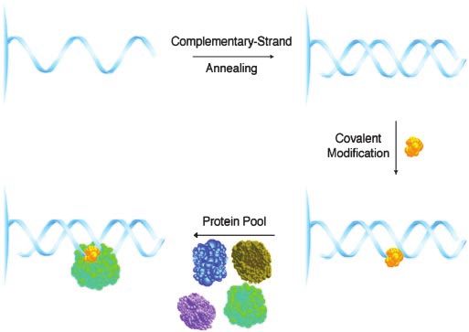
QADCN were synthesized by published procedures (12, 18). A as a probe (specific activity, 0.1 Ci; 1 Ci ⫽ 37 GBq), alone or
GAPDH small interfering RNA (siRNA) transfection kit was alkylated with one of the different small molecules of this study.
purchased from Ambion (Austin, TX). A CelLytic nuclear
extraction kit was purchased from Sigma. A CellTiter96AQ Nuclear Translocation Studies. Cytoplasmic and nuclear protein
nonradioactive cell proliferation assay kit was purchased from fractions (CelLytic nuclear extraction kit) derived from HeLa-S3
Promega. Frozen bovine brain was purchased from Pel Freez cells treated (0, 1, 2, or 3 days) with QADOH (17.5 nM) were
Biologicals (Rogers, AR). separately resolved by SDS兾PAGE (equivalent amounts of total
protein from each fraction were loaded onto the gel), and the
Construction of DNA–Small Molecule Affinity Reagents. The single- resolved protein bands were Western blotted for quantitative
stranded (ss) 26-mer oligonucleotide 5⬘-CCTTGGCCCGAGC- GAPDH detection. To ensure the integrity of the fractionation
CCGGTTCCTATT-3⬘ was synthesized on ToyoPearl Oligo- procedure, resolved protein bands from cytoplasmic and nuclear
Affinity Resin or on ToyoPearl Oligo-Affinity Resin fractions were separately Western blotted using mouse mono-
functionalized with a photocleaveable spacer phosphoramidite. clonal antibody to 14-3-3 (cytosolic protein) and mouse mono-
Hybridization to form a resin-bound duplex oligonucleotide was clonal antibody to Sp1 (nuclear protein).
achieved by annealing the resin-based 26-mer ss oligonucleotide Confocal microscopy experiments were performed by using
with the complementary 21-mer oligonucleotide 5⬘-GGAAC- control or drug-treated (17.5 nM of QADOH for 2 days)
CGGGCTCGGGCCAAGG-3⬘. Affinity resin displaying 1 nmol populations of HeLa-S3 cells. The cells were fixed, permeabil-
of double-stranded (ds) oligonucleotide was washed twice with ized, and immunolabeled according to a published procedure
buffer A (40 mM NaH2PO4, pH 5.8兾5 mM MgCl2兾0.1 mM (22). The immunolabeled cells were visualized by using a Zeiss
Na2EDTA) and to the washed resin was added 10 nmol of drug LSM510 confocal microscope.
(QAD, Pt650, and SafA) in buffer A. In the case of SafA, 1 mol
of DTT (1 M aqueous solution) was also added to the mixture GAPDH siRNA Transfection Studies. A549 cells were plated on a
of SafA and affinity resin to promote reductive activation. 24-well culture plate such that each well contained ⬇50,000 cells
Reaction suspensions were mixed on an Eppendorf Mixer 5432 in 1 ml of DMEM containing 10% FBS. Cell populations were
at 37°C for 15 h to complete formation of the DNA–small transfected with control siRNA or GAPDH siRNA according to
molecule affinity reagents. Literature procedures for the forma- the manufacturer’s recommended procedure. The cells were
tion of DNA–small molecule complexes were followed to pre- harvested and lysed after siRNA treatment for the indicated
pare MMC (19), cisplatin (20), and transplatin (20) affinity amount of time. Equal amounts of total protein from each
reagents with the modification that these reactions were mixed supernatant solution were resolved by SDS兾PAGE; resolved
on an Eppendorf Mixer 5432 at 37°C for 15 h. GAPDH was then quantified by Western blotting analysis.
MEDICAL SCIENCES
Affinity Chromatography Experiments. Bovine brain tissue (40 g) Effects of QAD or Cisplatin Treatment on the Growth of A549 Cells
was homogenized in 40 ml of buffer B (20 mM Tris䡠HCl, pH Transfected with GAPDH siRNA. A549 cells transfected with
7.4兾15 mM 2-mercaptoethanol兾0.3% Triton X-100兾0.015 mM GAPDH siRNA or a control siRNA (nontransfected cells were
pepstatin A兾0.025 mM leupeptin兾1 mM PMSF) at 4°C. The also examined as a further control) were cultured 6 h after
homogenate was centrifuged (100,000 ⫻ g) for 30 min at 4°C, and transfection in 1 ml of DMEM containing 10% FBS and a
the supernatant obtained was clarified by filtration through a defined concentration of QADCN or cisplatin in an atmosphere
Bio-spin column (Bio-Rad). Lysate from an A549 tumor-cell line of 95% air兾5% CO2 for 72 h at 37°C. Viable cells in each well
was prepared by incubating A549 cells (250-l packed cell were quantified by using the CellTiter96AQ nonradioactive cell
volume) with 1 ml of buffer C (50 mM Tris䡠HCl, pH 7.4兾1% proliferation assay kit following the manufacturer’s recom-
CHEMISTRY
NP-40兾0.25% sodium deoxycholate兾150 mM NaCl兾1 mM mended procedure. Percent cell survival versus log drug con-
Na3VO3兾1 mM NaF兾1 mM EGTA) for 1 h at 4°C on an centration was plotted in each case; GI50 values were calculated
end-over-end rotator. To reduce nonspecific binding of proteins, by interpolation using the cubic spline method.
protein pools from clarified bovine homogenate or A549 cell
lysate were preincubated for 1 h at 4°C with a guard column Results and Discussion
prepared by conjugation of Toyopearl AF-carboxy-650M resin Development of an Affinity Technique for the Identification of Pro-
with ethanolamine in the presence of 1-ethyl-3-(3⬘-dimethyl- teins Recognizing DNA–Small Molecule Adducts. To identify proteins
aminopropyl)carbodiimide hydrochloride. After this preincuba- that bound DNA–small molecule alkylation products, we devel-
tion step, treated bovine brain homogenate or A549 cell lysate oped a technique for the preparation of a series of DNA-linked
was filtered through a Bio-spin column to further reduce the affinity reagents that was initiated by the automated synthesis of
level of nonspecific protein binding. Bovine brain lysate or A549 a 26-mer ss oligonucleotide from a commercial solid support
cell lysate pretreated in this fashion was then incubated with bearing chemically or photochemically cleavable linkers, fol-
poly(dI-dC) (a nonspecific competitor) and a DNA–small mol- lowed by 21-mer complementary-strand hybridization (Scheme
ecule affinity reagent at 4°C for 15 h on an end-over-end rotator. 1). The DNA duplex that was produced contained multiple
After washing the resin three times with buffer D (20 mM known binding sites for the saframycin class of antiproliferative
Tris䡠HCl, pH 7.4兾15 mM 2-mercaptoethanol兾0.3% Triton agents as well as cisplatin, transplatin, and MMC, used as
X-100兾0.5 M NaCl), bound proteins were released from the controls, and was linked to the resin at the cleavable site by way
affinity resin by heat denaturation at 95°C for 5 min, the of an intervening, unpaired sequence TATTT. Conditions for
denatured proteins were resolved by SDS兾PAGE (4–20% gra- optimal binding of each drug to the duplex were then developed,
dient gels), and protein bands were visualized by silver staining guided by gel-shift characterization data for each DNA–small
or identified by protein mass spectrometry sequencing and molecule complex after photochemical cleavage from the resin.
Western blotting. Affinity columns prepared from the quinone-containing mole-
cules SafA and MMC used DTT for reductive alkylation,
Southwestern Blotting Experiments. Southwestern blotting exper- whereas no such activation was required for QAD, Pt650, or
iments were performed according to a published procedure (21) platinum-based alkylating agents. Exposure of the duplex DNA–
by using purified human GAPDH or human HMG1 in the assay. small molecule affinity columns to protein pools derived initially
Downloaded by guest on February 5, 2022
A 32P-labeled 21-mer dsDNA corresponding in sequence to the from bovine-brain lysate and, later, from human A549-cell
duplex region of DNA–small molecule affinity reagents was used lysate, followed by washing of nonbound proteins from the
Xing et al. PNAS 兩 April 20, 2004 兩 vol. 101 兩 no. 16 兩 5863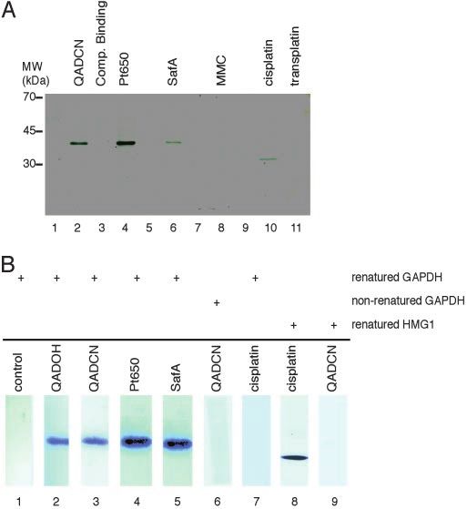
Scheme 1. DNA-linked affinity reagents for the isolation of proteins that
                                          bind small molecule-DNA adducts. The resin-bound 26-mer ss-oligonucleotide
                                          (5⬘-CCTTGGCCCGAGCCCGGTTCCTATTT-3⬘) was prepared by automated syn-
                                          thesis on Toyopearl Oligo-Affinity Resin. Complementary-strand annealing
                                          with the 21-mer ss-oligonucleotide 5⬘-GGAACCGGGCTCGGGCCAAGG-3⬘ fol-
                                          lowed by covalent modification of the resulting DNA duplex with DNA-
                                          reactive small molecules (represented in yellow) provided affinity reagents
                                          used to identify specific binding proteins within a pool derived from tissue
                                          lysate.

                                                                                                                         Fig. 2. Identification of proteins recognizing duplex DNA-small molecule
                                          column, release of bound proteins by heat denaturation, and                    adducts by a DNA-linked affinity chromatography technique and confirma-
                                          separation of the latter by SDS兾PAGE, allowed for the identi-                  tion of a direct binding interaction of the protein GAPDH by Southwestern
                                          fication of proteins that recognized each DNA–drug conjugate                   blot analysis. (A) SDS兾PAGE of proteins bound in affinity chromatography
                                          specifically. Preincubation of the protein pools with two guard                experiments using a protein pool derived from bovine-brain lysate. Lane 1,
                                          columns in series, incorporation of poly(dI-dC) during protein                 dsDNA resin with no drug modification: control matrix to lanes 2– 4; lane 2,
                                          binding to the affinity column, and the use of optimal salt and                QADCN-alkylated dsDNA resin plus 40 equivalents (equiv) poly(dI-dC); lane 3,
                                                                                                                         QADCN-alkylated dsDNA resin plus 40 equiv dsDNA-QADCN complex; lane 4,
                                          protein concentrations essentially abolished any nonspecific
                                                                                                                         Pt650-alkylated dsDNA resin plus 40 equiv poly(dI-dC); lane 5, dsDNA resin
                                          protein binding (identified by comparison with appropriate                     with no drug modification, control matrix to lane 6; lane 6, SafA-alkylated
                                          controls employing DNA duplexes that had not been exposed to                   dsDNA resin plus 40 equiv poly(dI-dC); lane 7, dsDNA resin with no drug
                                          drug). Specific protein-binding interactions were confirmed by                 modification, control matrix to lane 8; lane 8, MMC-alkylated dsDNA resin plus
                                          competitive binding studies with solutions of free duplex DNA                  40 equiv poly(dI-dC); lane 9, dsDNA resin with no drug modification, control
                                          alkylated by the appropriate drug. The identities of bound                     matrix to lanes 10 and 11; lane 10, cisplatin-alkylated dsDNA resin plus 40
                                          proteins were established by mass spectrometric protein se-                    equiv poly(dI-dC); lane 11, transplatin-alkylated dsDNA resin plus 40 equiv
                                          quencing of bands isolated by SDS兾PAGE, and were confirmed                     poly(dI-dC). (B) Results of Southwestern blotting experiments. Purified human
                                                                                                                         GAPDH or HMG1 was resolved by SDS兾PAGE, electroblotted onto nitrocellu-
                                          by Western blotting experiments.                                               lose, and then renatured (except for lane 6) on the membrane. The renatured
                                             Results obtained by using affinity reagents prepared from                   blots were probed with a 32P-labeled 21-mer dsDNA (sequence: Scheme 1)
                                          cisplatin and transplatin and a protein pool derived from bovine               unmodified (lane 1) or alkylated with the various compounds of study (as
                                          brain established the potential of DNA-linked, small-molecule                  indicated, lanes 2–9).
                                          modified affinity reagents to identify specific binding proteins, in
                                          that the cisplatin-derived reagent provided a single major pro-
                                          tein band, shown to be the 33-kDa (bovine) high-mobility group                 whose sequence was determined by mass spectrometry; com-
                                          protein HMG1, whereas no such binding of HMG1 (nor any                         parison of the sequence with a protein database showed the
                                          other protein) was observed to occur with the transplatin-                     38-kDa binding protein to be GAPDH. This identification was
                                          derived reagent (Fig. 2A). HMG1 binding was not observed to                    confirmed by Western blotting. Binding of GAPDH was specific
                                          occur with affinity reagents prepared from SafA, QAD, Pt650,                   (binding did not occur in the absence of small-molecule modi-
                                          nor MMC. HMG1 had previously been determined to be a                           fication of the DNA affinity reagent) and was subject to com-
                                          potential protein target of cisplatin–DNA adducts by sequence                  petitive displacement with the appropriate free dsDNA–drug
                                          analysis of cDNA isolated in an expression-library screening                   complexes. GAPDH of human origin was also shown to bind
                                          (23). Cisplatin–DNA adducts (but not transplatin–DNA ad-                       selectively to affinity columns derived from QAD and Pt650 (but
                                          ducts) were subsequently shown to bind HMG1 specifically (24);                 not cisplatin, transplatin, or mitomycin C) by using a protein pool
                                          this binding has been proposed to form the chemical basis of                   derived from the lysate of a human cancer cell line (A549) for
                                          cisplatin antitumor activity and, similarly, the lack of HMG1                  binding studies.
                                          binding to account for the poor clinical efficacy of transplatin in
                                          cancer therapy.                                                                Confirmation That Direct Binding Occurs Between GAPDH and Safra-
                                                                                                                         mycin–DNA Adducts by Southwestern Blotting Experiments. To es-
                                          Identification of GAPDH as a Protein Target of DNA Adducts of the              tablish that the binding of GAPDH to SafA–, QAD–, or
                                          Saframycin Antiproliferative Agents. Although affinity columns                 Pt650–DNA affinity columns was the result of a direct interac-
Downloaded by guest on February 5, 2022

                                          derived from SafA, QAD, and Pt650 did not bind HMG1, they                      tion, and had not been mediated by another, relatively less
                                          were found to bind a different, 38-kDa bovine protein (Fig. 2 A)               abundant protein that was not detected by SDS兾PAGE, we

                                          5864 兩 www.pnas.org兾cgi兾doi兾10.1073兾pnas.0307476101                                                                                                Xing et al.
MEDICAL SCIENCES
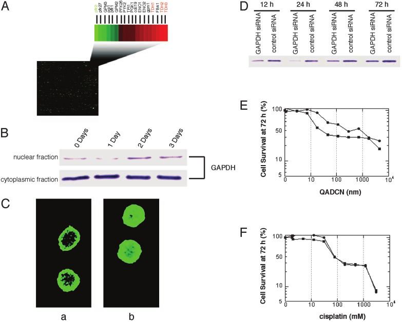
Fig. 3. The role of GAPDH in the response of eukaryotic cells to treatment with saframycin analogs (QAD). (A) Transcription-profiling studies performed in a
SafA-sensitive yeast strain show that all three isoforms of GAPDH (TDH1, TDH2, and TDH3) are substantially up-regulated after treatment with QADCN or SafA,
whereas the gene transcript for phosphfructokinase, pfk2, catalyzing the principle, rate-limiting step of glycolysis is down-regulated ⬎2-fold. Transcripts for each
of the histones were down-regulated ⬎2-fold (14). (B) Western blots for the determination of GAPDH levels in the nuclear and cytoplasmic fractions of HeLa-S3
cells treated with QADOH for the indicated times. (C) Confocal microscopy of HeLa-S3 cells treated (b) or untreated (a) with QADOH (17.5 nM, 48 h) and visualized
by using a standard FITC-conjugated antibody immunostaining protocol. Cell nuclei were visualized separately by Hoechst dye 33340 staining (not shown). (D)
GAPDH levels in A549 cells that had been transfected with GAPDH siRNA or control siRNA, respectively, as determined by immunoblotting. (E) Percentage of
viable cells transfected with GAPDH siRNA (filled circle) or control siRNA (filled square) after 72-h treatment with varying concentrations of QADCN. (F) As in E,
but treated with cisplatin in lieu of QADCN.
CHEMISTRY
conducted Southwestern blotting experiments using human catalyzes the conversion of glyceraldehyde-3-phosphate and
erythrocyte GAPDH (tetrameric form; molecular mass, 152 NAD⫹ into 3-bisphosphoglycerate and NADH. We found that
kDa) that was submitted to (denaturing) SDS兾PAGE, blotted duplex DNA–QAD adducts did not inhibit the ability of tet-
onto a nitrocellulose membrane, and renatured. Membranes rameric human erythrocyte GAPDH to catalyze the conversion
were probed with the alkylation products of QAD, SafA, Pt650, of glyceraldehyde-3-phosphate to 1,3-bisphosphoglycerate in an
or cisplatin and a 32P-labeled 21-mer dsDNA corresponding in in vitro assay, consistent with the idea that a different function
sequence to the duplex region of our affinity columns, and were of GAPDH is targeted by saframycin–DNA adducts. Consider-
subsequently visualized by autoradiography. QAD-, SafA-, and ing only its function in glycolysis, GAPDH may appear to be an
Pt650-conjugated radiolabeled oligonucleotides were shown to unlikely target for an anticancer drug. However, GAPDH has a
bind membrane-bound, renatured GAPDH, whereas cisplatin- complex and evolving role in the nucleus, where it has been
conjugated and nonmodified oligonucleotides did not (Fig. 2B). implicated to function as a monomer (25). GAPDH has been
Conversely, the cisplatin-conjugated oligonucleotide did bind identified as a component of a multiprotein nuclear complex that
membrane-bound human HMG1, but the QAD-conjugated
recognizes oligonucleotides incorporating the antileukemic
DNA probe did not (Fig. 2B). Also, the QAD-conjugated DNA
agent thioguanosine (26, 27), although GAPDH has not been
probe did not bind nonrenatured membrane-bound human
GAPDH, suggesting that only folded GAPDH is recognized. shown to bind directly to thioguanosine-modified DNA. Mono-
Interestingly, purified human erythrocyte GAPDH was found meric GAPDH has also recently been found to be the principle
not to bind saframycin DNA-linked affinity columns, but binding component of a multiprotein nuclear complex involved in tran-
did occur after the tetrameric protein was subjected to a scriptional coactivation of the histone 2B promoter, required for
denaturation–renaturation protocol. These findings, in conjunc- S phase progression in the cell cycle, whereas tetrameric
tion with results from Southwestern blotting experiments, lead us GAPDH does not function in transcriptional coactivation (28).
to conclude that a form of GAPDH other than the tetramer is In light of these findings, we note that our earlier transcription
recognized by saframycin–DNA adducts, perhaps the monomer. profiling experiments using a SafA-sensitive strain of yeast
exposed separately to SafA and QAD established in both cases
Saframycin–DNA Adducts Do Not Inhibit the Glycolytic Function of that the greatest degree of overexpression in response to drug
Downloaded by guest on February 5, 2022
GAPDH. Human GAPDH is most commonly associated with treatment occurred among transcripts for the three isoforms of
glucose metabolism. In its native tetrameric form, GAPDH GAPDH (1.7- to 3.8-fold), and that transcripts encoding each of
Xing et al. PNAS 兩 April 20, 2004 兩 vol. 101 兩 no. 16 兩 5865the histones were substantially down-regulated (⬎2-fold, Fig. levels (95% depletion versus control) was observed at 24 h;
3A) (14). thereafter, GAPDH levels partially recovered (50% versus
control at 72 h, Fig. 3D). GAPDH-depleted cells were found to
Exposure of Cancer Cells to Saframycins Leads to Translocation of be 8-fold resistant to growth inhibition by QADCN relative to
GAPDH to the Nucleus. To explore the potential role of nuclear controls with normal GAPDH levels (measured as percent cell
GAPDH in the antiproliferative activity of saframycin-like mol- survival at 72 h) (Fig. 3E). Furthermore, experiments using
ecules, we sought to determine whether exposure of cancer cells cisplatin in lieu of QADCN showed that there was no difference
to these agents would lead to translocation of GAPDH to the in sensitivity to growth inhibition by cisplatin in GAPDH-
nucleus. We conducted both confocal microscopy and cellular depleted and control cell populations (Fig. 3F). If GAPDH
fractionation experiments in a human cancer cell line (HeLa-S3) functioned as a general mediator of apoptosis, it might have been
exposed to the analog QADOH. Both types of experiments anticipated that the growth of cells treated with cisplatin would
showed that nuclear GAPDH levels were elevated relative to have been affected as well, although this need not necessarily
controls in which QADOH was not present (Fig. 3). Cellular have been the case, given the complexities of apoptotic signaling
fractionation studies showed that nuclear translocation of pathways.
GAPDH was maximal after ⬇48 h of drug exposure (2-fold
increase, Fig. 3B). Despite the increased translocation of Discussion. There is increasing evidence to suggest that GAPDH
GAPDH into the nucleus, it should be noted that by far the functions in one or more critical nuclear processes (25, 28). The
larger proportion of cellular GAPDH remains in the cytosol demonstration here that a specific binding interaction occurs
even after drug treatment (Fig. 3B).† Cellular microscopy studies between GAPDH, duplex DNA, and several known members of
further confirmed that translocation of GAPDH to the nucleus the saframycin class of antiproliferative agents both implicates a
occurred upon drug treatment (Fig. 3C). Visually, the cells previously unknown mechanism of action for the compound
exhibited morphological signs of apoptosis after drug treatment class and identifies GAPDH as a potential target for chemo-
(membrane blebbing). Additional evidence for an apoptotic therapeutic intervention. That the mechanism of action of the
pathway was obtained by the treatment of A549 cells with saframycins involves a direct interaction with GAPDH and DNA
QADOH, which produced a characteristic ladder of cleaved was not expected, but is supported herein by affinity experiments
chromosomal DNA. and Southwestern binding studies, by the fact that drug exposure
leads to an increased concentration of GAPDH in the nucleus,
Depletion of Cellular GAPDH by RNA Interference Confers Resistance and by the fact that depletion of cellular GAPDH levels confers
to Saframycins. We sought to determine whether altering the drug resistance. The latter finding may be of significance, in that
levels of GAPDH in human cancer cells would influence their many tumor cells have been shown to overexpress GAPDH (30),
sensitivity to saframycin compounds and so modulated GAPDH providing a potential basis for tumor-cell specificity in cell
levels by the reverse-genetic method of RNA interference killing. Exactly how a ternary complex of GAPDH, dsDNA, and
(siRNA). Treatment of human non-small cell lung cancer cells a small-molecule agent of the saframycin class is toxic to the cell
(A549) with a commercial siRNA transfection vector targeting is not evident. That it is becomes all of the more intriguing in
GAPDH led to time-dependent reduction of cellular GAPDH light of recent evidence implicating a role for GAPDH in
levels relative to a negative control cell population (treated with transcriptional coactivation necessary for S-phase progression of
siRNA of a similar base composition, but with no homology to the cell (28), but it is unclear at present whether or how these
any known coding sequence). Maximal diminution of GAPDH observations are related.
We thank Professor E. J. Corey for a generous gift of Pt650 and the
†Cytosolicconcentrations of GAPDH are typically ⬎1,000-fold higher than the concentra-
Harvard Microchemistry Facility for protein sequence analysis. We
tions of saframycins used in these studies (GI50 values of saframycins in most cancer cell
lines range from 1 to 100 nM; a typical measure of GAPDH in the cell is ⬇70 M) (29). These
gratefully acknowledge the National Institutes of Health for financial
calculations, plus the fact that saframycin–DNA adducts do not inhibit the glycolytic support, the Howard Hughes Medical Institute for a Predoctoral fel-
function of GAPDH, point toward a mechanism of GAPDH-mediated cytotoxicity that is lowship (to J.R.L.), and the American Cancer Society for a graduate
not related to an interference with glycolytic metabolism. research fellowship (to J.K.B.).
1. Arai, T. & Kubo, A. (1983) in The Alkaloids, ed. Brossi, A. (Academic, New 16. Gajate, C., An, F. & Mollinedo, F. (2002) J. Biol. Chem. 277, 41580–41589.
York), pp. 56–98. 17. Martinez, E. J., Owa, T., Schreiber, S. L. & Corey, E. J. (1999) Proc. Natl. Acad.
2. Rinehart, K. L., Holt, T. G., Fregeau, N. L., Keifer, P. A., Wilson, G. R., Perun, Sci. USA 96, 3496–3501.
T. J., Jr., Sakai, R., Thompson, A. G., Stroh, J. G., Shield, L. S., et al. (1990) 18. Myers, A. G. & Kung, D. W. (1999) J. Am. Chem. Soc. 122, 10828–10829.
J. Nat. Prod. 53, 771–792. 19. Tomasz, M., Chawla, A. K. & Lipman, R. (1988) Biochemistry 27, 3182–3187.
3. Aune, G., Furuta, T. & Pommier, Y. (2002) Anti-Cancer Drugs 13, 545–555. 20. Mansy, S., Rosenberg, B. & Thomson, A. J. (1973) J. Am. Chem. Soc. 95,
4. Ishiguro, K., Sakiyama, S., Takahashi, K. & Arai, T. (1978) Biochemistry 17, 1633–1640.
2545–2550. 21. Philippe, J. (2000) in The Nucleic Acid Protocols Handbook, ed. Rapley, R.
5. Ishiguro, K., Takahashi, T., Yazawa, K., Sakiyama, S. & Arai, T. (1981) J. Biol. (Humana, Totowa, NJ), pp. 773–782.
Chem. 256, 2162–2167. 22. Allan, V. J. (2000) in Protein Localization by Fluorescence Microscopy, ed. Allan,
6. Lown, J. W., Joshua, A. V. & Lee, J. (1982) Biochemistry 21, 428–436. V. J. (Oxford Univ. Press, Oxford), pp. 1–26.
7. Rao, K. E. & Lown, J. W. (1990) Chem. Res. Toxicol. 3, 262–267. 23. Toney, J. H., Donahue, B. A., Kellett, P. J., Bruhn, S. L., Essigmann, J. M. &
8. Rao, K. E. & Lown, J. W. (1992) Biochemistry 31, 12076–12082. Lippard, S. J. (1989) Proc. Natl. Acad. Sci. USA 86, 8328–8332.
9. Hill, G. C. & Remers, W. A. (1991) J. Med. Chem. 34, 1990–1998. 24. Pil, P. M. & Lippard, S. J. (1992) Science 256, 234–237.
10. Pommier, Y., Kohlhagen, G., Bailly, C., Waring, M., Mazumder, A. & Kohn, 25. Sirover, M. A. (1999) Biochim. Biophys. Acta 1432, 159–184.
K. W. (1996) Biochemistry 35, 13303–13309. 26. Krynetski, E. Y., Krynetskaia, N. F., Gallo, A. E., Murti, K. G. & Evans, W. E.
11. Martinez, E. J. & Corey, E. J. (1999) Org. Lett. 1, 75–77. (2001) Mol. Pharmacol. 59, 367–374.
12. Plowright, A. T. & Myers, A. G. (2001) J. Am. Chem. Soc. 123, 5114–5115. 27. Krynetski, E. Y., Krynetskaia, N. F., Bianchi, M. E. & Evans, W. E. (2003)
13. Martinez, E. J., Corey, E. J. & Owa, T. (2001) Chem. Biol. 8, 1151–1160. Cancer Res. 63, 100–106.
14. Plowright, A. T., Schaus, S. E. & Myers, A. G. (2002) Chem. Biol. 9, 607–618. 28. Zheng, L., Roeder, R. G. & Luo, Y. (2003) Cell 114, 255–266.
15. Takebayashi, Y., Pourquier, P., Zimonjic, D. B., Nakayama, K., Emmert, S., 29. Furfine, C. S. & Velick, S. F. (1965) J. Biol. Chem. 240, 844–855.
Ueda, T., Urasaki, Y., Kanzaki, A., Akiyama, S., Popescu, N., et al. (2001) Nat. 30. Vila, M. R., Nicolas, A., Morote, J., de Torres, I. & Meseguer, A. (2000) Cancer
Med. 7, 961–966. 89, 152–164.
Downloaded by guest on February 5, 2022
5866 兩 www.pnas.org兾cgi兾doi兾10.1073兾pnas.0307476101 Xing et al.You can also read