In Situ Study of Cave 98 Murals on Dunhuang Grottoes Using Portable Laser-Induced Breakdown Spectroscopy
←
→
Page content transcription
If your browser does not render page correctly, please read the page content below

ORIGINAL RESEARCH
                                                                                                                                               published: 11 March 2022
                                                                                                                                         doi: 10.3389/fphy.2022.847036

                                             In Situ Study of Cave 98 Murals on
                                             Dunhuang Grottoes Using Portable
                                             Laser-Induced Breakdown
                                             Spectroscopy
                                             Yaopeng Yin 1†, Zongren Yu 1*†, Duixiong Sun 2†, Zhongwei Shan 1, Qiang Cui 1, Yiming Zhang 2,
                                             Yaqi Feng 1, Biwen Shui 1, Zhuo Wang 1, Zhiyuan Yin 1, Bolong Chai 1, Wenyuan Zhang 1,
                                             Chenzhong Dong 2 and Bomin Su 1*
                                             1
                                               National Research Center for Conservation of Ancient Wall Paintings and Earthen Sites, Dunhuang, China, 2Key Laboratory of
                                             Atomic and Molecular Physics and Functional Materials of Gansu Province, College of Physics and Electronic Engineering,
                                             Northwest Normal University, Lanzhou, China

                                             The investigation of painted layers on murals at Cave 98 from Dunhuang Grottoes was
                                             carried out in situ using portable laser-induced breakdown spectroscopy (LIBS) for the first
                          Edited by:         time. The ablation effect of laser pulses on a mural surface was evaluated under fixed
                            Yufei Ma,        experimental parameters, and the results showed that the influence of laser ablation on
Harbin Institute of Technology, China
                                             ancient murals was acceptable. Then the pigments used in the red, green, and blue layers
                        Reviewed by:
                          Mingyin Yao,
                                             were indicated with the LIBS spectral data of the corresponding color coupled with a
 Jiangxi Agricultural University, China      classifiable model of pigments based on the principal component analysis (PCA) method.
                        Hongbin Ding,
                                             Finally, the depth profiling of the multilayer structure composed with overlapped painted
Dalian University of Technology, China
                                             layers was determined based on the pigment size information of the superficial green layer
                   *Correspondence:
                          Zongren Yu         and the relationship of laser shots and ablation depth, and the thickness of the superficial
                   13588975@qq.com           green layer was analyzed quantitatively, which fits well with the result of the cross-sectional
                           Bomin Su
                suboming@hotmail.com
                                             analysis. Therefore, this work can shed light on the great potential for ancient mural
     †
         These authors have contributed
                                             applications in LIBS.
                    equally to this work
                                             Keywords: Mogao Grottoes, Dunhuang murals, portable LIBS, in situ analysis, painted layers

                      Specialty section:
           This article was submitted to
                  Optics and Photonics,      INTRODUCTION
                 a section of the journal
                      Frontiers in Physics   As an important artistic crystallization of various cultural integrations on the ancient Silk Road, the
           Received: 01 January 2022         Mogao Grottoes is the largest, oldest, and the best-preserved Buddhist grottoes with the richest
           Accepted: 25 January 2022         contents in the world today, so it is also named “Library on the Wall” because it carries vast historical,
            Published: 11 March 2022         cultural, and scientific information. The UNESCO World Heritage Site (inscribed the List in 1987)
                             Citation:       contains 735 caves excavated into the 1.6 km of cliff face, encompassing approximately 45,000 m2 of
  Yin Y, Yu Z, Sun D, Shan Z, Cui Q,         chromatic murals and more than 2,000 painted sculptures [1].
   Zhang Y, Feng Y, Shui B, Wang Z,             The Mogao Grottoes is famous for its massive chromatic murals as shown in Figure 1, which were
Yin Z, Chai B, Zhang W, Dong C and           drawn using all kinds of colorful mineral pigments on dry ground according to the secco technique
Su B (2022) In Situ Study of Cave 98
                                             [2]. However, these precious murals are suffering from damage due to the influence of air, light,
 Murals on Dunhuang Grottoes Using
               Portable Laser-Induced
                                             humidity in grottoes, and human factors over a long time period [3]. The composition information of
            Breakdown Spectroscopy.          mineral pigments used on painted layers is the basis for revealing reasons for these different types of
              Front. Phys. 10:847036.        damage, such as flaking, detachment, net cracking, and paint loss; therefore, it can provide guidance
      doi: 10.3389/fphy.2022.847036          for selecting the most appropriate materials and conservation methods, along with determination of

Frontiers in Physics | www.frontiersin.org                                          1                                          March 2022 | Volume 10 | Article 847036
Yin et al.                                                                                                               In-Situ Murals Analysis With LIBS

  FIGURE 1 | Photograph of Mogao Grottoes and typical ancient murals in these caves.

suitable processes for possible restoration of the paintings. On the              utilizing the compact portable LIBS system. In fact, ancient
other hand, the information can give a rich source of exchanges                   murals are types of art painted on the wall around caves,
involved and cultural trade development from the perspective of                   which resulted in these immovable murals being not permitted
scientific investigations of ancient murals made from different                    to be taken to the laboratory for analysis, so the portable LIBS
pigments. Therefore, pigment research on Dunhuang ancient                         system was regarded as a useful tool to carry out painted layer
murals has become a focus of great concern.                                       research of murals in situ and real time in Mogao Caves.
    With the development of laser technology, the laser                               Therefore, in this work, a portable LIBS was used to analyze
measurement techniques have attracted a considerable amount                       the information of mural painted layers on-site in a cave for the
of research in the area due to its technical features, such as high               first time. For this study, the murals on the south wall of Cave 98
sensitivity, noninvasive, and in situ [4–7]. Laser-induced                        were chosen to be analyzed; the analytical area is approximately
breakdown spectroscopy (LIBS) technology has great                                1.8 m2 (about 2.0 m × 0.9 m). The laser ablation on the mural was
application potential in the field of cultural heritage analysis                   illustrated using optical microscope. Based on our previous
with the advantages of rapid, multi-element analysis, no                          experimental results, the different types of pigments were
sampling, minimal damage, and in situ analysis [8–10], which                      indicated and classified by combing the LIBS spectrum with a
meet the strict requirements for cultural objects. Based on the                   classifiable model. Furthermore, the depth profile of overlap
contribution significantly from ongoing advances in laser,                         painted layers was revealed, and the thickness of these layers
spectrometer, and detector technologies, the development of                       was also analyzed in Cave 98.
several mobile LIBS instruments provides a lot of
opportunities for analytical campaigns on site, such as at
museums, conservation laboratories and even outdoors at                           EXPERIMENTAL APPROACH AND MURAL
excavation sites [11, 12]. In a different investigation, several                  SAMPLES
Minoan bronze-age artifacts such as beads, vessels, and
decorative plaques and figurines made of faience were                              Portable Laser-Induced Breakdown
analyzed by mobile LIBS [13]. The main objective of different                     Spectroscopy Apparatus
types of stones, which are the main construction materials in this                The schematic and imaging of the portable LIBS setup are
class of historical buildings, was qualitatively discriminated                    presented in Figure 2. In our studies, all spectrum data were
among sandstone, limestone, and cement mortar [14–19]. An                         obtained using the portable LIBSCAN 100 ULTRA, which offers a
interesting analysis has enabled the identification of the main                    highly versatile, adaptable, and upgradeable product suitable for
minerals present in several building stones using a compact                       use either in a laboratory environment or in the field. The main
system based on a stand-off LIBS sensor [20]. The objectives                      components of the LIBSCAN 100 ULTRA are the LIBSCAN 100
of jewelry [21], glass artifacts [22], ceramics, and pottery [23, 24]             console and LIBSCAN head [31].
were investigated using LIBS technique. In some different studies                    The LIBSCAN 100 console contains the optical spectrometers
with a similar objective, painting [25, 26], pigments on                          with eight channels and electrical circuits for the laser safety
illuminated manuscripts [27] simulated murals [28], and wall                      interlock. The detection spectra were collected within the
painting fragments [29, 30] were carried out a quick survey                       wavelength range from 200 to 800 nm, with a maximum

Frontiers in Physics | www.frontiersin.org                                    2                                   March 2022 | Volume 10 | Article 847036
Yin et al.                                                                                                                  In-Situ Murals Analysis With LIBS

  FIGURE 2 | Schematic and imaging of the portable laser-induced breakdown spectroscopy (LIBS) setup.

  FIGURE 3 | Areas of ancient murals in the south wall of Cave 98 about 2.0 m × 0.9 m.

resolution of 0.07 nm in the range of 410 and 500 nm. The LIBS                      donor images were presented in the areas under the chamber wall,
spectra were recorded with a delay time of 1,800 ns and a gate                      as shown in Figure 3.
width of 3,000 ns with respect to the laser pulse.                                      There are 223 visible donor images included according to
   The LIBSCAN head contains the laser and associated optics                        statistical survey, and the species of pigments used in these areas
required to focus the laser beam onto a sample, and the plasma                      are particularly abundant, the red with different tones, and green,
light was collected for transmission to the spectrometers located                   white, and blue are all existing in these areas. So, the female donor
within the LIBSCAN 100 console. The head is designed to                             images in the south wall of Cave 98 were chosen to be analyzed. In
accommodate a miniature CCD camera, and associated                                  our current studies, the red, blue, and green mineral pigments
components are used with the optional imaging kit. A Quantel                        were selected to be investigated using LIBS. The test locations are
Big Sky CFR Ultra GRM and ICE 450 cooling group was used to                         illustrated in Figure 3 indicated by black circles.
produce laser light at 1,064 nm, generating 7-ns pulses, laser pulse
energy up to 100 mJ, repetition rate of 1–10 Hz, and in a 6.5 mm
diameter beam.                                                                      RESULTS AND DISCUSSIONS
Mural Samples                                                                       Laser Ablation Effect
Mogao Cave 98 is located on the first floor of the southern section                   Considering that a small amount of material is removed involving
of the South District. The cave was built in about 925 AD by a                      the laser ablated on the sample surface in an LIBS measurement
governor of the Return-to-Allegiance Army named Cao Yijin.                          (mass removal about a few nanograms), so the LIBS is termed as a
There are about 693.1 m2 murals on wall around the cave. The                        microdestructive technique. Therefore, it is necessary to analyze
outstanding feature of murals in the cave is that a large number of                 the influence of laser ablation on the surface ancient murals in

Frontiers in Physics | www.frontiersin.org                                      3                                    March 2022 | Volume 10 | Article 847036

Yin et al.                                                                                                                          In-Situ Murals Analysis With LIBS

  FIGURE 4 | Photographs of laser-ablated area after five laser pulses on the same spot at different distances from the surface.

situ before carrying out measurements. It is notable that the                         into two different parts, the penetrated area located inside the
energy of the laser pulse was fixed at 10 mJ according to the study                    area caused the Gaussian profile laser and the discoloration area
of laser ablation, which prevents excessive damage to the                             caused by the thermal effect with laser ablation. It is clear that the
murals [26].                                                                          diameter values of ablated areas are 246 ± 11 and 272 ± 25 μm on
    Under the optimized experimental parameters, the laser-                           the green and red layers, respectively. It is notable that the range
ablated areas in red- and green-painted layers were visualized                        of ablation area on the green layer is indicated as the penetrated
by digital photographs and images of optical microscopy.                              area. Actually, the color in the region of the laser ablation has
Figure 4 presents photographs of the laser ablated area after                         become black in the green-painted layer, the laser ablation area in
five laser pulses ablated on the same spot, and these pictures were                    the red layer penetrated in the center, and the edge range of the
taken at different distances from the mural surface. As shown in                      ablation crater started to turn in color from red to black in the
Figure 4, the values of d indicate the distances from camera to                       diameter range of 655 ± 50 μm. Furthermore, the edge
mural surface, for example, d = 0.5 m meant that the picture was                      morphology of the ablated crater was analyzed, and the
recorded at the location of 0.5 m from the surface. The picture at                    microscopy pictures showed that the interaction of laser pulse
the position of d = 0.5 m is a key reference to evaluate the effect of                and painted layers did not cause significant changes in the depth
laser ablation because most visitors and researchers are                              profile structure of the painted layer. This meant that the effect of
prohibited from going into this range [32]. From the picture,                         laser ablation on the stability of ancient murals was negligible.
the trace left on the object surface is hardly visible with the naked                    Based on the above statement, the portable LIBS technique
eye, so the laser-ablated crater cannot be observed outside of this                   was, therefore, feasible in the field of mural pigment analysis from
distance range at all. So, the minor damage was regarded as                           the views of mural appreciation and stability of painted layers.
acceptable and agreed on from the perspective of appreciation of
ancient murals.                                                                       Pigments Indication and Classification
    From the images of optical microscopy, which were taken at                        Most of the pigments used in Dunhuang murals were
200× magnification, there are obvious differences in laser-ablated                     manufactured from colored mineral materials, such as red
areas between red and green layers. As can be seen from Figure 4,                     pigments of cinnabar, red lead, and hematite, green pigments
the ablation effect in the green and red layer is different due to the                of malachite and atacamite, and blue pigments of lapis lazuli and
various physicochemical properties of the two kinds of mineral                        azurite. In order to obtain stable spectral data and improve the
pigments, that is, the red-painted layer was not penetrated after                     accuracy of the measurement on mural-painted layers with
laser ablation under the fixed parameters; only the discoloration                      different colors, the average method of cumulative pulse
was observed on the red layer, so, the ablation area is defined as                     ablation was employed to weaken the impact of sample
the range of black area. However, the green layer was penetrated                      surface irregularities on spectral fluctuation. The cumulative
with the same laser ablation; in fact, the ablation area was divided                  number means that one single shot ablated on a fresh location

Frontiers in Physics | www.frontiersin.org                                        4                                          March 2022 | Volume 10 | Article 847036
Yin et al.                                                                                                                        In-Situ Murals Analysis With LIBS

  FIGURE 5 | Stable LIBS spectra of cinnabar, lapis lazuli, and malachite pigments.

and different locations were used to acquire the average spectra;
the value of the cumulative number was fixed as 10 to obtain the
stable spectra in this part work. The stable LIBS spectra of the
three typical types of pigments are presented in Figure 5. In order
to compensate for spectral signal changes due to matrix effects
and influence in experimental conditions, the intensities of these
two selected lines subtracted the values of background signals and
then were normalized by using the min–max normalization
method. As can be seen from the figure, the LIBS spectra of
various color pigments were different and were shown to be
dominated by several characterized elements, including Hg and
Ca in red cinnabar, Mg, Al, Ca, and Na in blue lapis lazuli, Fe, Cu,
Mg, and Ca in green malachite pigment, with the use of
spectroscopic data from the National Institute of Standards
and Technology (NIST) Atomic Spectra Database (ASD) [33].
From the figure, it is also found that the quality of the LIBS
                                                                                       FIGURE 6 | Score plots of PC1 and PC2 for the variance between 250
spectra is excellent and the signal-to-noise, especially for the                       and 600 nm of R1–R7 areas on the mural.
characteristic lines, is high. It means that the LIBS spectral data
could illustrate the elemental information of these mineral
pigments.
   As shown in Figure 3, the red, green, and blue pigments are                        show any evidence of clusters or other relevant information. Then
commonly utilized in different areas according to the contents of                     100 spectral data points from the simulated samples of different
the mural surface. For the purpose of indication of the pigments                      colorful paintings were used to build a classifiable model of the
on the murals, a classifiable model of pigments was established                        murals coupling with the PCA method. In caves, considering the
coupled with principal component analysis (PCA). Before                               sensitivity and fragility of the murals, only 40 spectral data were
forming a PCA model, the optimum number of principal                                  collected in each area and were used to classify the pigments.
components (PCs) must be determined so as not to under- or                               It is a very interesting phenomenon that different shades of the
over-represent datasets. The appropriate way to determine the                         red hues (dark red, light red, or pink red) were presented on
rank is to analyze the number of features captured by each                            necklace, skirts, and shawl as indicated with the label of R1 to R7.
principal component against the principal component number.                           Figure 6 presents the plots of the scores for PC1 and PC2 for the
The two PC models (more than 80% variance) was optimum for                            spectral data variance obtained from the simulated painted layers
capturing all the important information buried in the dataset. The                    (hollow symbols) and in situ murals (solid circle points) of the red
other high-order PCs were not included because they did not                           pigments over the range of 250–600 nm. As can be seen from the

Frontiers in Physics | www.frontiersin.org                                        5                                       March 2022 | Volume 10 | Article 847036
Yin et al.                                                                                                                         In-Situ Murals Analysis With LIBS

  FIGURE 7 | X-ray diffraction (XRD) patterns of pigments in red (A–D), green (E), and blue (F) areas on murals in caves.

figure, the 400 spectral data from different pigments of red lead,                     of R6 and R7. As can be seen from the figure, the data points of R6
cinnabar, orpiment, and hematite were classified into four                             and R7 were not closely overlapping each other among them, and
distinct groups as calibration sets according to the scores in                        the unexpected phenomena were revealed using the further x-ray
PC1 and PC2. As presented in the figure, PC1 and PC2                                   diffraction (XRD) analysis by obtaining some pigment powders
explain 64.1% and 19.3%, respectively, 83.4% of the total                             presented on these areas.
variance in the data matrix. As shown in Figure 6, the scores                            Figure 7 presents the XRD patterns of red, blue, and green
of PC1 varied from 1.2 to 3.6 for the hematite group, and −1.3 to 0                   pigments in different areas on murals in caves. The patterns in
for the red lead, cinnabar, and orpiment groups; the scores of PC1                    Figure 7A illustrates that the components of pigments were the
meant that the hematite data can be distinguished clearly from                        same as hematite in the R1, R2, and R3 areas, and as cinnabar in
other three pigment data according to the first principal                              the R4 and R5 areas in Figure 7B, and it can be seen that these
component. The scores of PC2 varied from −1.3 to −0.8, −0.5                           XRD analytical results were in good agreement with the results
to −0.5, and 0.8 to 2.5 for cinnabar, orpiment, and red lead,                         from the classifiable model based on the combination of LIBS and
respectively, and it demonstrated that these spectral data of the                     PCA method for the R1 to R5 case. However, the XRD result of
three pigments could be clustered into three separate groups. In                      the dark red pigment in the R6 areas in Figure7C shows that the
short, the classifiable model of these four kinds of pigments was                      components of the painted layer were a mixture of red lead and
established according to the two-dimensional principal                                lead dioxide (PbO2); the lead dioxide was mainly from the
component analysis.                                                                   products of lead red discoloration, and this resulted in the
    Actually, considering the composition of these painted layer,                     spectral data including the mixture information, which caused
coupled with the loading vectors at some characteristic lines in                      the fluctuation of data points in the R6 area among the lead
the spectra, it could be concluded that the feature elements of                       red group.
each mineral pigment provided the dominant contributions to                              Similar results were also observed in the R7 area in Figure 7D;
the first and second principal components. Then the classifiable                        the materials of the painted layer was a mixture of orange lead
model was applied to identify and classify the unknown pigments                       (PbO) and hematite, but the lead-based pigment was not lead red
on the murals; the spectral data of the red pigments from seven                       because the orange lead pigment is not commonly used in the
sites on the murals, which are labeled R1 to R7 in the picture                        murals existing in caves, and actually the orange was usually
presented in Figure 3, were analyzed under the classifiable model.                     mistaken for lead red according to the elemental analysis of lead
    As shown in Figure 6, it is obvious that the 40 spectral data for                 by the x-ray fluorescence (XRF) analysis in our previous
each area were located in different clusters; the points of R1, R2,                   investigation. So, it is the first time to find orange lead in the
and R3 tended to be located in the hematite cluster, the points of                    view of scientific testing. The indication of different lead-
R4 and R5 tended to distribute in the cinnabar group, and the                         containing pigments using the LIBS technique will be further
spectral data of R6 and R7 tend to locate near the same area of                       studied in our later work.
red lead.                                                                                The same method was used to achieve pigment
    The score results of painted layers indicated that the pigments                   classification for the green- and blue-painted layers. As
of R1, R2, and R3 were hematite, the cinnabar pigment was used                        shown in Figure 3, the green color was presented on
in the R4 and R5 areas, and red lead was used in the areas of R6                      decoration in clothing, and the spectral data from these
and R7. It is notable that the points were closely distributed in the                 area (G1, G2) tended to locate the cluster of malachite
cluster of the hematite and cinnabar groups except for the points                     pigment sets in Figure 8A, which has been reported in our

Frontiers in Physics | www.frontiersin.org                                        6                                         March 2022 | Volume 10 | Article 847036
Yin et al.                                                                                                                  In-Situ Murals Analysis With LIBS

  FIGURE 8 | Score plots of PC1 and PC2 for the variance from 250 to 530 nm of green (A) and blue (B) areas mural.

  FIGURE 9 | Photo of overlapped painted layer in mural.

previous work about indication of green pigments [26]; the                         and protection of the ancient murals. Therefore, in this part work,
distribution of points in this figure demonstrated that the                         the LIBS technique was used to achieve the depth profiling
malachite pigment was used to present the green color in the                       analysis of murals with multilayers.
murals. Figure 8B gives the classifiable results for blue                              Figure 9 presents an example of multilayer murals, as shown in
pigments; the results show that the blue pigment was lapis                         the areas indicated with the black box in the figure. As can be seen in
lazuli mineral powder. Furthermore, the XRD analysis on the                        the figure, the red layer was applied with a green layer in the labeled
green- and blue-painted layers was carried out, and the results                    area. Because the components of the red and green layers have been
are included in Figures 7E,F, respectively. The validation                         indicated as hematite and malachite pigments in previous research,
results of XRD were very well fitted with the results from                          respectively, the lines of iron (Fe I 385.99 nm) and copper (Cu I
the classifiable model based on LIBS coupled with the PCA                           324.75 nm) element were considered to be indicators of red- and
method.                                                                            green-painted layers, respectively. The background of these
                                                                                   characteristic lines was subtracted from the corresponding signal,
Depth Profiling Analysis of Murals                                                  and the min–max method was used to complete the intensity
Dunhuang murals are the typical secco according to the painting                    normalization for the aim of canceling the spectral signal
technique. The painted layers of these murals are often composed                   fluctuation result of matrix effects and surface roughness of murals.
of several layers, so the painted layers are actually a multilayer                    In fact, the superficial green layer was irradiated with the laser
structure. The profiling information of these layers is the basis of                shots in the initial ablation, and then the intensities of copper lines
establishing appropriate conservation procedures for restoration                   presented a significant decreasing trend when the green layer was

Frontiers in Physics | www.frontiersin.org                                     7                                     March 2022 | Volume 10 | Article 847036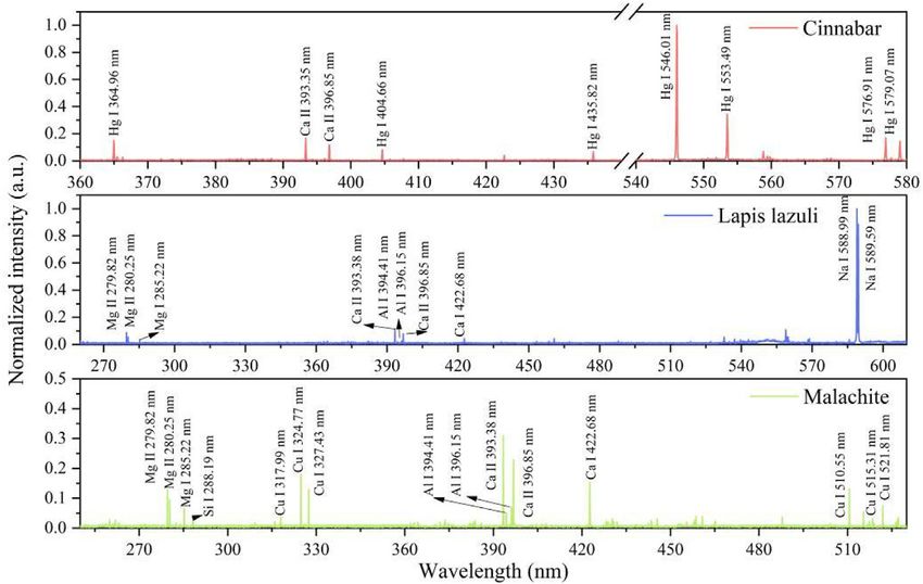
Yin et al.                                                                                                                                 In-Situ Murals Analysis With LIBS

  FIGURE 10 | (A) Normalized intensities of Cu and Fe lines versus number of laser shots used to irradiate a single spot in overlapped painted areas and (B) cluster
  classification of green pigments in four types of particle size fractions.

penetrated by the successive laser pulse with an increasing number of                     pulse was influenced significantly by the pigment size of the green-
ablation shots in the same spot; in contrast, the signal of iron lines                    painted layer, the AAR value gradually became larger with the
seemed to intensify at the same time. Therefore, the depth profiling                       decreasing pigment sizes on green layers [28]. Therefore, the
structure of murals was determined using varied trends of indicator                       pigment size in the green area labeled in Figure 9 was estimated
element lines for the corresponding painted layer.                                        first by using a classifiable model of green pigment sizes from our
    Figure 10 presents the varied trends of characteristic line                           reported results [30]. In the current work, the pigment size in this
intensities for copper and iron with different numbers of laser                           green area was estimated with 20 spectral data by employing the
pulse shots at the same point, which can give stratigraphic                               classifiable model of green pigment, and the result is included in
information in this area. From Figure 10, the normalized                                  Figure 10B. The 20 spectral points tended to be located in the P3
intensity trends of characteristic lines clearly indicated that the                       group, where the mean diameter was about 25 μm.
green-painted layers could be penetrated using six laser pulses.                              On the whole, the thickness of the green layer was determined
That is, the penetration numbers of laser shots on the overlapped                         quantitatively based on the information of green pigment size and the
painted area were determined as six by means of this method.                              relationships of laser pulses and ablation depth for corresponding
    It is necessary to get the average ablation rate (AAR) of laser pulse                 pigment size fractions. The expression of fitting relationship was
ablated on the green layers in order to obtain the thickness of the                       concluded as Y = 18.5X + 4.8 on the painted layers for the group
painted layer. From our previous research about the AAR of laser                          (~22.6 μm), where Y indicates the laser ablation depth, and X
pulse on the green layer, the results showed that the AAR of laser                        indicates the penetration numbers of laser pulses. The thickness

  FIGURE 11 | Microscopy images of cross-sections for the mural pieces.

Frontiers in Physics | www.frontiersin.org                                            8                                           March 2022 | Volume 10 | Article 847036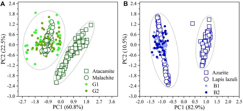
Yin et al.                                                                                                                                  In-Situ Murals Analysis With LIBS

was calculated at approximately 115.8 µm. To confirm our result, a                        DATA AVAILABILITY STATEMENT
cross-sectional analysis of this area was prepared in our laboratory, as
shown in Figure 11. The microscope image shows that the thickness                        The raw data supporting the conclusion of this article will be
of the layer was 108.8 ± 5.1 µm, which was consistent with the                           made available by the authors, without undue reservation.
estimated results from our evaluated results employing the LIBS
technique.
                                                                                         AUTHOR CONTRIBUTIONS
CONCLUSION                                                                               YY, ZY, and DS contributed to the design of the study and wrote
                                                                                         the first draft of the manuscript. YY, ZY, and DS contributed
In this paper, a portable LIBS system was used to analyze the                            equally as first authors in this paper. ZS, QC, YZ, and WZ
mural-painted layers in the cave for the first time. First, the laser                     completed the profiling analysis with optical microscopy. YF
ablation effect on the mural surface using microscopy images, the                        and BSh performed the XRD analysis of the mural-painted
size of the ablation crater, and the stable ablation edge suggested                      layers. BC provided all mural pictures in the caves. YZ and
that the microdestructive effect was acceptable from the view of                         WZ organized the spectral data. CD and BSu contributed to
mural conservation. Second, the mural pigments in Cave 98 were                           the idea and conception of the research. All authors contributed
indicated by using the pigment classifiable model, and the                                to the manuscript revision, and read and approved the submitted
pigments commonly used in red, green, and blue areas were                                version.
determined. Finally, the multilayer overlapped painted areas were
analyzed in situ by the portable LIBS, and the thickness of the
superficial green layer was calculated quantitatively according to                        FUNDING
the reported studies.
   The above research shows that the portable LIBS technique is a                        This work was supported by the National Key Research and
potential way to achieve in situ accurately illustrated information                      Development Program of China (Grant No. 2019YFC1520701),
about ancient murals. On the one hand, the information is                                the National Natural Science Foundation of China (Grant No.
significant for possible restoration and conservation of murals.                          61965015), the Industrial Support Program for Colleges of Gansu
On the other hand, the accurate spectral data of mural-making                            Province (Grant No. 2020C-17), and the Science and Technology
materials can provide an opportunity for in-depth illustration of                        Project of Gansu Province (Grant Nos. 21JR7RA759 and
the historical information.                                                              21JR7RA131).

                                                                                               Analysis and Applications to Different Fields. Appl Spectrosc (2012) 66(4):
REFERENCES                                                                                     347–419. doi:10.1366/11-06574
                                                                                         11.   Siano S, Salimbeni R. Advances in Laser Cleaning of Artwork and Objects of
 1. Fan J. The Conservation and Management of the Mogao Grottoes. Dunhuang                     Historical Interest: The Optimized Pulse Duration Approach. Acc Chem Res
    Res (2000) 63:1–4. doi:10.13584/j.cnki.issn1000-4106.2000.01.001                           (2010) 43(6):739–50. doi:10.1021/ar900190f
 2. Duan X. Study on the Making Materials of the wall Paintings at the Mogao             12.   Paraskevi P, Alexandros S, Savas G, Costas F. Recent Studies of Laser Science in
    Grottoes. Dunhuang Res (1988) 3:41–59.                                                     Paintings Conservation and Research. Acc Chem Res (2010) 43(6):771–81.
 3. Sun M, Zhang D, Wang Z, Ren J, Chai B, Sun J. What’s Wrong with the Murals                 doi:10.1021/ar900224n
    at the Mogao Grottoes: A Near-Infrared Hyperspectral Imaging Method. Sci             13.   Brysbaert A, Siozos P, Vetters M, Philippidis A, Anglos D. Materials Analyses of
    Rep (2015) 5:14371. doi:10.1038/srep14371                                                  Pyrotechnological Objects from LBA Tiryns, Greece, by Means of Laser-Induced
 4. Liu X, Qiao S, Ma Y. Highly Sensitive Methane Detection Based on Light-Induced             Breakdown Spectroscopy (LIBS): Results and a Critical Assessment of the Method.
    Thermoelastic Spectroscopy with a 2.33 Μm Diode Laser and Adaptive Savitzky-               J Archaeological Sci (2017) 83:49–61. doi:10.1016/j.jas.2017.06.007
    Golay Filtering. Opt Express (2022) 30:1304–13. doi:10.1364/oe.446294                14.   Giurato L, Candura A, Grasso G, Spoto G. In Situ identification of Organic
 5. Ma Y, Hong Y, Qiao S, Lang Z, Liu X. H-shaped Acoustic Micro-resonator-                    Components of Ink Used in Books from the 1900s by Atmospheric Pressure
    based Quartz-Enhanced Photoacoustic Spectroscopy. Opt Lett (2022) 47:                      Matrix Assisted Laser Desorption Ionization Mass Spectrometry. Appl Phys A
    601–4. doi:10.1364/ol.449822                                                               (2009) 97(2):263–9. doi:10.1007/s00339-009-5390-0
 6. Liu X, Ma Y. Sensitive Carbon Monoxide Detection Based on Light-Induced              15.   Kautek K, Qujja M, Castillejo M, Ferrence S, Betancourt P, Anglos D. Analysis
    Thermoelastic Spectroscopy with a Fiber-Coupled Multipass Cell [Invited].                  of Archaeological Objects with LMntI, a New Transportable LIBS Instrument.
    Chin Opt Lett (2022) 20:031201. doi:10.3788/col202220.031201                               In Lasers in the Conservation of Artworks. Berlin, Heidelberg: Springer (2005)
 7. Ma Y, Hu Y, Qiao S, Lang Z, Liu X, He Y, et al. Quartz Tuning forks Resonance              100:443–9. doi:10.1007/3-540-27176-7_56
    Frequency Matching for Laser Spectroscopy Sensing. Photoacoustics (2022) 25:         16.   Bertolini A, Carelli G, Francesconi F, Francesconi M, Marchesini L, Marsili P,
    100329. doi:10.1016/j.pacs.2022.100329                                                     et al. Modì: a New mobile Instrument for In Situ Double-Pulse LIBS Analysis.
 8. Cremers A, Radziemski L. Handbook of Laser-Induced Breakdown Spectroscop.                  Anal Bioanal Chem (2006) 385(2):240–7. doi:10.1007/s00216-006-0413-6
    Wiley (2006).                                                                        17.   Cuñat J, Palanco S, Carrasco F, Simón MD, Laserna JJ. Portable Instrument
 9. Hahn DW, Omenetto N. Laser-induced Breakdown Spectroscopy (LIBS), Part                     and Analytical Method Using Laser-Induced Breakdown Spectrometry for In
    I: Review of Basic Diagnostics and Plasma-Particle Interactions: Still-                    Situ Characterization of Speleothems in Karstic Caves. J Anal Spectrom (2005)
    Challenging Issues within the Analytical Plasma Community. Appl                            20(4):295–300. doi:10.1039/b417161f
    Spectrosc (2010) 64(12):335–66. doi:10.1366/000370210793561691                       18.   Martin M, Castillejo M, Torres R, Guerra F, Silva D. LIBS Spectra of
10. Hahn DW, Omenetto N. Laser-Induced Breakdown Spectroscopy (LIBS), Part                     Polychromes with a Low Cost CCD Camera Based Detector. J Cult
    II: Review of Instrumental and Methodological Approaches to Material                       Heritage (2000) 1:293. doi:10.1016/s1296-2074(00)00172-2

Frontiers in Physics | www.frontiersin.org                                           9                                             March 2022 | Volume 10 | Article 847036
Yin et al.                                                                                                                                           In-Situ Murals Analysis With LIBS

19. Castillejo M, Martín M, Oujja M, Silva D, Torres R, Domingo C, et al.                               Breakdown Spectroscopy. J Cult Heritage (2021) 47:109–16. doi:10.1016/j.
    Spectroscopic Analysis of Pigments and Binding Media of Polychromes by                              culher.2020.10.006
    the Combination of Optical Laser-Based and Vibrational Techniques. Appl                       29.   Giussani B, Monticelli D, Rampazzi L. Role of Laser Ablation-Inductively
    Spectrosc (2001) 55(8):992–8. doi:10.1366/0003702011953135                                          Coupled Plasma-Mass Spectrometry in Cultural Heritage Research: a Review.
20. De Giacomo A, Dell’Aglio M, Casavola A, Colonna G, De Pascale O, Capitelli                          Analytica Chim Acta (2009) 635(1):6–21. doi:10.1016/j.aca.2008.12.040
    M. Elemental Chemical Analysis of Submerged Targets by Double-Pulse                           30.   Yin Y, Yu Z, Sun D, Su M, Wang Z, Shan Z, et al. A Potential Method to
    Laser-Induced Breakdown Spectroscopy. Anal Bioanal Chem (2006) 385:                                 Determine Pigment Particle Size on Ancient Murals Using Laser Induced
    303–11. doi:10.1007/s00216-006-0323-7                                                               Breakdown Spectroscopy and Chemometric Analysis. Anal Methods (2021)
21. Chen YQ, Mo JY, Zhou Q, Lou Y, Li RH. Quantitative Analysis of Copper                               13(11):1381–91. doi:10.1039/d0ay01546f
    Impurity in Silver Jewellery by Laser-Ablation Laser-Induced Breakdown                        31.   Sorauf K, Bauer A, Miziolek A, Frank C. Spectral Data Analysis Approaches for
    Spectroscopy. Guang Pu Xue Yu Guang Pu Fen Xi (2015) 35(3):782–6.                                   Improved Provenance Classification. Next-generation Spectroscopic Tech VIII,
    doi:10.3964/j.issn.1000-0593(2015)03-0782-05                                                        SPIE Proc. (2015) 9482(12):01–10. doi:10.1117/12.2177913
22. Carmona N, Oujja M, Rebollar E, Römich H, Castillejo M. Analysis of                           32.   Shigeo A, Yoko T, Stephen R, Michiyo M, Takayasu K, Su B, et al. Conservation
    Corroded Glasses by Laser Induced Breakdown Spectroscopy.                                           and Painting Techniques of Wall Paintings on the Ancient Silk Road. Singapore:
    Spectrochimica Acta B (2005) 60(7-8):1155–62. doi:10.1016/j.sab.2005.05.016                         Springer Nature (2021).
23. Colao F, Fantoni R, Lazic V, Spizzichino V. Laser-induced Breakdown Spectroscopy              33.   NIST Atomic Spectra Database: Available from: http://www.nist.gov/pml/data/
    for Semi-quantitative and Quantitative Analyses of Artworks-Application on Multi-                   asd.cfm (Accessed February 15, 2021).
    Layered Ceramics and Copper Based Alloys. Spectrochimica Acta B: At Spectrosc
    (2002) 57(7):1219–34. doi:10.1016/s0584-8547(02)00054-x                                       Conflict of Interest: The authors declare that the research was conducted in the
24. Resano M, García-Ruiz E, Vanhaecke F. Laser Ablation-Inductively Coupled                      absence of any commercial or financial relationships that could be construed as a
    Plasma Mass Spectrometry in Archaeometric Research. Mass Spectrom Rev                         potential conflict of interest.
    (2010) 29(1):55–78. doi:10.1002/mas.20220
25. Kaszewska EA, Sylwestrzak M, Marczak J, Skrzeczanowski W, Iwanicka M,                         Publisher’s Note: All claims expressed in this article are solely those of the authors
    Szmit-Naud E, et al. Depth-Resolved Multilayer Pigment Identification in                       and do not necessarily represent those of their affiliated organizations, or those of
    Paintings: Combined Use of Laser-Induced Breakdown Spectroscopy (LIBS)                        the publisher, the editors, and the reviewers. Any product that may be evaluated in
    and Optical Coherence Tomography (OCT). Appl Spectrosc (2013) 67:960–72.                      this article, or claim that may be made by its manufacturer, is not guaranteed nor
    doi:10.1366/12-06703                                                                          endorsed by the publisher.
26. Yin Y, Sun D, Su M, Yu Z, Su B, Shui B, et al. Investigation of Ancient wall Paintings
    in Mogao Grottoes at Dunhuang Using Laser-Induced Breakdown Spectroscopy.                     Copyright © 2022 Yin, Yu, Sun, Shan, Cui, Zhang, Feng, Shui, Wang, Yin, Chai,
    Opt Laser Technol (2019) 120:105689. doi:10.1016/j.optlastec.2019.105689                      Zhang, Dong and Su. This is an open-access article distributed under the terms of the
27. Duchene S, Bruder R, Sirven JB, au J. Chemometrics and Laser Induced                          Creative Commons Attribution License (CC BY). The use, distribution or
    Breakdown Spectroscopy (LIBS) Analyses for Identification of Wall Paintings                    reproduction in other forums is permitted, provided the original author(s) and
    Pigments. Cac (2010) 6(1):60–5. doi:10.2174/157341110790069600                                the copyright owner(s) are credited and that the original publication in this journal is
28. Yin Y, Sun D, Yu Z, Su M, Shan Z, Su B, et al. Influence of Particle Size                      cited, in accordance with accepted academic practice. No use, distribution or
    Distribution of Pigments on Depth Profiling of Murals Using Laser-Induced                      reproduction is permitted which does not comply with these terms.

Frontiers in Physics | www.frontiersin.org                                                   10                                             March 2022 | Volume 10 | Article 847036
You can also read