In vitro establishment of two cultivars released from strawberries: Revista Mexicana de Ciencias ...
←
→
Page content transcription
If your browser does not render page correctly, please read the page content below
Revista Mexicana de Ciencias Agrícolas volume 9 number 4 May 16 - Jun 29, 2018

                                                                                                     Article

       In vitro establishment of two cultivars released from strawberries:
                           strawberry and raspberry

Teresita Ruíz Anchondo1, 2§
Julio Adriano Martínez2
Teresa Carrillo Castillo2
Rafael Ángel Parra Quezada1
Dámaris Leopoldina Ojeda Barrios1
Adriana Hernández Rodríguez1
1
 Graduate in Horticultural Sciences, Faculty of Agrotechnological Sciences-Autonomous University of
Chihuahua. Street Escorza num. 900, Col. Centro, Chihuahua, CP. 31100. (aernande@uach.mx;
raparra@uach.mx; dojeda@uach.mx). 2Plant Biotechnology Laboratory-Faculty of Agrotechnological
Sciences-Autonomous University of Chihuahua. (julio-adriano@hotmail.com; teresadarlen@hotmail.com).

§
Corresponding author. truiz@uach.mx.

Abstract
The development of protocols for the production of healthy plants in vitro is important to satisfy
the growing demand for strawberries in northern Mexico as an alternative to traditional fruit trees
such as walnut and apple trees. In vitro strawberry and raspberry establishment techniques were
evaluated from meristematic explants, apices and internodes. A convective disinfection method
was used, where disinfecting agents were used as a 70% ethanolic solution, then they were
immersed in a 3% commercial solution solution of sodium hypochlorite: water (1:6), for 15 min.
Finally, in 0.05% timerosal® solution for 15 min. Apices were evaluated in the case of strawberry
apices, and meristems and internodes in the raspberry. For the establishment, Murashige and Skoog
salts added with cytokinin 6-benzylaminopurine (BAP), gibberellic acid (GA) and indole butyric
acid (AIB) in different concentrations were used for the strawberry case. In raspberry, MS salts
added with BAP, thidiazuron (TDZ), GA and AIB were used. The percentages of establishment,
necrosis and contamination were evaluated in both cases. The planting of apices in the case of
strawberry and its disinfection system, allowed its establishment in vitro, the variables were
analyzed by nonparametric tests showed that the best treatment was TT1 with 70% effectiveness.
For raspberries, the case of internodes was better during establishment than in the case of
meristems, and the best treatment was TT4, with BAP + GA without auxin, with 57% effectiveness.

Keywords: berries, 6-benzylaminopurine, meristems, micropropagation, thidiazuron.

Reception date: March 2018
Acceptance date: May 2018

                                                                                                         799
Rev. Mex. Cienc. Agríc. vol. 9 num. 4 May 16 - Jun 29, 2018

                                          Introduction
The term tissue culture refers to the development of plant cells under in vitro conditions, said cells
are cultured aseptically in a culture medium of defined chemical composition and incubated under
controlled environmental conditions; encompassing a heterogeneous group of techniques to carry
out such an end (Roca and Mroginski, 1991; Purohit, 2012). Within these techniques,
micropropagation is found, which is widely used in the mass production of high quality genetic
material for large scale cultivation, genetic improvement, genetic conservation and research
(Jaakola et al., 2001; Gajdošová et al., 2006; Alanagh et al., 2014).

Among the benefits of micropropagation are: a single explant can be multiplied thousands of times,
the propagation periods are shorter, there is homogeneity in the propagated crops, it allows the
production of superior quality plants with resistance capacities and tolerance to biotic or abiotic
stress factors, among others (Yesil et al., 2010; Hussain et al., 2012). Micropropagation via
meristems or axillary shoots is highly recommended to obtain genetic homogeneity in the mass
production of fruit plants, with characteristics identical to the mother plant (Song and Sink, 2004;
Ostrolucká et al., 2007).

The traditional propagation of the commercial strawberry plant (Fragaria x ananassa Duch)
was carried out through stolons, which is not always adequate due to the vulnerability and
susceptibility of these to pathological agents, such as Botrytis cinerea, Fusarium spp. and
Penicilum (Clavijo et al., 2010). Therefore, after the propagation and establishment of the crop,
large losses occur, up to 50%) of the production, caused by the affectations caused by
microorganisms (Guédez et al., 2009; Ying et al., 2009). In 1974, Boxus described a method
for the mass micropropagation of strawberry plants by means of the technique of axillary
shoots, since then the production of strawberry cultivated in vitro has evolved, taking place
nowadays in many countries of the world.

These micropropagated plants are used to establish plantations, and it has been found that they
show more flowers, higher fruit yield per hectare, more stolons per plant and greater vigor, in
comparison with conventionally propagated plants (Tan et al., 2003). In addition, this method
has been successful in the prevention of diseases transmitted in soil and plants (Moradi et al.,
2011).

On the other hand, the propagation of raspberry Rubus idaeus sp.) Is carried out by means of
root staking, with which a high propagation percentage and a good quality of plants is achieved.
However, it is essential that the quality and age of the mother plants be monitored, since after
two or three years the percentage of rootedness decreases considerably, while the risk of
diseases and contamination by viruses increases; therefore, this method does not ensure the
production of disease free plants. In addition, sometimes the staking method has very low
multiplication rates that do not meet the needs of the nursery industry (González et al., 2000;
Wu et al., 2009; García et al., 2014). Previously, micropropagation of vegetative raspberry
material has been achieved with good results, using nodal segments or meristemati c tissue as
explants and thus obtaining disease-free plants (Reed, 1990; Minas and Neocleous, 2007;
Arencibia et al., 2013).

                                                                                                         800
Rev. Mex. Cienc. Agríc. vol. 9 num. 4 May 16 - Jun 29, 2018
The objective of this study was to develop methodologies and protocols for the in vitro
establishment of commercial strawberry berries cv Aromas and raspberry cv Heritage, using
different types of explants in different culture media formulated with plant hormones.
Materials and methods
The test was carried out in the Biotechnology laboratory with materials from the experimental
greenhouse of the Faculty of Agrotechnological Sciences of the Autonomous University of
Chihuahua, from where strawberry, cv Aromas, and raspberry cv Heritage, from which the
vegetative material used in the present investigation was collected to be established in vitro, in
2015.
For the disinfection of the vegetative material in strawberry (Fragaria x annanasa Duch. cv
Aromas)
In this case, for the disinfection of strawberry plants apical buds were selected that did not
show symptoms of disease. They were cut and stored in sterile distilled water with 150 mgL -1
of ascorbic acid prior to handling. After 1 h, the shoots were immersed in aqueous solution of
5% alkaline soap with 2 drops of Tween-80 for 15 min, in constant agitation. Afterwards, they
were rinsed under the water tap for 20 min. Under aseptic conditions in a laminar flow hood,
the vegetative material was immersed in 70% ethanol for 30 s, then rinsed three times with
sterile distilled water.
The explants were then submerged in a commercial sodium hypochlorite solution and distilled
water in a ratio of 1:6 for 10 min, shaking gently, and then rinsing again three times with sterile
distilled water. Next, the internodes were immersed in 0.05% solution of mercury salts, Thimerosal
(Sigma Aldrich®, CAS 54-64-8) for 10 min, then rinsing with sterile distilled water in three
occasions.
For in vitro establishment from apical strawberry shoots (Fragaria x annanasa Duch. cv
Aroma)
The already disinfected apical buds were seeded in 4 different formulations of culture media, three
of them with micro and macro elements, added with organic according to the modified MS
formulation, see table 1 (Murashige and Skoog, 1962), supplemented with 1.11 μM benzyl
aminopurine (BAP), 0.87 μM gibberellic acid (GA) and 2.95 μM indole butyric acid (IBA) for the
first control treatment (TT0); the first treatment (TT1) was supplemented with 4.44 μM of BAP,
0.29 μM GA and 4.92 μM of IBA; also, the second treatment (TT2), with 2.22 μM BAP, 0.29 μM
GA and 0.49 μM of IBA. Finally, another culture medium formulated with 50% MS salts and
vitamins, supplemented with 0.44 μM BAP, 0.29 μM GA and 4.92 μM IBA was identified as the
third treatment (TT3), using the suggested growth regulators. by several authors who have reported
success in their formulations, such as (Boxus, 1974; de Lopez, 2001; Beltran, 2002; Caboniet et
al., 2008) (Table 1).
801Rev. Mex. Cienc. Agríc. vol. 9 num. 4 May 16 - Jun 29, 2018
Table 1. Formulation of culture media for strawberry cv Aromas, supplemented with cytokinin
(benzylaminopurine BAP), auxins (indolbutyric acid AIB) and gibberellins (GA),
according to previous propagation reports. TT1 by Caboniet et al., 2008; TT2, Beltran,
2002; de Lopez, 2001; TT3 and TT4 Boxus, 1974.
Treatment Formulation (sales MS) BAP (µM) GA (µM) IBA (µM)
TT1 100% 1.11 0.87 2.95
TT2 100% 4.44 0.29 4.92
TT3 100% 2.22 0.29 0.49
TT4 50% 0.44 0.29 4.92
In addition, 1 g L-1 of activated charcoal, 5 g L-1 of Phyta-gel (Sigma®) and 30 g L-1 of sucrose
brand Sigma® CAS 71010-52-1 were added to all treatments. The pH was adjusted to 5.8 in all
cases and then sterilized at 1.5 kg cm2 of pressure at 120 °C for 15 min. After sowing the explants,
they were incubated at a temperature of 24 ±2 °C, with a luminosity of 86 µmol m-2 s-1 supplied by
LED lamps model T8 TecnoLite® and a photoperiod of 16/8 h during 15 days. After that time, the
ex-plantes that showed growth were transferred in the same formulation, for two more weeks. After
this lapse of time, the variables evaluated were percentage of contamination, necrosed plants and
established explants.
For the disinfection of vegetative material from nodal explants of raspberry (Rubus idaeus L.
cv Heritage)
For the disinfection of raspberry, healthy mother plants were selected. Stem segments 30 cm in
length were sectioned with vegetative axillary buds. These segments were sheltered and placed in
moistened plastic bags prior to disinfection. The explants were sectioned leaving a vegetative bud
per section, and were immersed in 5% alkaline soap solution with two drops of Tween-80 for 15
min. Then, they were rinsed under the tap for 30 min.
They were then transferred to aseptic conditions in a laminar flow hood by immersing the explants
in 70% ethanol for 30 s, then rinsing three times with sterile distilled water. They were then
immersed in a solution of mercury salts, Thymerosal (Sigma Aldrich®) at 0.05% for 15 min, then
rinse three times with sterile distilled water. Subsequently, the explants were immersed in a solution
of commercial sodium hypochlorite with sterile distilled water in relation (1:6) for 10 min, agitating
gently, to immediately rinse three times. Finally, the explants were immersed in ascorbic acid
solution (150 mgL-1) and stored for 24 h at 4 °C.
For the in vitro establishment of the vegetative material in raspberry (Rubus idaeus cv
Heritage)
Two micropropagation techniques were used to introduce vegetative raspberry material into in vitro
conditions, nodal segments (internodes) and axillary meristems.
802Rev. Mex. Cienc. Agríc. vol. 9 num. 4 May 16 - Jun 29, 2018
In the case of the internodes, after 24 h, they were sectioned in 2-3 cm explants and planted in four
different culture media. The first treatment (TT1) consisted of a modified MS medium, added with
19.75 μM BAP and 0.04 μM of IBA, according to Minas and Neocleus (2007). The second one
(TT2) was formulated equal to the previous one, but with the difference that 20.2 μM of Tidiazuron
(TDZ) was added instead of BAP, since this cytokinin has been widely used in the propagation of
vegetative raspberry tissue (Gajdošová et al., 2006; Dai et al., 2006; Cappeletti et al., 2006). The
third (TT3) was formulated with MS salts and LS vitamins (Linsmaier and Skoog, 1965), in
addition to 4.4 μM of BAP, 0.29 μM of GA and 0.49 μM of IBA (Poothong and Reed, 2015).
Finally, the fourth culture medium (TT4) was formulated according to Sigarroa and García (2011),
which consisted of an MS medium with modified vitamins plus 4.44 μM of BAP and 1.44 μM of
GA, as shown in Table 2 In the case of the meristems, these were extracted with the help of sterile
clamps and scalpel and using an optical microscope of the VANGUARD® brand-stethoscope type
at 2x magnification.
Table 2. Formulation of culture media supplemented with cytokinins, auxins and gibberellins.
According to previous propagation reports for raspberry cv Heritage. TT1 Minas and Eoclus,
2007; TT2 Gadjosova et al., 2006; TT3 salts by Linsmaier and Skoog, 1965; hormones by
Poothong and Reed, 2015; TT4 Sigarroa, 2011.
Formulation BAP/TDZ
Treatment (Sales MS) Vitamins GA (µM) IBA (µM)
(µM)
TT1 100% Modified MS 19.75 BAP - 0.04
TT2 100% Modified MS 20.2 TDZ - 0.04
TT3 100% LS 4.4 BAP 0.29 0.49
TT4 100% Modified MS 4.44 BAP 1.44 -
TT1M 100% Modified MS 4.44 BAP 1.44 -
The bracts were removed from the axillary buds and the meristematic tissue was seeded in modified
MS culture medium added with 4.44 μM BAP and 1.44 μM GA (TT1M), according to Sigarroa
and García (2011). In all cases, 1 g L-1 of activated carbon, 5 gL-1 of Phytagel (Sigma® CAS (71010-
52-1) as a gelling agent and 30 g L-1 of sucrose, brand SIGMA® (CAS 57-50-1) were added. The
pH of the culture was adjusted to 5.7 in the case of internodes and six for meristems (Sigarroa and
García, 2011; Poothong and Reed, 2015) and then sterilized to 1.5 kg cm2 of pressure at 120 °C by
15 min.
After sowing, the vegetative material was transferred to a transfer chamber under controlled
temperature conditions of 24 ±2°C under conditions of total darkness. After seven days, they were
transferred to brightness conditions of 86 µmol m-2 s-1 supplied by LED T8 TecnoLite® lamps and
a photoperiod of 16/8 h. After 21 days, the percentages of contamination, necrosed plants and
established explants were evaluated.
Statistical analysis
In the case of strawberry micropropagation, the qualitative variables evaluated were the percentage
of contamination, which evaluates the success of the disinfection treatment, the percentage of
necrotic plants, which evaluates the formulation of the establishment medium and the percentage
803Rev. Mex. Cienc. Agríc. vol. 9 num. 4 May 16 - Jun 29, 2018
of explants established (Marulanda et al., 2000; Sigarroa and García, 2011), the repetitions were
10 units for each of the four treatments, and these were analyzed using a completely randomized
experimental design under homogeneous conditions.
These variables were analyzed by the non-parametric Kruskal-Wallis tests due to the small number
of repetitions per treatment with which chi2 is not valid for proportions test, the Dunn test was
applied for the separation of Neave means and Worthington (1988), using the Minitab® statistical
package version 17.1.0
In the case of the raspberry, the number of necrotic plants, and the established explants, both of the
internodes were evaluated. The repetitions were 65 units per treatment. The data were analyzed
using the logistic regression model, using as an independent variable the treatments and the
response to necrosis and establishment. All data was processed in the IBM SPSS Statistics 20
software.
Results and discussion
Necrosis
Most plants produce phenolic compounds after passing through stages of stress, such as that
produced by contact with disinfectants, so caution must be exercised in their concentration when
applying them to plant tissues. This procedure is essential to be able to establish vegetative material
under in vitro conditions (Rostami and Reza, 2012). These compounds affect the growth and
survival of the explants, oxidizing the tissues and leading to necrosis and death.
To avoid this, it is advisable to add antioxidants in the environment, as well as increase the
frequency of subcultures (Swartz and Lindstrom, 1986). Other options to avoid the oxidation
processes that lead to the darkening of the tissues and their subsequent death are the use of a liquid
culture medium, change of the gelling agent, use of activated carbon, addition of ascorbic acid and
polyvinyl polypyrrolidone; however, since the problem is complex, a comprehensive solution is
required that involves a greater number of variables (Azofeifa, 2009, García et al., 2010).
In the present investigation, we sought to counteract the oxidation of tissues by applying activated
carbon to the culture medium and placing the explants in dark conditions for a period of time.
Activated carbon is distinguished from elemental carbon by its absence of impurities and almost
graphitic form, a fine network of pores, an extraordinarily large surface area and a volume that
gives it a unique absorption capacity, thus helping to reduce effects of tissue oxidation. In the same
way, decreasing light intensity prevents oxidation and necrosis of tissues (Swartz and Lindstrom,
1986; Thomas, 2008). In this trial, in the case of strawberry, the treatments that presented the
highest percentage of oxidation and subsequent necrosis were TT3, with 40% and TT4, 70%; on
the other hand, TT1 and TT2 only presented 10%; that is, a low percentage of necrotic tissue. In
the test for raspberry, the percentage of necrosis in general was high, with an average of 78.1%,
despite the addition of activated carbon to the culture medium and having placed the explants under
dark conditions for 7 days.
804Rev. Mex. Cienc. Agríc. vol. 9 num. 4 May 16 - Jun 29, 2018
The treatment with the lowest percentage of necrosis was TT1, with 27.69% and according to the
Mann-Wittney test, the TT1 of meristems showed a statistically significant difference with respect
to the other treatments, with a percentage of high necrosis (Table 3). In this technique millimeter
portions of tissues from axillary or apical buds are cultured, each portion consists of a fragment of
the meristematic region with or without leaf primordia, therefore, these tiny fragments of tissue are
more sensitive to oxidation (García et al., 2011; George et al., 2008).
Table 3. Frequencies calculated for the response from explant to establishment (0); disinfection
(1) and necrosis (2); where it can be seen that TT1 provides the best treatment for the
three responses, calculated by analyzing the frequency of the SPSS® IBM v20 statistical
package.
Treatment Frequency Percent Valid percent Cumulative percent
TT1 Valid 0 7 70 70 70
1 2 20 20 90
2 1 10 10 100
Total 10 100 100
TT2 Valid 0 6 60 60 60
1 3 30 30 90
2 1 10 10 100
Total 10 100 100
TT3 Valid 0 5 50 50 50
1 1 10 10 60
2 4 40 40 100
Total 10 100 100
TT4 Valid 0 2 20 20 20
1 1 10 10 30
2 7 70 70 100
Total 10 100 100
Contamination
The most important process prior to the establishment of the crop is the ideal formulation in the
application of disinfectants and antioxidants, as well as the times in which the explants are
subjected to the disinfection process. Since in vitro conditions some microorganisms such as
bacteria and fungi find an optimal environment to develop, an unsuccessful disinfection hinders
the progress of propagation, so the application of substances and the use of disinfecting techniques
are focused on eliminating said unwanted microorganisms, including mercury salts in percentages
of 0.05-0.15%. (Mikropavai-Rošana et al., 2009; Yildiz, 2012).
805Rev. Mex. Cienc. Agríc. vol. 9 num. 4 May 16 - Jun 29, 2018
Regarding the evaluation of the contamination variable strawberry, cv Aromas, it was found that
TT 2 showed a higher percentage of explants with the presence of microorganisms (30%), whereas
TT 3 and TT4 only showed 10% of contaminated explants. In general, the disinfection protocol
presented acceptable values in treatments TT3 and TT4, as shown in Table 3.
Establishment
In the case of strawberry, cv Aromas, the four treatments were statistically different when analyzed
by the Kruskal-Wallis and Dunn tests, by comparing the differences between treatments. The
treatment that behaved the best was the TT4, with a greater number of explants established
successfully (70%). In Figure 1 and Figure 2, it is illustrated that effectively the treatment
formulated according to Caboni et al. (2008), by using the combination of BAP+GA+IBA,
hormonal activity is more efficiently potentiated, obtaining a more efficient vegetative response
than in the other cases, with a percentage of 70% of established explants.
Figure 1. Comparative of the response in establishment, contamination and necroses to the different
formulations or treatments evaluated in strawberry, cv Aromas. It is appreciated that in the
TT4 treatment, the difference is smaller. Nonparametric Kruskal-Wallis tests were used for the
separation of means and Dunn’s multiple comparisons test, from the SPSS® statistical package
version 20.
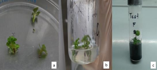
Figure 2. Strawberry vegetative material cv Aromas. a. Explants of strawberry apical buds. b.
Explants established in culture medium. c. Explante in culture medium with activated
carbon.
806Rev. Mex. Cienc. Agríc. vol. 9 num. 4 May 16 - Jun 29, 2018
Raspberry
In the case of raspberry, cv Heritage, Kruskal-Wallis and Dunn (reference), to analyze the statistical
differences in the use of meristems against internodes, was found to be better internodal tissue to
settle, but not in the case of meristems, being that, in this tissue, even though it was the one with
the lowest percentage of contamination (1.54%), it had a very low percentage of establishment with
respect to intermodal tissue. Figure 3 shows the marked difference between treatments using
cuttings against meristem cells.
80
Percent of establishment of explants
70
60
50
40
30
20
10
0
T1-Entrenudos t1-Meristemos
Treatments
Figure 3. Statistical difference in the establishment of internodes and meristems ex-pressed in
percentage, raspberry cv Heritage. Using nonparametric Kruskal-Wallis tests and the
Mann-Whitney two-proportions test of the Minitab® Statistical Package version 17.1.0.
In the case of the response to treatments with different formulations reported, it was found that the
logistic regression analysis, in response to treatments, in the case of the formulation of TT4 (4.44
µM of BAP + 1.44 µM of GA) (Anderson, 1980; Sigarroa, 2011) was the best response to
establishment with respect to other treatments, since it had much lower necrosis. It has been
reported that the micropropagation through meristems allows not only obtaining high quality plants
and more uniform, but also the spread of free specific clones pathogens, achieving reduce
endogenous contamination to a minimum level without to apply more aggressive disinfection
protocols (Sigarroa and García, 2011). But also, the vegetative response to nutritive formulations,
determine to a great extent success, in the prompt response to the culture medium (Sincha et al.,
1987; Reed, 1990; Reed et al., 2013).
Even though it has been reported that for raspberry, the medium formulated with MS salts, express
poor growth and in vitro development problems Greenwayet et al. (2012), in this case the Figure 4
shows the statistical results of ranges between differences in formulations or treatments evaluated,
being effectively TT4 held difference statistically significant at internodes used com vegetative
material is establishment and the combination of hormones 44.4 µM of BAP + 1.44 of GA
(Sigarroa and García, 2011).
807Rev. Mex. Cienc. Agríc. vol. 9 num. 4 May 16 - Jun 29, 2018
Figure 4. Effect of the formulation on contaminated, necrotic and established raspberry leaflets
cv Heritage for evaluated treatments. Analyzed using the logistic regression model, using
as independent variables the effect of the treatments, to the necrosis by establishment,
obtaining Χ2. Made in the IBM SPSS Statistics 20 statistical package.
In raspberry only in two treatments it was possible to observe the establishment of the explants.
The TT4 of internodes presented the highest percentage of success, reaching 56.92% of established
explants (Figure 5). According to the test of two proportions, it was found that there are statistically
significant differences between said treatment and the explants of the meristem technique.
a) b
)
c) d
Figure 5. Raspberry vegetative material cv Heritage; a) meristematic tissue observed in 2x stereoscope
microscope; b) raspberry internode, disinfected and ready to be planted; c) explant from
meristematic tissue in full growth; d) explant in growth from nodal segment.
Conclusions
In strawberry cv Aromas, in vitro establishment, based on apices, was successful in the four culture
media evaluated, since it was possible to establish them, although TT1 proved to have the greatest
viability, as it had the highest percentage of vegetative tissue. stable with 70% and the lowest
percentage with TT4, with only 20%. The combination of hormones in the formulation allowed the
successful development of the tissue for its in vitro establishment of strawberry (Folta, et al., 2006).
808Rev. Mex. Cienc. Agríc. vol. 9 num. 4 May 16 - Jun 29, 2018
In all treatments, the percentage of contamination did not exceed 30%, considering, as well as
acceptable for an in vitro propagation system.
For the case of raspberry, cv Heritage, the formulation proposed by Anderson (1980) and Sigarroa
and García (2011) was the only one that allowed the development of the explants, there being a
greater percentage of establishment by the technique where internodes schemes were used, but not
by medium of meristems, where there was a high rate of necrosation and death of the established
tissue, in the other treatments. The formulation with the combination of BAP + GA hormones,
without the presence of auxin AIB, was more successful in the establishment of nodal segments in
raspberry.
On the other hand, when meristem cells were extracted, it showed a much lower percentage of
contamination compared to the explants obtained by sowing internodes, because the disinfection
system is more successful when it comes to extracting tissue of the cambium (Yildiz, 2012). When
the meristems were evaluated, a high percentage of tissue necrosis was obtained, but the percentage
of contamination was very low. Therefore, it is suggested to evaluate this technique again, using
another type of disinfectant agents and tissue exposure times before them.
Acknowledgments
The authors thank the National Council of Science and Technology (CONACYT) for the financing
of the PRO-INNOVA 2015 project with num. 221265: fruit-innovation using tissue culture and
organic agriculture in alternative high-value fruit trees in the state of Chihuahua.
Cited literature
Abobkar, I. 2012. Plant tissue cultura media, Recent advances in plant in vitro culture. Leva, A.
(Ed.). INTECH Open Access Publisher. http://www.intechopen.com/books/recent-
advances-in-plant-in-vitro-culture/plant-tissue-culture-media.
Alanagh, E. N.; Garoosi, G. A.; Haddad, R.; Maleki, S.; Landín, M. and Gallego, P. P. 2014. Design
of tissue culture media for efficient Prunus rootstock micropropagation using artificial
intelligence models. Plant Cell, Tissue and Organ Culture (PCTOC). 117(3):349-359.
Ali, A.; Ahmad, T.; Akhtar, N. and Ahmed, I. 2009. Effect of different media and growth regula
tors on in vitro shoot proliferation of olive cultivar “Moraiolo”. Pak. J. Bot. 41(2):783-795.
Anderson, W. C. 1980. Tissue culture propagation of red and black raspberries, Rubus idaeus
and R. occidentalis. In: Symposium on Breeding and Machine Harvesting of Rubus.
112:13-20.
Arencibia, A.; Vergara, C.; Quiroz, K.; Carrasco, B. and García, R. 2013. Establishment of
photomixotrophic cultures for raspberry micropropagation in Temporary immersion
bioreactors (TIBs). Scientia Hortic. 160(1):49-53.
Ashrafuzzaman, M.; Faisal, S.; Yadav, D.; Khanam, D. and Raihan, F. 2013. Micropropagation of
strawberry (Fragaria ananassa) through runner culture. Bangladesh J. Agril. Res.
38(3):467-472.
Azofeifa, A. 2009. Problemas de oxidación y oscurecimiento de explantes cultivados in vitro.
Agronomía mesoamericana. 20(1):153-175.
809Rev. Mex. Cienc. Agríc. vol. 9 num. 4 May 16 - Jun 29, 2018
Balachandran, S.; Bhat, S. and Chandel, K. 1990. In vitro clonal multiplication of turmeric
(Curcuma spp.) and ginger (Zingiber officinale Rosc.). Plant Cell Reports. 8(9):521-524.
Biswas, M.; Hossain, M.; Ahmed, M.; Roy, U.; Karim, R.; Razvy, M.; Salahin, M. and Islam, R.
2007. Multiple shoot regeneration of strawberry under various colour illuminations. Am.
Eur. J. Sci. Res. 2(2):133-135.
Boxus, P. 1974. The production of strawberry plants by in vitro micropropagation. J. Horti. Sci.
49(3):209-210.
Broome, O. C. and Zimmerman, R. H. 1978. In vitro propagation of Blackberry. HortSci.
13(2):151-153.
Caboni, E.; Condello, E.; Meneghini, M.; Palombi, M. A.; Frattarelli, A. and Damiano, D. 2008.
Progresses in cryopreservation of Pyrus spp. and evaluation of genetic stability of the
recovered shoots. Cryopreservation of cropspecies in Europe: cryoplanet cost action
87120th- 23rd of February 2008, Oulu, Finland/Jaana Laamanen, Marjatta Uosukainen, Hely
Häggman, Anna Nukari and Saija Rantala. (Eds.). 36 p.
Cappelletti, R.; Sabbadini, S. and Mezzetti, B. 2016. The use of TDZ for the efficient in vitro
regeneration and organogenesis of strawberry and blueberry cultivars. Sci. Hortic. 207:
(1):117-124.
Clapa, D.; Fira, A. and Pacurar, I. 2008. The in vitro propagation of the raspberry cultivar citria.
Bulletin of the University of Agricultura Sciences. 65(1):99-103.
Clavijo, R.; Beltrán, A.; Llauger, R.; Rodríguez, A.; Farrés, E.; García, M. E. y Rodríguez, M.
2010. Apuntes sobre el cultivo de la fresa (Fragaria x ananassa Duch.). Rev. Citrifrut,
27(2):67-71.
Dai, W.; Magnusson, V.; Hatterman, H. and Carter, J. 2006. Micropropagation of ‘Amethyst’
purple raspberry (Rubus occidentalis L. x R. Idaeus L. ‘Amethyst’). J. Environ. Hort.
4(1):35-38.
Folta, K. M.; Dhingra, A.; Howard, L.; Stewart, P. J. and Chandler, C. K. 2006. Characterization
of LF9, an octoploid strawberry genotype selected for rapid regeneration and
transformation. Planta. 224(5):1058-1067.
Gajdošová, A.; Ostrolucká, M.; Libiaková, G.; Ondrušková, E. and Šimala, D. 2006. Microclonal
propagation of Vaccinium sp. and Rubus sp. and detection of genetic variability in culture
in vitro. J. Fruit Ornamental Plant Res. 14(1):103-119.
García, E.; Lorente, P.; Marín, J.; Arbeloa, A. y Andre, P. 2010. Micropropagación e injerto in
vitro de pistacho. Información Técnica Económica Agraria. 106(4):294-302.
García, E.; Lorente, P.; Marín, J. A.; Andreu, P. y Arbeloa, A. 2011. Factores que afectan a la
necrosis apical de brotes de Pistacia vera L. Cultivados in vitro. ITEA. 107(4):315-323.
García, J.; González, G. y Ciordia, M. 2014. El cultivo del frambueso. Servicio regional de
investigación y desarrollo agroalimentario. España. 33 p.
George, E.; Hall, Michael and De Klerk, Geert-Jan. 2008. Plant propagation by tissue culture. 3ra
Edición. 1 p.
Greenway, M. B.; Phillips, I. C.; Lloyd, M. N.; Hubstenberger, J. F. and Phillips, G. C. (2012). A
nutrient medium for diverse applications and tissue growth of plant species in vitro. In Vitro
Cel. Develop. Biol. Plant. 48(4):403-410.
Guédez, C.; Cañizález, L.; Castillo, C. y Olivar, R. 2009. Efecto antagónico de Trichoderma
harzianum sobre algunos hongos patógenos postcosecha de la fresa (Fragasia spp.). Revista
Soc. Venezolana Microbiol. 29(1):34-38.
González, M.; López, M.; Valdés, A. and Ordas, R. 2000. Micropropagation of three berry fruit
species using nodal segments from field-grown plants. Ann. Appl. Biol. 137(1):73-78.
810Rev. Mex. Cienc. Agríc. vol. 9 num. 4 May 16 - Jun 29, 2018
Haddadi, F.; Abd-Aziz, M.; Saleh, G.; Abd-Rashid, A. and Kamaladini, H. 2010. Micropropagation
of strawberry cv Camarosa: prolific shoot regeneration from in vitro shoot tips using
Thidiazuron with N6-benzylamino-purine. Hortic. Sci. 45(3):453-456.
Hussain, A.; Ahmed, I.; Nazir, H. and Ullah, I. 2012. Plant tissue culture: current status and
opportunities, Recent Advances in Plant In Vitro Culture. Leva, A. (Ed.). INTECH.
http://www.intechopen.com/books/recent-advances-in-plant-in-vitro-culture/plant-tissue-
culture-current-status-and-opportunities.
Jaakola, L.; Pirttilä, A. M.; Halonen, M. and Hohtola, A. 2001. Isolation of high quality RNA from
bilberry (Vaccinium myrtillus L.) fruit. Mol. Biotechnol. 19(2):201-203.
Kessel, D. A. 2012. Mejora genética de la fresa (Fragaria ananassa Duch.), a través de métodos
biotecnológicos. Cultivos Tropicales. 33(3):34-41.
Linsmaier, E. and Skoog, F. 1965. Organic growth factor requirements of tobacco tissue cultures.
Physiol Plant. 18(1):100-127.
Litwińczuk, W.; Okołotkiewicz, E. and Matyaszek, I. 2009. Development of in vitro shoot cultures
of strawberry (Fragaria x ananassa Duch.) “Senga Sengana” and “Elsanta” under the
influence of high doses of gibberellic acid. Folia Hortic. 21(2):43-52.
Minas, G. and Neocleous, D. 2007. A protocol for rapid clonal micropropagation in vitro of
primocane-fruiting red rasberry cultivars. Agricultural Research Institute, Ministry of
Agriculture, Natural Resources and the Environment. Lefkosia, Cyprus. Miscellaneous
Report: 95(1):3-7 http://news.ari.gov.cy/publications/mr95-minas.pdf.
Moradi, K.; Otroshy, M. and Azimi, M. 2011. Micropropagation of strawberry by multiple shoots
regeneration tissue cultures. J. Agric. Technol. 7(6):1755-1763.
Murashige, T. and Skoog, F. 1962. A revised medium for rapid growth and bio assays with tobacco
tissue cultures. Physiol. Plantarum. 15(3):473-497.
Neave, H. R. and Worthington, P. L. 1988. Distribution-Free Tests. Editorial Unwin Hyman Ltd.
London, UK. 430 p.
Nitsch, J. and Nitsch, C. 1969. Haploid plants from pollen grains. Science. 163(3862):85-87.
Ostrolucká, M.; Libiaková, G.; Ondrušková, E. and Gajdošova A. 2004. In vitro propagation of
Vaccinium species. Acta Universitatis Latviensis. 676(1):207-2012.
Ostrolucká, M. G.; Gajdošová, A.; Libiaková, G.; Hrubíková, K. and Bežo, M. 2007. Protocol for
micropropagation of selected Vaccinium spp. In: protocols for micropropagation of woody
trees and fruits. Springer Netherlands. 445-455 pp.
Pérez, A.; Nápoles, L.; Concepción, O. y Trujillo, R. 2002. Multiplicación in vitro de brotes de
guayaba (Psidium guajava L.) var. enana roja cubana EAA 18-40 obtenidos a partir de
semillas. Cultivos tropicales. 23(3):57-61.
Poothong, S. and Reed, B. 2015. Increased CaCl2, MgSO4, and KH2PO4 improve the growth of
micropropagated red raspberries. In vitro Cel. Develop. Biol. Animal. 51(6):648-658.
Purohit, S. 2012. Introduction to plant cell, tissue and organ culture. PHI Learning Pvt Ltd, New
Delhi. 1 p.
Roca, W. and Mroginsky, L. 1991. Cultivo de tejidos en la agricultura. Fundamentos y
aplicaciones. (No. 151). Ciat.
Reed, B. M. 1990. Multiplication of Rubus germplasm in vitro: a screen of 256 accessions. Fruit
Var J. 44(1):141-148.
Reed, B. M.; Wada, S.; DeNoma, J. and Niedz, R. P. 2013. Improving in vitro mineral nutrition for
diverse pear germplasm. In Vitro Cel. Develop. Biol. Plant. 49(3):343-355.
Rostami, A. A and Reza, A. 2012. In vitro propagation of Olive (Olea europaea L.) by nodal
segments. J. Biol. Environ. Sci. 6(17):155-159.
811Rev. Mex. Cienc. Agríc. vol. 9 num. 4 May 16 - Jun 29, 2018
Singha, S.; Oberly, G. H. and Townsend, E. C. 1987. Changes in nutrient composition and pH of
the culture medium during in vitro shoot proliferation of crabapple and pear. Plant Cell,
Tissue and Organ Culture. 11(3):209-220.
Sigarroa, A. y García, C. 2011. Establecimiento y multiplicación in vitro de mora de castilla (Rubus
glaucus Benth.) variedad sin espinas, mediante ápices meristemáticos. Acta Agronómica,
60(4):347-354.
Song, G. Q. and Sink, K. C. 2005. Optimizing shoot regeneration and transient expression factors
for Agrobacterium tumefaciens transformation of sour cherry (Prunus cerasus L.) cultivar
Montmorency. Sci. Hortic. 106(1):60-69.
Ostrolucká, M. G.; Gajdošová, A.; Libiaková, G.; Hrubíková, K. and Bežo, M. 2007. Protocol for
micropropagation of selected Vaccinium spp. In: protocols for micropropagation of woody
trees and fruits Springer Netherlands. 445-455 pp.
Swartz, H. and Lindstrom, J. 1986. Small fruit and grapes tissue culture from 1980 to 1985:
Commercialization of the technique. In: tissue culture as a plan production system for
horticultural crops. Springer Netherlands. 201-220 pp.
Tan, D.; Takamura, T.; Watanabe, H.; Okamoto, K. and Tanaka, M. 2003. Responses of strawberry
plantets cultured in vitro under superbright red and blue light-emitting diodes (LEDs). Plant
Cell, Tissue Organ Culture. 73(1)43-52.
Thomas, T. D. 2008. The role of activated charcoal in plant tissue culture. Biotechnol. Adv.
26(6):618-631.
Yesil, O.; Gurel, A. and Vardar, F. 2010. Large scale cultivation of plant cell and tissue culture in
bioreactors. Transworld Research Network, Kerala. 3 p.
Yildiz, M. 2012. The prerequisite of the success in plant tissue culture: high frequency shoot
regeneration, recent advances in plant in vitro Culture. Leva, A. (Ed.). INTECH.
http://www.intechopen.com/books/recent-advances-in-plant-in-vitro-culture/the-
prerequisite-of-the-success-in-plant-tissue-culture-high-frequency-shoot-regeneration.
Ying, C.; Al-Abdulkarim, A.; Al-Jowid, S. and Al-Baiz, A. 2009. An effective disinfection
protocol for plant regeneration from shoot tip cultures of strawberry. Afr. J. Biotechnol.
8(11):2611-2615.
Reed, B. 1990. Multiplication of Rubus germplasm in vitro: a screen of 256 accessions. Fruit
Varieties J. 44(1):141-148.
Wu, J.; Miller, S.; Hall, H. and Mooney, P. 2009. Factors affecting the efficiency of
micropropagation from lateral buds and shoot tips of Rubus. Plant Cell, Tissue and Organ
Culture. 99(1):17-25.
812You can also read