Inflammatory reactions in rainbow trout fins and gills exposed to biocides
←
→
Page content transcription
If your browser does not render page correctly, please read the page content below
Vol. 146: 9–21, 2021                 DISEASES OF AQUATIC ORGANISMS
                                                                                             Published online August 26
 https://doi.org/10.3354/dao03617                    Dis Aquat Org

                                                                                                 OPEN
                                                                                                 ACCESS

        Inflammatory reactions in rainbow trout fins
               and gills exposed to biocides
      Heidi Mathiessen1, Moonika Haahr Marana1, Rozalia Korbut1, Boqian Wu2,
       Azmi Al-Jubury1, Asma M. Karami1, Per Walter Kania1, Kurt Buchmann1,*
         1
          Laboratory of Aquatic Pathobiology, Faculty of Health and Medical Sciences, University of Copenhagen,
                                            1870 Frederiksberg C., Denmark
                                     2
                                         Sundew ApS, 2200 Copenhagen N., Denmark

       ABSTRACT: Several biocides are widely used in rainbow trout aquaculture against various ecto-
       parasites and ectobionts, but the inflammation induced in treated fish is less well described. Dose-
       response studies were conducted to elucidate the effects on rainbow trout (gills and fins) induced
       by a series of biocides including formalin, hydrogen peroxide (H2O2), peracetic acid (PAA) and the
       surfactant SPH6, which was isolated from the bacterium Pseudomonas H6. The compounds have
       documented antiparasitic effects, but the specific effects on fish needs further documentation.
       This study was performed over 24 h, and inflammatory reactions were evaluated in gills and fins.
       A dose-dependent effect was noted for expression of immune genes encoding for IL-1β, TNFα,
       IFNγ, IL-10, IL-8, lysozyme, serum amyloid A (SAA), hepcidin, precerebellin and complement fac-
       tor C3. PAA induced the strongest upregulation of cytokine and acute phase reactant genes fol-
       lowed by H2O2 and formalin. SPH6 showed a lower effect, and in several cases the compound
       induced downregulation of several genes. Gills showed a stronger response compared to fins. The
       mucous cell density in fins showed a range of changes which varied by compound. PAA, and to a
       lesser degree H2O2 and formalin, initially induced mucous cell hyperplasia, whereas SPH6 imme-
       diately decreased the number of cells containing mucus.

       KEY WORDS: Biocides · Inflammation · Cytokines · Gene expression

                  1. INTRODUCTION                             al. 2020). Fish tank water in RAS may also show ele-
                                                              vated levels of bacteria (Becke et al. 2020) and occur-
   Aquaculture production worldwide is challenged             rence of harmful algae (Moestrup et al. 2014). Colo-
by a range of viral, bacterial and parasitic diseases         nizing protozoans can reduce oxygen uptake by the
(Woo et al. 2020), as systemic infections caused by           gill and elicit inflammation in mucosal fish surfaces
microorganisms can elicit significant morbidity and           (Buchmann et al. 2004a, Jørgensen et al. 2009, Chet-
mortality (Assefa & Abunna 2018). Even subacute               tri et al. 2014). For example, the infective stages
infections may be associated with stress and thereby          (theronts) of the intradermal parasitic protozoan
reduced growth and suppressed immunity in farmed              Ichthyophthirius multifiliis can invade the fish sur-
fish (Tort 2011). In addition, freshwater fish produc-        face and cause severe inflammation (Jørgensen et al.
tion, in both classical ponds/raceways as well as             2018). This is a highly pathogenic parasite, causing
modern recirculated aquaculture systems (RAS), can            high mortality at high infection rates, which empha-
suffer from epibiont colonization of fish surfaces            sizes the need for reduction of microorganisms in
including amoebae, flagellates and ciliates (Buch-            rearing water. Even non-viable and inert particles in
mann & Bresciani 1997, Dyková et al. 2010, Jensen et          the fish tank water elicit inflammatory reactions in
                                                              © The authors 2021. Open Access under Creative Commons by
*Corresponding author: kub@sund.ku.dk                         Attribution Licence. Use, distribution and reproduction are un-
                                                              restricted. Authors and original publication must be credited.
                                                              Publisher: Inter-Research · www.int-res.com
10 Dis Aquat Org 146: 9–21, 2021
fish gills (Lu et al. 2018). This is the basis for frequent clinical signs be taken out from the study and eutha-
use of biocides (formalin, hydrogen peroxide [H2O2] nized immediately.
and peracetic acid [PAA]) at the farm level (Polinski
et al. 2013, Lieke et al. 2020), aiming at reducing the
concentration of infective stages in rearing water 2.2. Experimental fish
(Rintamäki-Kinnunen et al. 2005). The effect of some
of the biocides on the microorganisms, such as cili- Rainbow trout Oncorhynchus mykiss were hatched
ates (Buchmann et al. 2003, Meinelt et al. 2009, (at 7°C) from iodophore disinfected eyed eggs ori-
Bruzio & Buchmann 2010), oomycetes (Jussila et al. ginating from the Hallesø trout farm (Jutland, Den-
2011, Straus et al. 2012), flagellates (Jaafar et al. mark) and subsequently reared (at 12°C) at the
2013) and amoebae (Jensen et al. 2020) is well docu- disease-free recirculated facility (Bornholm Salmon
mented, but the use has been questioned due to en- Hatchery) (Xueqin et al. 2012). When the fish
vironmental and occupational health concerns. The reached an average body weight of 1 g, they were
compounds are degraded in the environment (Peder- transported to the experimental fish facility at the
sen et al. 2007, 2013), but depending on exposure University of Copenhagen, Frederiksberg, and ac-
time and concentration, usage may induce injuries in commodated for 14 d acclimatization at 15°C in two
surfaces of fish or induce stress and thereby chal- 200 l tanks containing internal biofilters (20 l min−1,
lenge the welfare of the fish (Buchmann et al. 2004b, EHEIM) with continuous aeration. Feeding was con-
Jørgensen & Buchmann 2007, Liu et al. 2017). PAA ducted with commercial pelleted feed, at a rate of
and H2O2 are considered to be more environmentally 1% of the biomass per day (Inicio Biomar), up until
safe than formalin, since degradation of PAA results experimentation. No feed was offered during the
in acetic acid and H2O2, which eventually degrades 24 h experimental period.
to water, oxygen and carbon dioxide. The search for
novel and less environmentally detrimental com-
pounds has recently revealed that a novel lipopep- 2.3. Biocide preparation
tide biocide, isolated from a bacteria Pseudomonas
H6 strain (SPH6), is effective against both I. multifiliis Aqueous solutions of different biocide concentra-
(Al-Jubury et al. 2018), amoebae (Jensen et al. 2020) tions for fish exposure were prepared based on mu-
and the oomycete Saprolegnia (Liu et al. 2015). This nicipal tap water (pH 7.6, CaCO3 450 mg l−1, Fred-
biologically derived compound may be applied as a eriksberg municipality). Formaldehyde solutions were
control agent in farm settings, but before implement- made by dilution of a 37% aqueous solution (cat. no.
ing large-scale usage, any effect on fish health, sur- 10.005.000, Hounisen Laboratorieudstyr). PAA solu-
vival and fish welfare must be fully documented. The tions were prepared from Aqua-Oxides Super 15%
inflammation induced in fish surfaces exposed to bio- (cat. no. 241525, www.s-sorensen.dk) and H2O2 from
cides can be measured by determining the expres- 30% H1009 (cat. no. 16911-250ML-F, Sigma-Aldrich).
sion levels of genes encoding pro-inflammatory cyto- The surfactant SPH6 was isolated by the company
kines and acute phase reactants. The present study Sundew, Copenhagen, from Pseudomonas H6 bacte-
examined the effects of SPH6, PAA, H2O2 and forma- ria as previously described by Liu et al. (2015). The
lin on the expression of these innate immune genes freeze-dried compound (appearing as a white powder)
in rainbow trout gills and skin and the mucous cell was pre-dissolved into 100 ml water. Stock solutions
density in fins over a 24 h exposure period. at different concentrations of the different compounds
were prepared in 100 ml beakers. The contents were
then dripped into the experimental fish tank (water
2. MATERIALS AND METHODS volume 15 l) with fish over 60 s. Continuous aeration
ensured mixing and the near 100% oxygen content
2.1. Ethics and legislation in the tank. Control tanks similarly received 100 ml
water.
The experiments were conducted under license
2019-15-0201-00388 issued by the Experimental
Animal Inspectorate, Veterinary and Food Adminis- 2.4. Experimental design and exposure
tration, Denmark. The general welfare laboratory
guidelines at the University of Copenhagen were fol- Experiments to evaluate the effect of biocides on
lowed, which require that fish showing any abnormal rainbow trout, in comparison to untreated time pointMathiessen et al.: Biocide effects on trout 11
controls, were performed in duplicate over 24 h. Five 2.6. Gene expression analysis
different concentrations were tested for PAA, formalin
and H2O2 and 4 concentrations for SPH6 (see Fig. 1 The gene expression analysis was performed as
key). The experimental procedure was performed previously described by Jaafar et al. (2020). The gills
over 2 d (2 × 24 h). At Day 1 fish were exposed for 24 h and fins were homogenized using the Tissue-lyser II
to different concentrations of PAA (2 × 5 tanks) and (cat. no. 85300, Qiagen). RNA from the gills were
formalin (2 × 5 tanks) in parallel. At Day 2 the remain- then extracted by GenEluteTM mammalian RNA kit
ing groups were exposed similarly but to H2O2 (2 × (cat. no. RTN350, Sigma-Aldrich). The fins were pre-
5 tanks) and SPH6 (2 × 4 tanks). Separate time point treated with Proteinase K (cat. no. P4850-1ML, P4850,
controls with fish exposed only to water were used for Sigma-Aldrich), due to the high collagen content, be-
each of these experiments (2 × 2 tanks). One tank was fore being processed for RNA extraction. Samples
kept for pre-experimental sampling. Samples of fish were then treated with DNAse kit (AMPD1, Sigma-
were taken at 3 time points following exposure: 2, 12 Aldrich) to remove genomic DNA contamination. The
and 24 h post exposure (hpe) including 5 control fish RNA concentration was determined by measuring op-
in duplicate. At the start of the experiment a total of tical density at 260/280 nm on a NanoDrop 2000 (cat.
645 fish with an average weight of 1.46 g and average no. ND-2000, Saveen & Werner ApS) and the quality
total length of 5.23 cm were used. Fish were randomly and integrity assessed visually by running 2 μl of each
allocated to 43 tanks (volume 15 l) each with 15 fish. sample in a 1.5% agarose gel electrophoresis. The ex-
Ten fish from 1 tank (pre-exposure control tank at tracted RNA was then stored at −80°C. Subsequently,
Day 0) were taken as the basis before experimenta- cDNA was synthesized in T100 thermocycler (Biorad)
tion. Fish in 38 tanks were exposed to biocide, and (10 min at 25°C, 60 min at 37°C and 5 min at 95°C) us-
fish in 4 tanks served as non-treated time point con- ing TaqMan® Reverse Transcriptase Reagents (cat.
trols. All treatments and controls were run in duplicate. no. N8080127, Thermo Fisher Scientific), Oligo d(T)
Water was aerated continuously, kept at 15°C (ther- primers and up to 1000 ng sample adding up to a total
mostat-controlled room), and water quality parame- volume of 20 μl in each well. The cDNA was then
ters were monitored daily for NH3 mg l−1, NO2 mg l−1, diluted using 80 μl of RNA/DNA-free water (cat.
NO3 50 mg l−1 and pH 7.6. The fish were kept in the ex- no. 10977-035, Thermo Fisher Scientific) and stored at
posure tanks 24 h prior to initiation of the experiment. −20°C. To perform quantitative PCR (qPCR), we used
an AriaMx Real-Time PCR machine (cat. no. G8830A-
04R-010, AH diagnostics AS) with a set-up of 1 cycle
2.5. Sampling at 94°C for 10 min followed by 40 cycles at 95°C for
10 s and 60°C for 15 s. A total volume of 12.5 μl reac-
Samples of 5 fish were taken from each tank at 3 tion was added to each well. It consisted of 2.5 μl
timepoints following exposure: 2, 12 and 24 h hpe. At cDNA, 6.25 μl Brilliant III Ultra-Fast QPCR Master
each time point the fish were captured by a hand-net Mix (cat. no. 600881, AH Diagnostics AS), 1.0 μl
and immediately euthanized by immersion into an primer-probe mixture (10 μM forward primer and re-
overdose (300 mg l−1) of tricaine methane sulphonate verse primer 5 μM TaqMan probe) and 2.75 μl
(MS-222) (cat. no. A5040, Sigma-Aldrich). For gene RNA/DNA-free water. Reverse transcriptase and
expression analyses all fins, except the caudal fin, negative controls were used for each gene set up. The
and the gill arches, from one side of the fish, were genes investigated in this study were related to in-
sampled and placed in separate 2.5 ml cryotubes flammatory responses (cytokines IL-1β, TNFα, IFNγ,
(cat. no. GR-122277, In Vitro) containing 0.5 ml the regulatory cytokine IL-10, a chemokine IL-8) and
RNAlater (cat. no. R0901, Sigma-Aldrich). The tubes other genes associated with the innate immune re-
were held at 5°C for 24 h before being stored at sponse (lysozyme, the acute phase proteins serum
−20°C until processing according to the standard amyloid A [SAA], hepcidin, precerebellin and com-
procedures. For recording mucous cell density, the plement factor C3). For reference genes, ARP, β-actin
caudal fin from the fish was excised and formalin and elongation factor α (ELF-1α) were applied. Se-
fixed, stained and mounted on a microscope slide. All quence details for primer and probes were used ac-
caudal fin samples from one tank were pooled in 4 ml cording to Jaafar et al. (2020). By using the software
Narrow-Mouth HDPE bottles (cat. no. 02-923-6A, NormFinder (Andersen et al. 2004), evaluating the
Thermo Fisher Scientific), containing 4% neutral applicability of reference genes and combinations of
formaldehyde for fixation, and stored at room tem- these, an average of the 3 genes were found to be the
perature until processing (staining and mounting). most suitable as reference.12 Dis Aquat Org 146: 9–21, 2021
2.7. Mucous cell density ferences in gene expression between in fish ex-
posed to various compounds and the corresponding
In order to determine the mucous cell density, the time point controls was determined by an ordinary
excised caudal fins were fixed in neutral formalin 1-way ANOVA with Dunnett’s multiple compar-
and stained with Alcian Blue (Buchmann et al. isons test. Only gene expressions fulfilling both
2004b). In brief, the fixed fins were rinsed using dis- p < 0.05 and a minimum of 2-fold regulations were
tilled water (dH2O) and placed in filtered Alcian considered significant. All genes considered had
Blue: 1 g Alcian Blue (C74240, Gurr), 3 ml glacial enough valid Cq-values to be tested quantitatively.
acetic acid (ARK2183, Sigma-Aldrich), 97 ml dH2O Please note that as folds are exponential data, they
and pH adjusted to 2.6. After 20 min staining, the fins are presented as geometrical means with geomet-
were rinsed twice in dH2O to remove excess Alcian rical standard deviations (GSD). The histogram
Blue, mounted (whole mounts) on microscope slides bars are to be divided/multiplied by GSD rather
in AQUATEX® (cat. no. HC568794, Merck) and cov- than added/subtracted.
ered by a coverslip. Mucous cell densities (the num-
ber of stained goblet cells per unit area in 3 different
locations on the caudal fin from each fish) were 2.8.3. Mucous cell density
counted under a light microscope (magnification
200×) (Leica DM5000B). Pictures covering a fixed The ordinary 1-way ANOVA with Dunnett’s multi-
fin surface area were taken by photo software LAS ple comparisons test was used to compare mucous
V 4.12 (Leica Microsystems), and the number of cells cell density in fish exposed to different biocide con-
was counted by use of the ImageJ programme centrations with a corresponding time point control.
(https://imagej.nih.gov). Positive superficial mucous
cells were stained by Alcian Blue (indicating pres-
ence of mucus in the cell) within a total area of 2.8.4. Software
0.4032 mm2 per fish (3 different tail fin locations,
each with an area of 0.1344 mm2). For all analyses data were analyzed using Micro-
soft Office Excel and GraphPad Prism 9. Differences
were considered statistically significant at a proba-
2.8. Data analysis bility level of 5% (p < 0.05).
2.8.1. Testing for normality
3. RESULTS
The Shapiro-Wilk test was used to test for normal-
ity (p > 0.05). The Brown-Forsythe test (p > 0.05) was 3.1. Reactions of fish
used to test for homogeneity of variances. Gene
expression folds and levels were calculated by 2−ΔΔCq No mortality occurred during the experiment. The
and 2−ΔCq (see Section 2.8.2). These are exponential biocide concentrations applied in the study did not
data and by nature did not follow a normal distribu- induce significant adverse gross lesions or clinical
tion and did not pass tests for normality and homo- signs in the fish. No balance disturbances or erratic
geneity of variances. Log2 transformation was there- swimming were observed during the 24 h observa-
fore performed resulting in −ΔΔCq and −ΔCq, re- tion period.
spectively, which have equal distributions and SD.
Transformed data (ΔCq-values) was then used for the
tests. 3.2. Gene expression analysis — general
A full summary with data of all gene expression
2.8.2. Gene expression analyses (with statistical details) are available in
Table S1 in the Supplement at www.int-res.com/
Data were analyzed using the 2−ΔΔCq method articles/suppl/d146p009_supp.xlsx. The regulation
(Livak & Schmittgen 2001, Schmittgen & Livak of immune genes in relation to exposure time and
2008) as all qPCR assays had efficiencies within concentration (Fig. 1) indicated that the genes in the
100 ± 5%. This standard efficiency value provides gills were mainly upregulated (except for those en-
a measure of variation and possible inhibition. Dif- coding for IL-10, IFNγ and TNFα), and to a higherMathiessen et al.: Biocide effects on trout 13
extent compared to the fins (Fig. 1B). SPH6 induced 3.3. Expression in gills
some increased expression at early time points
(2 hpe) for genes encoding IL-10, IFNγ (Fig. 1B), 3.3.1. Exposure time
lysozyme (Fig. 1C), and C3 (Fig. 1D). However, ex-
posure to this compound resulted in most cases in a A significant upregulation associated with PAA ex-
downregulation or no regulation at various time posure was recorded at 12 and 24 hpe for the genes
points. encoding IL-1β, IL-8, IFNγ, TNFα, hepcidin, precere-
Fig. 1. Relative fold change in expression of inflammatory immune cytokine genes encoding for (A) IL-1β and IL-8; (B) IL-10,
IFNγ and TNFα; (C) innate effector molecules hepcidin, precerebellin and lysozyme; and (D) complement factor C3 and serum
amyloid A (SAA) in rainbow trout over 24 h post exposure (hpe) to different biocides (at 4 or 5 different concentrations). Data
are geometrical means with GSD (geometric standard deviation). NA: not applicable. 쐓Significantly different from the time
point control (Dunnett’s, p < 0.05) and fold change ≥ 2. Table S1 in the Supplement contains the gene expression results in a
tabular form, which includes number of achieved quantification cycle (Cq)-values and the ANOVA F-statistics
(Fig. 1. continued on next pages)14 Dis Aquat Org 146: 9–21, 2021
Fig. 1. (continued)Mathiessen et al.: Biocide effects on trout 15
Fig. 1. (continued)16 Dis Aquat Org 146: 9–21, 2021
Fig. 1. (continued)
bellin and SAA. At 2 hpe the IL-8 and C3 genes were tion with the formalin concentration, but downregu-
also up-regulated. For fish exposed to H2O2, time sig- lation was seen for IL-1β, precerebellin and lysozyme
nificant upregulations were measured for genes en- genes. The H2O2 concentration had a lesser effect,
coding IL-1β, IL-8, IL-10, IFNγ, TNFα, hepcidin, pre- but the expression of genes encoding IL-8, IL-10,
cerebellin, lysozyme and SAA. Formalin exposure IFNγ, TNFα, hepcidin, precerebellin, lysozyme and
resulted in an upregulation of genes encoding IL-8 SAA were positively correlated with this parameter.
(weakly at 12 hpe), IL-10, IFNγ, TNFα, hepcidin and SPH6 appeared to be the group with the lowest num-
C3. Some genes were downregulated at later time ber of genes affected by concentration (IL-1β, IFNγ,
points (IL-1β, precerebellin, lysozyme). SPH6 expo- C3, hepcidin, lysozyme, precerebellin and SAA).
sure time was associated with an increased expression
of hepcidin, precerebellin, lysozyme and SAA genes.
3.4. Expression in fins
3.3.2. Concentration 3.4.1. Exposure time
A significant effect of increasing the PAA concen- An effect of time on fish exposed to PAA was seen
tration was recorded for most of the genes investi- for genes encoding IL-8, IL-10, IFNγ, hepcidin and
gated in this study, but the effect differed between lysozyme. Prolonged H2O2 exposure merely affected
time points. Several genes showed a positive correla- the gene encoding IL-8, whereas IFNγ and TNFαMathiessen et al.: Biocide effects on trout 17
genes showed decreased expression over time. Ex- reacting at 2 h) mainly after 24 h, where genes
tended formalin exposure induced a higher expres- encoding acute phase reactants hepcidin, lyso-
sion of genes encoding for IL-β, IL-8, IL-10, IFNγ, zyme, precerebellin and SAA were upregulated.
hepcidin, lysozyme and SAA. Increased SPH6 expo-
sure time did not lead to elevated expression of any
of the investigated genes in fins. 3.7. Specific actions of formalin
Formalin induced a strong reaction (for genes
3.4.2. Concentration encoding IL-8, IL-10, IFNγ, TNFα, hepcidin, lyso-
zyme, C3, SAA), but gills and fins differed in their
An increasing PAA concentration affected genes response. In fins mainly TNFα and IFNγ genes
encoding IL-1β and hepcidin. An elevated concentra- showed an early upregulation. With regard to the
tion of H2O2 influenced 4 genes positively (IL-1β, IL- acute phase reactants the expression of the C3 gene
10, IFNγ and lysozyme). An increased formalin con- was high at 2 and 12 hpe in gills. In fins the reaction
centration elevated expression of genes encoding was also seen at 24 hpe, at which time point hepcidin
IL-10, IFNγ, hepcidin and SAA. No effect of an and SAA genes were clearly regulated as well.
increasing SPH6 concentration was reflected by an
increasing expression of genes.
3.8. Specific actions of SPH6
3.5. Specific actions of peracetic acid The surfactant exposure showed no or minimal
effect at early time points. The IL-1β gene was down-
PAA showed the overall strongest effect on most regulated to some extent at 2 hpe, whereas IL-10,
cytokine and acute phase reactant gene expression IFN and C3 genes were upregulated. At 24 hpe the
after short- or long-term exposure. In a few cases a acute phase reactant genes encoding hepcidin,
downregulation was induced in fins for SAA and pre- lysozyme, precerebellin and SAA were upregulated
cerebellin. in gills.
3.6. Specific actions of hydrogen peroxide 3.9. Mucous cell density
Elevation of cytokine expression was evident (IL- 3.9.1. Peracetic acid
10, IFNγ, TNFα) in fins at early time points,
whereas the reaction occurred later in gills. The A significant increase in mucous cell density
reaction to H2O2 in gills was evident (except for C3 (Fig. 2) was observed at 2 hpe in all concentrations
Fig. 2. Mucous cell densities in fins of rainbow trout exposed to different biocides (different concentrations) over 24 h. Each
column represents the number (mean and SD) of mucous cells mm−2 (caudal fin) in 10 fish. NA: not applicable. x-axis shows
hours post exposure (hpe). Filled circles: time point control; histogram bars with increasing level of shading: increasing con-
centrations. Five concentrations were tested for each compound except for SPH6 (4 concentrations) using the ordinary 1-way
ANOVA. *Significantly different from the time point control (p < 0.05)18 Dis Aquat Org 146: 9–21, 2021
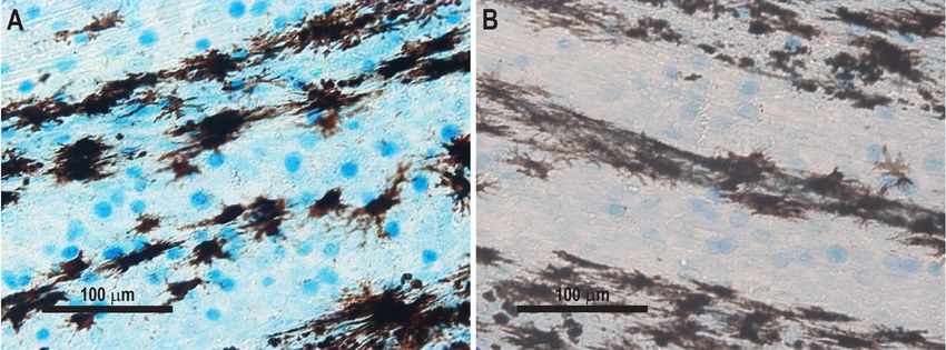
Fig. 3. Superficial mucous cells in rainbow trout caudal fins stained by Alcian Blue. (A) Non-stimulated mucus-filled mucous
cells; (B) stimulated cells with partial exhaustion of mucus
of PAA (Fig. 3A). At 12 hpe, this response was 4. DISCUSSION
seen only for 0.5 mg l−1, where cell densities in
fish fins exposed to the other concentrations had The need for auxiliary compounds and biocides is
decreased to control levels. At 24 hpe, significantly positively correlated with the content of organic mat-
lower mucous cell densities (Fig. 3B) were ob- ter in fish culture systems. Recirculated production fa-
served for the 2 highest concentrations of PAA cilities reuse the water containing dissolved organic
(2 and 1 mg l−1). matter and particles released from biofilters and fish
(Becke et al. 2020, Schumann & Brinker 2020), which
3.9.2. Hydrogen peroxide is associated with a risk of propagation of several
types of microorganisms (bacteria, virus, amoebae,
At 2 hpe a significantly higher mucous cell density flagellates, ciliates) (Jørgensen et al. 2009, Moestrup
was observed in the groups receiving 3.125 mg l−1 of et al. 2014). Even if some of these microorganisms are
H2O2 whereas the groups receiving 12.5 mg l−1 relatively benign epibionts, their abundance and
showed a significantly lower density. At 12 hpe a sig- overgrowth in gills may create respiratory problems
nificant decrease in cell density was recorded in all for the fish. Others are primary pathogens (Ichthy-
concentrations. At 24 hpe lower cell densities were obodo, Ichthyophthirius) and may cause severe prob-
observed in the groups receiving 1.56, 12.5 and lems even at lower intensities. The organisms may
25 mg l−1 of H2O2. also aggregate to particles with direct effects on fish
gills (Lu et al. 2018). Future development of filtration
3.9.3. Formalin techniques may be a solution to some of these prob-
lems, but at present elimination of these organisms is
Slight but non-significant decreases of mucous cell usually achieved by use of certain biocides. Among
densities were seen after treatment with formalin at these, PAA, H2O2 and formalin are commonly used
2 hpe. At 12 hpe a significantly lower cell density was (Straus & Meinelt 2009, Straus et al. 2012, Jaafar et al.
observed in all concentrations except 3.125 mg l−1. A 2013). The present study has documented that these
significantly higher mucous cell density was seen for biocides, when used for bath exposure of rainbow
the lowest concentration (3.125 mg l−1) at 24 hpe. trout, induce strong inflammatory reactions especially
in gills and to some extent in fins. This may explain
3.9.4. Surfactant why farmers and researchers have recognized ad-
verse reactions after exposure to auxiliary compounds
A significantly lower mucous cell density was ob- such as PAA (Liu et al. 2017, Straus et al. 2018, Soleng
served in all groups treated with SPH6 at 2, 12 and et al. 2019), H2O2 (Jia et al. 2021) and formalin (Buch-
24 hpe. mann et al. 2004b). We measured expression of genesMathiessen et al.: Biocide effects on trout 19
encoding inflammatory cytokines and acute phase re- cells in the fish epidermis, may assist the elimination
actants in gills and fins of rainbow trout. Some differ- of epibionts on fish surfaces and act as a biological
ences between compounds were noted, but PAA ex- cleaning process. With a relatively benign effect on
hibited a marked effect at almost all levels. Bath the fish host, and a significant lethal effect on various
treatments of rainbow trout, using solutions of these parasites, the novel biocide SPH6 may show promise
compounds, may therefore induce some irritation or for future use in aquaculture. It is not expected that
pain in the skin of the fish. This question should be biocides of any kind will have no side effects on fish,
further elucidated. In some cases, the compounds but the SPH6 surfactant may challenge the welfare of
(e.g. formalin and PAA) downregulated expression of the fish to a lesser degree than other commonly used
some cytokine and acute phase reactant genes, which biocides (formalin, H2O2, PAA). It is unknown if this
calls for a study on the implications for fish health. We compound will be commonly applied in the future,
also investigated effects on fish of a novel biocide but history has shown that all microorganisms ex-
SPH6, which is a lipopeptide with a surfactant effect posed to various antimicrobials may achieve some re-
(Liu et al. 2015) able to eliminate pathogenic ciliates sistance due to selection of resistant organisms (White
(Al-Jubury et al. 2018) and amoebae (Jensen et al. et al. 2002, Miller & Harbottle 2018). It is therefore
2020). Although the compound has promising effects recommended that this issue is also elucidated in any
on parasites in vitro, it is necessary to determine its ef- future study or use of the compound.
fects on fish gills and fins if it is to be applied at the
farm level. Other auxiliary compounds hitherto used
were shown to affect fish adversely. Tissue injuries 5. CONCLUSIONS
and physiological disturbances occur following expo-
sure to similar concentrations of formalin (Buchmann A range of parasites and epibionts which are se-
et al. 2004b, Jørgensen & Buchmann 2007), PAA verely challenging freshwater aquaculture can be con-
(Straus et al. 2018) and H2O2 (Polinski et al. 2013, trolled by water treatments using various biocides or
Henriksen et al. 2015, Chalmers et al. 2018, Jia et al. auxiliary substances. We have shown that many of the
2021). It was therefore noteworthy that the surfactant compounds applied induce an inflammatory reaction
SPH6 had a relatively low stimulatory effect on ex- in the surfaces (skin and gills) of the fish present in the
pression of inflammatory genes in rainbow trout, and fish tank. Gene expression studies showed that genes
in some cases it downregulated some genes. We also encoding inflammatory cytokines and acute phase re-
recorded the presence and mucus content of mucous actants become upregulated to various degrees by use
cells in the fin epidermis. The density of superficial of formalin, H2O2 and PAA. A novel biological biocide,
mucous cells in the fish epidermis is flexible and sen- a surfactant lipopeptide isolated from Pseudomonas
sitive to environmental disturbances. The cells are H6, was shown to affect fish surfaces to a considerably
recruited from the lower epidermal cell layers and lower degree. No regulation or downregulation of the
increase in numbers a few hours after formalin stimu- genes was demonstrated in several cases. The com-
lation, but extended exposure may stimulate mucus pound stimulated immediate release of mucus from su-
expulsion from cells (Buchmann et al. 2004b). The perficial mucous cells. This action may together with its
process is highly temperature dependent (Quiniou et direct antimicrobial effect call for further studies on its
al. 1998), but the present study was performed at a application in aquaculture enterprises.
stable temperature. We documented that especially
PAA, and to a lesser extent H2O2 and formalin, in-
Acknowledgements. The present study was supported by
duced an increase in superficial mucous cells in the the Danish Ministry of Environment and Food by grant no.
fin, but extended stimulation resulted in an elevated 34009-19-1578 (BIOKOS — Biological control of ectopara-
release of mucus. This was shown as a reduction or sites in freshwater fish) under the GUDP Green Develop-
absence of Alcian Blue-stained mucus in the cells. ment and Demonstration Programme. The authors are
indebted to the staff at the Bornholm Salmon Hatchery for
Mucous cell densities in caudal fins following SPH6
rearing fish.
exposure decreased within 2 h. This suggests that this
biocide stimulates mucous cells to release their con-
tent of mucus, but no evidence of elevated recruitment LITERATURE CITED
of new cells to the fin surface was found. The implica-
Al-Jubury A, Lu C, Kania PW, Jørgensen LvG and others
tions of this for practical use in farms should be further (2018) Impact of Pseudomonas H6 surfactant on all exter-
investigated. We cannot rule out the possibility that a nal life cycle stages of the fish parasitic ciliate Ichthyoph-
strong expulsion of mucus, from superficial mucous thirius multifiliis. J Fish Dis 41:1147−115220 Dis Aquat Org 146: 9–21, 2021 Andersen CL, Jensen JL, Ørntoft TF (2004) Normalization of sues of common carp (Cyprinus carpio). Sci Total Environ real-time quantitative reverse transcription-PCR data: a 757:143831 model-based variance estimation approach to identify Jørgensen TR, Buchmann K (2007) Stress response in rain- genes suited for normalization, applied to bladder and bow trout during infection with Ichthyophthirius multifil- colon cancer data sets. Cancer Res 64:5245−5250 iis and formalin bath treatment. Acta Ichthyol Piscat 37: Assefa A, Abunna F (2018) Maintenance of fish health in 25−28 aquaculture: review of epidemiological approaches for Jørgensen TR, Larsen TB, Buchmann K (2009) Parasite prevention and control of infectious disease of fish. Vet infections in recirculated rainbow trout (Oncorhynchus Med Int 2018:5432497 mykiss) farms. Aquaculture 289:91−94 Becke C, Schumann M, Geist J, Brinker A (2020) Shape char- Jørgensen LvG, Korbut R, Jeberg S, Kania PW, Buchmann K acteristics of suspended solids and implications in differ- (2018) Association between adaptive immunity and neu- ent salmonid aquaculture production systems. Aqua- trophil dynamics in zebrafish (Dania rerio) infected by a culture 516:734631 parasitic ciliate. PLOS ONE 13:e0203297 Bruzio M, Buchmann K (2010) The effect of peracetic acid Jussila J, Makkonen J, Kokko H (2011) Peracetic acid (PAA) products on parasites causing white spot diseases. Fish treatment is an effective disinfectant against crayfish Farmer 33:25−27 plague (Aphanomyces astaci) spores in aquaculture. Buchmann K, Bresciani J (1997) Parasitic infections in pond- Aquaculture 320:37−42 reared rainbow trout Oncorhynchus mykiss in Denmark. Lieke T, Meinelt T, Hoseinifar SH, Pan B, Straus DL, Stein- Dis Aquat Org 28:125−138 berg CEW (2020) Sustainable aquaculture requires envi- Buchmann K, Jensen PB, Kruse KD (2003) Effects of sodium ronmental-friendly treatment strategies for fish diseases. percarbonate and garlic extract on Ichthyophthirius Rev Aquacult 12:943−965 multifiliis theronts and tomocysts: in vitro experiments. Liu Y, Rzeszutek E, van der Voort M, Wu CH and others N Am J Aquaculture 65:21−24 (2015) Diversity of aquatic Pseudomonas species and Buchmann K, Nielsen T, Sigh J, Bresciani J (2004a) Amoebic their activity against the fish pathogenic oomycete gill infections of rainbow trout in freshwater ponds. Bull Saprolegnia. PLOS ONE 10:e0136241 Eur Assoc Fish Pathol 24:87−91 Liu D, Pedersen LF, Straus DL, Kloas W, Meinelt T (2017) Buchmann K, Bresciani J, Jappe J (2004b) Effects of forma- Alternative prophylaxis/disinfection in aquaculture — lin treatment on epithelial structure and mucous cell adaptable stress induced by peracetic acid at low con- densities in rainbow trout, Oncorhynchus mykiss (Wal- centration and its application strategy in RAS. Aquacul- baum), skin. J Fish Dis 27:99−104 ture 474:82−85 Chalmers L, Vera LM, Taylor JF, Adams A, Migaud H (2018) Livak KJ, Schmittgen TD (2001) Analysis of relative gene Comparative ploidy response to experimental hydrogen expression data using real-time quantitative PCR and peroxide exposure in Atlantic salmon (Salmo salar). Fish the 2−ΔΔCT method. Methods 25:402−408 Shellfish Immunol 81:354−367 Lu C, Kania PW, Buchmann K (2018) Particle effects on fish Chettri JK, Kuhn JA, Jafaar RM, Kania PW, Møller OS, gills: an immunogenetic approach for rainbow trout and Buchmann K (2014) Epidermal response of rainbow trout zebrafish. Aquaculture 484:98−104 to Ichthyobodo necator: Immunohistochemical and gene Meinelt T, Matzke S, Stüber A, Pietrock M, Wienke A, expression studies indicate a Th1-/Th2-like switch. Mitchell AJ, Straus DL (2009) Toxicity of peracetic acid J Fish Dis 37:771−783 (PAA) to tomonts of Ichthyophthirius multifiliis. Dis Dyková I, Kostka M, Wortberg F, Nardy E, Pecková H (2010) Aquat Org 86:51−56 New data on aetiology of nodular gill disease in rainbow Miller RA, Harbottle H (2018) Antimicrobial drug resistance trout, Oncorhynchus mykiss. Folia Parasitol 57:157−163 in fish pathogens. Microbiol Spectr 6:501−520 Henriksen MMM, Kania PW, Buchmann K, Dalsgaard I Moestrup Ø, Hansen G, Daugbjerg N, Lundholm N and oth- (2015) Evaluation of the immune response in rainbow ers (2014) The dinoflagellates Pfiesteria shumwayae and trout fry, Oncorhynchus mykiss (Walbaum), after water- Luciella masanensis cause fish kills in recirculation fish borne exposure to Flavobacterium psychrophilum and/ farms in Denmark. Harmful Algae 32:33−39 or hydrogen peroxide. J Fish Dis 38:55−66 Pedersen LF, Pedersen PB, Sortkjær O (2007) Temperature- Jaafar RM, Kuhn JA, Chettri JK, Buchmann K (2013) Com- dependent and surface specific formaldehyde degrada- parative efficacies of sodium percarbonate, peracetic tion in submerged biofilters. Aquacult Eng 36:127−136 acid, and formaldehyde for control of Ichthyobodo neca- Pedersen LF, Meinelt T, Straus DL (2013) Peracetic acid tor — an ectoparasitic flagellate from rainbow trout. Acta degradation in freshwater aquaculture systems and pos- Ichthyol Piscat 43:139−143 sible practical implications. Aquacult Eng 53:65−71 Jaafar R, Ødegård J, Mathiessen H, Karami AM and others Polinski MP, Jensen NR, Foltz J, Ireland SC, Cain KD (2013) (2020) Quantitative trait loci (QTL) associated with re- Hydrogen peroxide treatments administered to hatch- sistance of rainbow trout Oncorhynchus mykiss against ery-reared burbot: assessing treatment regimes from the parasitic ciliate Ichthyophthirius multifiliis. J Fish Dis embryonic development through juvenile rearing. N Am 43:1591−1602 J Aquaculture 75:50−56 Jensen HM, Karami AM, Mathiessen H, Al-Jubury A, Kania Quiniou MA, Bigler S, Clem LW, Bly JE (1998) Effects of PW, Buchmann K (2020) Gill amoebae from freshwater water temperature on mucous cell distribution in chan- rainbow trout (Oncorhynchus mykiss): in vitro evaluation nel catfish epidermis: a factor in winter saprolegnia. Fish of antiparasitic compounds against Vannella sp. J Fish Shellfish Immunol 8:1−11 Dis 43:665−672 Rintamäki-Kinnunen P, Rahkonen M, Mannermaa-Keränen Jia R, Du J, Cao L, Feng W, He Q, Xu P, Yin G (2021) AL, Suomalainen LR, Mykrä H, Valtonen ET (2005) Treat- Immune, inflammatory, autophagic and DNA damage ment of ichthyophthiriasis after malachite green. I. Con- responses to long-term H2O2 exposure in different tis- crete tanks at salmonid farms. Dis Aquat Org 64:69−76
Mathiessen et al.: Biocide effects on trout                                       21

Schmittgen TD, Livak KJ (2008) Analyzing real-time PCR               Straus DL, Meinelt T, Liu D, Pedersen LF (2018) Toxicity of
   data by the comparative CT method. Nat Protoc 3:                     peracetic acid to fish: variation among species and impact
   1101−1108                                                            of water chemistry. J World Aquacult Soc 49:715−724
Schumann M, Brinker A (2020) Understanding and manag-                Tort L (2011) Stress and immune modulation in fish. Dev
   ing suspended solids in intensive salmonid aquaculture:              Comp Immunol 35:1366−1375
   a review. Rev Aquacult 12:2109−2139                               White DG, Zhao S, Simjee S, Wagner DD, McDermott PF
Soleng M, Johansen LH, Johnsen H, Johansson GS and oth-                 (2002) Antimicrobial resistance of foodborne pathogens.
   ers (2019) Atlantic salmon (Salmo salar) mounts systemic             Microbes Infect 4:405−412
   and mucosal stress responses to peracetic acid. Fish              Woo PTK, Leong AL, Buchmann K (2020) Climate change
   Shellfish Immunol 93:895−903                                         and infectious fish diseases (CCIFD), Sections I, II, III.
Straus DL, Meinelt T (2009) Acute toxicity of peracetic acid            CAB International, Wallingford
   (PAA) formulations to Ichthyophthirius multifiliis the-           Xueqin J, Kania PW, Buchmann K (2012) Comparative
   ronts. Parasitol Res 104:1237−1241                                   effects of four feed types on white spot disease sus-
Straus DL, Meinelt T, Farmer BD, Mitchell AJ (2012) Per-                ceptibility and skin immune parameters in rainbow
   acetic acid is effective for controlling fungus on channel           trout, Oncorhynchus mykiss (Walbaum). J Fish Dis 35:
   catfish eggs. J Fish Dis 35:505−511                                  127−135

Editorial responsibility: Stewart Johnson,                           Submitted: April 19, 2021
 Nanaimo, British Columbia, Canada                                   Accepted: June 23, 2021
Reviewed by: 3 anonymous referees                                    Proofs received from author(s): August 21, 2021
You can also read


