Knifefish's suction makes water boil - Nature
←
→
Page content transcription
If your browser does not render page correctly, please read the page content below
www.nature.com/scientificreports
OPEN Knifefish’s suction makes water
boil
Victor M. Ortega‑Jimenez* & Christopher P. J. Sanford
We discovered that knifefish (Apteronotus albifrons) during suction feeding can produce millimeter-
sized cavitation bubbles and flow accelerations up to ~ 450 times the acceleration of gravity. Knifefish
may use this powerful suction-induced cavitation to cause physical damage on prey hiding in narrow
refuges, therefore facilitating capture.
Cavitation bubbles in a liquid are induced by intense and sudden changes of local pressure1. These collapsing
bubbles can produce intense compressional waves and extremely high temperatures, as well as serious damage
on structures. In nature, besides mantis2 and pistol s hrimps3, there is no other taxon known that uses cavitation
for prey capture. This is particularly intriguing because feeding in several aquatic o rganisms4,5, such as fishes,
rely on generating rapid low pressures inside the buccal c avity6,7.
Knifefish are nocturnal freshwater fishes that thrive in Central and South American streams and rivers. They
are well known for their remarkable electrical senses, slender morphology, and anal fin propulsion8,9 (Fig. 1a).
In the wild, knifefish feed mostly on aquatic invertebrates, such as insects10, which can perform rapid evasive
maneuvers. Furthermore, prey can hide inside tiny holes of submerged logs, between rocks or in the mud, mak-
ing it difficult for a predator to reach, detach and extract them.
Results
We discovered that knifefish while generating suction at the tip of an 8 mm long capillary tube (1.5 mm diameter),
open to the air, induces a rapid jet of water toward the mouth which in turn can generate cavitation bubbles
(Video S1). Approximately 3 ms after the onset of suction, we registered a maximum flow speed and acceleration
(amax) of up to ~ 7 m/s and ~ 4500 m/s2 respectively (Fig. 1). In the open tube experiments we confirmed cavita-
tion bubbles appearing close to the mouth in three individuals and four of the 16 sampled trials. It is noteworthy
that in all trials we observed marked flow instabilities appearing in the air–water interface inside the open tube
during suction (Video S1), which may be related to cavitation.
It has recently been demonstrated that cavitation induced by an impulsive system can be better defined by
acceleration rather than speed1. Here we calculated the cavitation number as Ca = (pr − pv)/(ρLamax)1, where p r is
the atmospheric pressure, p v is the vapor pressure, ρ is the water density, and L is the distance between the start
and end positions of water level in the tube, respectively. We found that knifefish suction feeding generates Ca of
0.65 (± 0.20 SD, N = 4), which sufficiently meets the criteria to generate cavitation (Ca < 1). We excluded the extra
pressure required to overcome resistance in the capillary tubes (i.e., pressure drop), which could result in even
lower cavitation numbers. Remarkably, when fish suction feed from a tube sealed at one end, several cavitation
bubbles appeared, grew, and then collapsed during a 22 ms period (Fig. 2). All four individuals produced cavita-
tion bubbles in the sealed tube. A hammer-like sound during suction was also heard (Video S1) that seemed to
coincide with collapsing of the bubble (Video S2).
To investigate the fluid dynamics produced by the knifefish suction feeding between two parallel plates
(1 mm separation) we performed 2D particle image velocimetry, filming at ~ 7000 frames/s. We found that a
knifefish generates very close-range suction (~ 3 mm) in front of the mouth. Flow speed in this region increased
from ~ zero to ~ 2.5 m/s in one millisecond (i.e., an acceleration of ~ 2500 m/s2, Fig. 3).
Discussion
This study demonstrates that black ghost knifefish can produce extreme suction resulting in a powerful jet, a
hammer-like sound, and cavitation bubbles in a capillary tube. Therefore, we suggest that this nocturnal and
riverine fish may use cavitation to facilitate prey extraction, capture and intake of small prey, hiding inside
the matrix of submerged vegetation or between microcracks of rocks. We observed that knifefish were able to
extract the air inside a bamboo stick by producing suction at the tip (Video S2 [00:10 s]). It is known that snap-
ping shrimp can also use a powerful jet, followed by cavitation bubbles to stun and kill prey3. Thus, knifefish
Department of Ecology, Evolution, and Organismal Biology, Kennesaw State University, Kennesaw, GA 30144, USA.
*
email: ornithopterus@gmail.com
Scientific Reports | (2020) 10:18698 | https://doi.org/10.1038/s41598-020-75788-x 1
Vol.:(0123456789)www.nature.com/scientificreports/
Figure 1. (a) Black ghost Knifefish (Apteronotus albifrons). (b) Flow acceleration (blue) and (c) speed (black)
over time produced by suction in a capillary tube open to the air. Lines represent time series of each sampled
individual. Starting time is when acceleration increases from zero. Shadows represent average value and one SD
(N = 4). (d) Drawing represents the experimental setup. This figure was created using MATLAB R2017b (https://
www.mathworks.com/), GIMP 2.8 (free available at: https://www.gimp.org/), and Inkscape 0.92.3 (free available
at: https://inkscape.org/).
Figure 2. Frame sequence filmed at 500 frames/s of a fish suction feeding a the tip of a sealed tube, showing
large cavitation bubbles. This figure was created using GIMP 2.8 (free available at: https://www.gimp.org/) and
Inkscape 0.92 (free available at: https://inkscape.org/).
can generate cavitation using a different mechanical system from invertebrates. This finding represents the first
example of cavitation we are aware of in vertebrate systems.
Furthermore, cavitation may facilitate prey immobilization, dislodgement and capture. Invertebrates, such as
copepods, exposed to ultrasonic cavitation can experience damage and can even break apart11. Another possibil-
ity is that sound waves produced during bubble collapse (Video S2) may be useful to pinpoint and evaluate prey
located in confined spaces. Vapor bubbles can be detected using a rebound shock wave during c ollapse12. This
has the potential to be a useful mechanism of prey detection as electroreception in knifefishes can be impaired
by cluttered e nvironments13.
It is important to consider the negative effects produced by cavitation on those organisms that generate it.
Mantis shrimps, for example, produce cavitation via a powerful strike, which can damage their own appendages2.
However, their appendages are well designed to resist such impact forces14. Thus, we can expect that cavitation
bubbles produced in the oral cavity of knifefish could also cause certain damage. Future work on knifefishes in
their natural habitat will reveal important clues regarding the role of cavitation in their ecology.
Particle image velocimetry demonstrates that knifefish suction-feeding can generate flow speeds of 2.5 m/s
in ~ 1 ms, and an average flow acceleration of 2500 m/s2. This aligns well with the measurements of knifefish
when generating suction on an open capillary tube (Fig. 1). Our results suggest that higher flow speeds and lower
Scientific Reports | (2020) 10:18698 | https://doi.org/10.1038/s41598-020-75788-x 2
Vol:.(1234567890)www.nature.com/scientificreports/
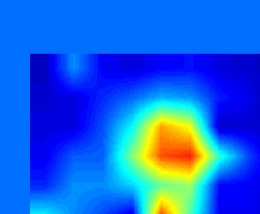
Figure 3. Flow velocity fields using PIV produced by a fish generating suction between two transparent plates,
at the beginning (top) and after ~ 1 ms (bottom). Drawing represents the experimental setup. For details see
Supplementary information and video S1. This figure was created using MATLAB R2017b (https://www.mathw
orks.com/) and Inkscape 0.92.3 (free available at: https://inkscape.org/).
pressures are likely generated inside the mouth of a knifefish. Seahorses for example, can reach flow speeds inside
the mouth cavity up to 3.5 m/s15. When compared to other fishes, knifefish generate suction flow speeds that are
two to three times higher than those generated by sunfish (max 0.8 m/s in 2 ms16) and largemouth bass (1.4 m/s
in 4 m s17), respectively. However, in comparison to pistol shrimps, flow speed and acceleration in knifefish are
modest. A computational model indicates that pistol shrimp can generate a jet of water with a peak speed up to
30 m/s in 0.3 m s18, which corresponds to an acceleration of 1 05 m/s2. Nevertheless, knifefish are still capable of
producing cavitation at lower speeds.
In conclusion, we found that knifefish under laboratory conditions can produce cavitation bubbles during
suction feeding. To our knowledge this is the first reported example of induced cavitation in a vertebrate system.
It is possible that this capability may serve an important role for immobilization, dislodgement, and capture of
prey. Furthermore, it could be used as a method of prey detection in narrow refuges. Future comparative work
among suction feeding organisms, such as carnivorous plants, amphibians and other fishes will establish if this
ability to cavitate is more widespread than has been previously thought.
Methods
Ethics statement. All methods were carried out in accordance with relevant federal guidelines and regu-
lations. All training and experimental procedures were approved by Kennesaw State University’s Institutional
Animal Care and Use Committee (ACUP #20-005), and no animals were sacrificed for this study.
Fish suction on an open tube. Four black ghost knifefish (Apteronotus albifrons) were sampled. Fish were
acquired from a local aquatic pet store at Kennesaw, Georgia. Each individual was housed in a 151 L tank with
a PH of 7–8 and a water temperature of 25–26 °C. Animals were fed frozen blood worms ad-libitum. During
experiments, each individual was transferred to a small experimental tank (19 L). They were trained for two days
to feed on blood worms introduced to a plastic clear tube (internal diameter 1.5 mm, external diameter 2.5 mm
and 8 cm long) (Fig. 1b).
One high speed camera (Fastec HiSpec 1) was used for recording at 1000 frames/s each fish feeding at the
capillary tube. The tube was filled with water and introduced vertically by hand into the tank. The top end of the
tube was open to the air. Air pockets inside the tube were used to track the distance traveled during suction. A
Scientific Reports | (2020) 10:18698 | https://doi.org/10.1038/s41598-020-75788-x 3
Vol.:(0123456789)www.nature.com/scientificreports/
sheet of white paper placed outside the tank was used to provide contrast. Two LED lights illuminated the capil-
lary tube. In this study, because we were interested in maximal performance of knifefish, only one sequence for
each individual was analyzed in which flow acceleration was maximal. Variation between each sampled fish (4
trials per fish) with body size are shown in Figure S1.
For each sequence (one for each fish) we digitized the vertical position of the water level (i.e., the air pocket
interface) in the tube using the DLTdv5.m digitizing tool for Matlab (https://biomech.web.unc.edu/dltdv/). Flow
speed and acceleration induced by suction were calculated from the first and second derivatives of the MSE-
quintic spline f unction19.
Cavitation number. We calculated the cavitation number as follows Ca = (pr − pv)/(ρLamax), where pr is the
atmospheric pressure (100 kPa), pv is the vapor pressure at 25 ºC (3.17 kPa), ρ is the water density (1 × 10³ kg/
m3), L is the distance between the start and end positions of water level in the tube (6 ± 1 cm), and a max is the
maximal flow acceleration (2800 ± 1200 m/s2), respectively. We found that Ca was 0.65 ± 0.20. Data is presented
as average value ± one SD (N = 4).
Suction on sealed tube. We performed similar experiments to those described above. However, in this
case the fish was allowed to suction feed at the tip of a capillary tube (internal diameter 1.5 mm, external diam-
eter 2.5 mm and 4 cm long) filled with water, and hermetically sealed on one end (Fig. 1c). Similarly as above, we
filmed each fish while suction feeding at the tip of the tube at 500 frames/s.
Particle image velocimetry. A class-4 laser (Opto Engine LLC, 532 nm, 5 W) was used to produce a hori-
zontal light sheet to illuminate plastic beads (~ 50 µm) introduced between two parallel plastic plates. Each plate
was 2 × 3 cm with a thickness of 0.5 mm. Separation between plates was 1 mm. A fish was trained to suction feed
on blood worms placed between both plates (Fig. 1d). We filmed the top view using a high-speed camera (Fastec
HiSpec 4) at 6754 frames/s and used paired frames from the recorded video sequence to calculate velocity fields
using PIVlab (https://pivlab.blogspot.com/2017/07/pivlab-direct-download.html)20. A ROI of 163 × 121 pixels
was sampled. An interrogation window from 64 × 64 pixels to 32 × 32 pixels, excluding those vectors with stand-
ard deviation greater than 5, was used.
Data availability
The datasets supporting this article have been uploaded as part of the electronic supplementary material.
Code availability
The PIV and Digitization software that support the findings of this study are available in https://pivlab.blogspot.
com/2017/07/pivlab-direct-download.html and https://biomech.web.unc.edu/dltdv/ respectively.
Received: 23 June 2020; Accepted: 6 October 2020
References
1. Pan, Z. et al. Cavitation onset caused by acceleration. Proc. Natl. Acad. Sci. USA 114, 8470–8474 (2017).
2. Patek, S. N. et al. Deadly strike mechanism of a mantis shrimp. Nature 428, 819–820 (2004).
3. Versluis, M. et al. How snapping shrimp snap: through cavitating bubbles. Science 289, 2114–2117 (2000).
4. Deban, S. M. & Olson, W. M. Suction feeding by a tiny predatory tadpole. Nature 420, 41–42 (2002).
5. Vincent, O. et al. Ultra-fast underwater suction traps. Proc. Roy Soc. B 278, 2909–2914 (2011).
6. Westneat, M. W. & Olson, A. M. How fish power suction feeding. Proc. Natl Acad. Sci. USA 112, 8525–8526 (2015).
7. Gemmell, B., Sheng, J. & Buskey, E. Morphology of seahorse head hydrodynamically aids in capture of evasive prey. Nat. Commun.
4, 2840 (2013).
8. Blake, R. W. Swimming of electric eels and knifefishes. Can. J. Zool. 61, 1432–1441 (1983).
9. Turner, C. R. et al. Phylogenetic comparative analysis of electric communication signals in ghost knifefishes (Gymnotiformes:
Apteronotidae). J. Exp. Biol. 210, 4104–4122 (2007).
10. Winemiller, K. O. & Adite, A. Convergent evolution of weakly electric fishes from floodplain habitats in Africa and South America.
Environ. Biol. Fishes 49, 175–186 (1997).
11. Svendsen, E. et al. Effect of ultrasonic cavitation on small and large organisms for water disinfection during fish transport. Aquac.
Res. 49, 1166–1175 (2018).
12. Leighton, T. G. The Acoustic Bubble 640 (Academic Press, Cambridge, 1994).
13. Babineau, D. et al. Spatial acuity and prey detection in weakly electric fish. PloS Comput. Biol. 3, e38 (2007).
14. Currey, J. D. et al. Calcified cuticle in the stomatopod smashing limb. J. Mater. Sci. 17, 1939–1944 (1982).
15. Roos, G. et al. Kinematics of suction feeding in the seahorse Hippocampus reidi. J. Exp. Biol. 212, 3490–3498 (2009).
16. Day, S. W. et al. Spatial and temporal patterns of water flow generated by suction-feeding bluegill sunfish Lepomis macrochirus
resolved by particle image velocimetry. J. Exp. Biol. 208, 2661–2671 (2005).
17. Higham, T. E. et al. Multidimensional analysis of suction feeding performance in fishes: fluid speed, acceleration, strike accuracy
and the ingested volume of water. J. Exp. Biol. 209, 2713–2725 (2006).
18. Koukouvinis, P. et al. Unveiling the physical mechanism behind pistol shrimp cavitation. Sci. Rep. 7, 13994 (2017).
19. Walker, J. A. Estimating velocities and accelerations of animal locomotion: a simulation experiment comparing numerical dif-
ferentiation algorithms. J. Exp. Biol. 201, 981–995 (1998).
20. Thielicke, W. & Stamhuis, E. J. PIVlab—towards user-friendly, affordable and accurate digital particle image velocimetry in MAT-
LAB. J. Open Res. Softw. 2, e30 (2014).
Author contributions
V.M.O.-J. conceived the project, V.M.O.-J. and C.P.J.S. designed the experiments, V.M.O.-J. collected and analyzed
data, V.M.O.-J. and C.P.J.S. wrote the manuscript.
Scientific Reports | (2020) 10:18698 | https://doi.org/10.1038/s41598-020-75788-x 4
Vol:.(1234567890)www.nature.com/scientificreports/
Competing interests
The authors declare no competing interests.
Additional information
Supplementary information is available for this paper at https://doi.org/10.1038/s41598-020-75788-x.
Correspondence and requests for materials should be addressed to V.M.O.-J.
Reprints and permissions information is available at www.nature.com/reprints.
Publisher’s note Springer Nature remains neutral with regard to jurisdictional claims in published maps and
institutional affiliations.
Open Access This article is licensed under a Creative Commons Attribution 4.0 International
License, which permits use, sharing, adaptation, distribution and reproduction in any medium or
format, as long as you give appropriate credit to the original author(s) and the source, provide a link to the
Creative Commons licence, and indicate if changes were made. The images or other third party material in this
article are included in the article’s Creative Commons licence, unless indicated otherwise in a credit line to the
material. If material is not included in the article’s Creative Commons licence and your intended use is not
permitted by statutory regulation or exceeds the permitted use, you will need to obtain permission directly from
the copyright holder. To view a copy of this licence, visit http://creativecommons.org/licenses/by/4.0/.
© The Author(s) 2020
Scientific Reports | (2020) 10:18698 | https://doi.org/10.1038/s41598-020-75788-x 5
Vol.:(0123456789)You can also read