Locomotor activity and histological changes observed in a mouse model of knee osteoarthritis - J-Stage
←
→
Page content transcription
If your browser does not render page correctly, please read the page content below
J. Phys. Ther. Sci. 32: 370–374, 2020
The Journal of Physical Therapy Science
Original Article
Locomotor activity and histological changes
observed in a mouse model of knee osteoarthritis
Satoshi Kojima, RPT, PhD1)*, Masanori Watanabe, RPT, PhD2), Keiji Asada, RPT, PhD3)
1) Graduate School of Rehabilitation, Course of Rehabilitation, Kinjo University: 1200 Kasama-machi,
Hakusan-city, Ishikawa 924-8511, Japan
2) Department of Physical Therapy, Faculty of Rehabilitation Science, Nagoya Gakuin University,
Japan
3) Department of Rehabilitation, Faculty of Health Science, Suzuka University of Medical Science,
Japan
Abstract. [Purpose] This study aimed to elucidate the changes in locomotor activity in a mouse model of knee
osteoarthritis (OA). [Materials and Methods] Fourteen 20-week-old mice were divided into control and OA groups.
Knee OA was surgically induced under anesthesia by destabilizing the meniscus. The OA group was reared normal-
ly for 8 weeks following surgery, during which OA was induced. Locomotor activity was measured every hour for
8 weeks using an infrared locomotor activity measurement device. Histological changes were evaluated according
to the classification-system of Glasson. [Results] Locomotor activity in the OA group significantly decreased up to
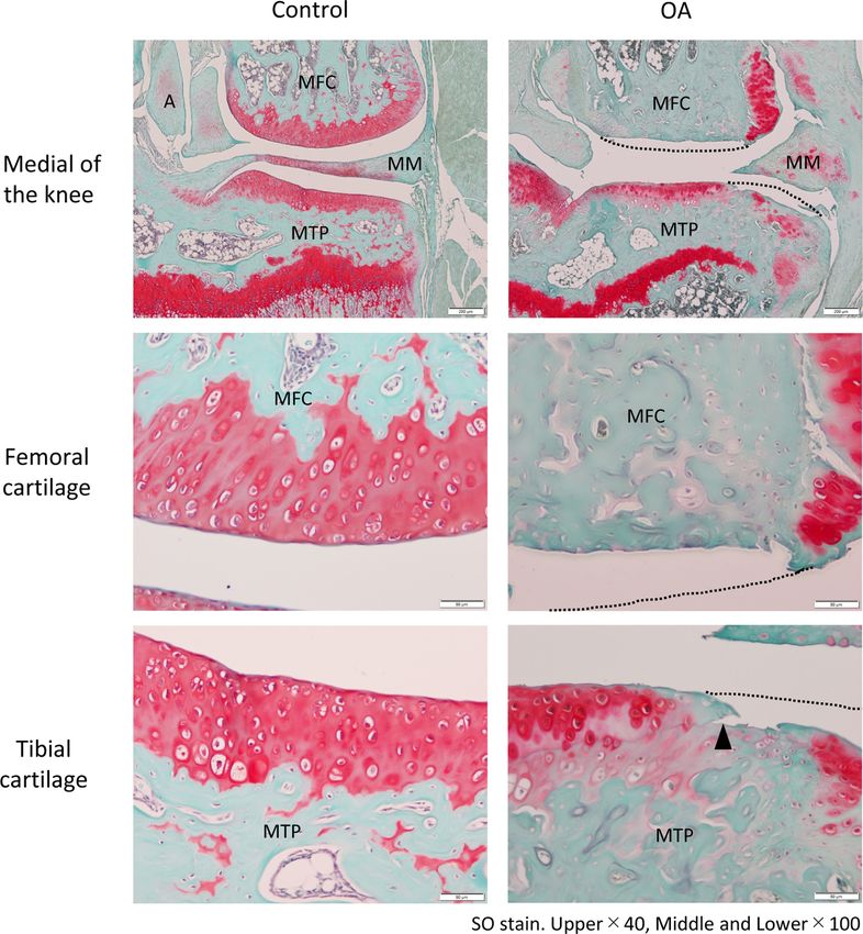
2 weeks after surgery. Histological findings in the control group revealed an irregular cartilage surface in a portion
of the tibia with no other abnormalities. Contrastingly, those in the OA group had eburnation of the medial femoral
condyle, as well as fibrillation and fissures in the medial tibial plateau. Histological scores in the OA group were
significantly higher than the control group. [Conclusion] Locomotor activity evaluations, in addition to histological
scores and findings, are imperative for studies aiming to clarify the disease state and effect of interventions using
mice models.
Key words: Mouse osteoarthritis, Locomotor activity, Histology
(This article was submitted Jan. 20, 2020, and was accepted Mar. 1, 2020)
INTRODUCTION
Knee osteoarthritis (KOA) is a multifactorial disease associated with aging, obesity, and mechanical stress. Considering
that minute damage generated in the cartilage gradually progresses to degeneration and joint destruction, this disease has
been characterized by difficulties in structural improvement. According to the Research on Osteoarthritis Against Disability
report, over 30 million Japanese patients aged >40 years suffer from KOA, with its prevalence being expected to increase in
the future1).
The Japanese Guidelines for the Physical Therapy of KOA2) have recommended muscle strengthening and walking for
better stabilization of the knee joint. Muscle weakness due to decreased activity increases the amount of stress placed on the
knee and decreases activity and quality of life, resulting in a vicious negative cycle. Therefore, maintaining and improving
muscle strength and activity are imperative. Treatment guidelines have recommended aerobic exercise, including walking
(Grade A), which is supported by Level 1 evidence.
Conversely, considering the mechanism of KOA onset, increasing the activity volume of patients with KOA may lead
to excessive mechanical stress, whereas limiting the same may delay the progression of KOA depending on the degree of
*Corresponding author. Satoshi Kojima (E-mail: kojima@kinjo.ac.jp)
©2020 The Society of Physical Therapy Science. Published by IPEC Inc.
This is an open-access article distributed under the terms of the Creative Commons Attribution Non-Commercial No Deriva-
tives (by-nc-nd) License. (CC-BY-NC-ND 4.0: https://creativecommons.org/licenses/by-nc-nd/4.0/)
370progression. However, only few clinical or basic studies have evaluated the relationship between KOA progression and activ-
ity volume. If changes in locomotor activity affect KOA progression, it is necessary to investigate activity volume in addition
to histological findings. Therefore, this study aimed to elucidate the changes in locomotor activity in a mouse model of KOA.
MATERIALS AND METHODS
A total of 14 ICR male mice aged 20 weeks (body weight 31.5 ± 10.3 g) were included. All mice were individually housed
throughout the experimental period and were allowed free access to water and food in a 12-h light and dark cycle. Animal
Care and Use Committee for Kinjo University approved this study (Approval no. 0012).
Following environmental acclimatization for 4 weeks, mice were randomly allocated to an OA group (n=7) and a con-
trol group (n=7) reared in a typical manner throughout the experimental period. After placing the mice under anesthesia
via inhalation of isoflurane, KOA was surgically induced in the OA group based on previous studies3, 4). Accordingly, the
articular capsule, medial patellar retinaculum, and vastus medialis were longitudinally incised 1 cm from the inside of the
patellar ligament. The patella was laterally inverted, and the knee joint was opened. The medial meniscotibial ligament was
then incised to establish a model for destabilization of the meniscus (DMM). Thereafter, the joint was thoroughly cleaned,
and the articular capsule, muscle, and skin were sutured. Surgical manipulation was performed on both knees. Following the
surgery, normal rearing was performed for 8 weeks in accordance with a previous study, during which KOA was induced in
both knee joints.
Locomotor activity was measured every hour for 24 consecutive hours over 8 weeks using an infrared (IR) locomotor
activity measurement device (LE8825, PanLab, Barcelona, Spain). This devise contains 32 IR beam sensors (16 each in
length and width) in a 45 cm square.
After the experimental period, the mice were euthanized, and the hip joint was transected to collect the hind leg. The
collected hind legs underwent tissue fixation for 72 h in 10% neutral buffered formalin solution, followed by decalcification
for 72 h using Plank-Rychlo’s solution. After decalcification, the knee joint was excised by cutting along the center of the
frontal plane. The sample was then neutralized with 5% anhydrous sodium sulfate solution for 72 h, washed in running water,
and immersed in 100% alcohol for 3 h for degreasing. Paraffin embedding was performed using paraffin blocks that were
sliced thinly (approximately 3 μm) with a microtome (TU-213, Yamato, Saitama, Japan). Samples were then stained with
safranin-O fast green to create tissue specimens, which were observed under an optical microscope (BX53, Olympus, Tokyo,
Japan). The right knee was used for histological findings and left knee was used for histological scoring. Histological changes
in the knees were classified using the semi-quantitative scoring system of Glasson et al.5) wherein a score of 0 represents
normal cartilage; 0.5, loss of safranin-O without structural changes; 1, small fibrillations without loss of cartilage; 2, vertical
clefts down to the layer immediately below the superficial layer and some loss of surface lamina; 3, vertical clefts/erosion to
the calcified cartilage extending 75% of the articular surface. The scores were evaluated by
blinded person who was not involved in this study.
The changes of locomotor activity between both groups was statistically tested by two-way repeated measure analysis of
variance after Shapiro-Wilk test and F-test using statistical software R (version 3.3.2). The main effect was then analyzed as
a post-hoc test using Welch’s t-test with Bonferroni correction. And histological scores were compared using Mann-Whitney
U test. The level of significance was set as 0.05.
RESULTS
The locomotor activity value of the mice before surgery (at the age of 24 weeks) was 19,449 ± 373 times. In the OA group,
the value decreased significantly only up to 2 weeks after surgery (pTable 1. Locomotor activity
Contol OA p value
Before surgery 19,449 ± 373
1 week after surgery 20,555 ± 269 9,040 ± 551Table 2. Histological scores
Femoral Number of sumple
1 2 3 4 5 6 7 p value
Control 0 0 0 0.5 0.5 0.5 0.5
OA 2 4 4 4 4 5 6 0.0017**
Tibial Number of sumple
1 2 3 4 5 6 7 p value
Control 0 0 0 0 0.5 0.5 0.5
OA 1 2 3 4 4 3 4 0.0477*
**Significant difference from control (pREFERENCES
1) Yoshimura N: Epidemiology of musculoskeletal diseases in Japan: the ROAD study. Bone, 2010, 24: 39–42 (in Japanese).
2) Japanese Physical Therapy Association: Japanese Guidelines for the Physical Therapy, 1st ed. Tokyo: Japanese Physical Therapy Association, 2011 (in Japa-
nese).
3) Kamekura S, Hoshi K, Shimoaka T, et al.: Osteoarthritis development in novel experimental mouse models induced by knee joint instability. Osteoarthritis
Cartilage, 2005, 13: 632–641. [Medline] [CrossRef]
4) Glasson SS, Blanchet TJ, Morris EA: The surgical destabilization of the medial meniscus (DMM) model of osteoarthritis in the 129/SvEv mouse. Osteoarthri-
tis Cartilage, 2007, 15: 1061–1069. [Medline] [CrossRef]
5) Glasson SS, Chambers MG, Van Den Berg WB, et al.: The OARSI histopathology initiative - recommendations for histological assessments of osteoarthritis
in the mouse. Osteoarthritis Cartilage, 2010, 18: S17–S23. [Medline] [CrossRef]
6) Kubo T, Saito M: The clinical basis of osteoarthritis. Jpn J Rehabil Med, 2015, 52: 256–264 (in Japanese). [CrossRef]
7) Iijima H, Aoyama T, Ito A, et al.: Exercise intervention increases expression of bone morphogenetic proteins and prevents the progression of cartilage-
subchondral bone lesions in a post-traumatic rat knee model. Osteoarthritis Cartilage, 2016, 24: 1092–1102. [Medline] [CrossRef]
8) Hashimoto K, Akagi M: The produce of experimental non-operative osteoarthritis model for forced running. Med J Kinki Univ, 2012, 32: 11–19 (in Japanese).
9) Kim BJ, Kim DW, Kim SH, et al.: Establishment of a reliable and reproducible murine osteoarthritis model. Osteoarthritis Cartilage, 2013, 21: 2013–2020.
[Medline] [CrossRef]
J. Phys. Ther. Sci. Vol. 32, No. 6, 2020 374You can also read