MDEmic in a use case for microscopy metadata harmonization: Facilitating FAIR principles in practical application with metadata annotation tools
←
→
Page content transcription
If your browser does not render page correctly, please read the page content below
MDEmic in a use case for microscopy metadata
                                                harmonization: Facilitating FAIR principles in
                                             practical application with metadata annotation tools
                                              Susanne Kunis1,* , Sebastian Hänsch2 , Christian Schmidt3 , Frances Wong4 , Caterina
                                                          Strambio-De-Castillia5 , and Stefanie Weidtkamp-Peters2
arXiv:2103.02942v3 [q-bio.QM] 28 Jul 2021

                                               1
                                                 Department of Biology/Chemistry and Centre for Cellular Nanoanalytics, University Osnabrueck, 49076
                                                                                         Osnabrueck, Germany
                                                           2
                                                             Centre for Advanced Imaging, University Duesseldorf, 40225 Duesseldorf, Germany
                                                     3
                                                       Bioimaging Centre, Department of Biology, University of Konstanz, 78457 Konstanz, Germany
                                            4
                                              Division of Computational Biology, Centre for Gene Regulation and Expression, University of Dundee, Dundee
                                                                                                   UK
                                               5
                                                 Program in Molecular Medicine, University of Massachusetts Medical School, Worcester MA 01605, USA

                                            Abstract
                                            While the FAIR principles are well accepted in the scientific community, the implementa-
                                            tion of appropriate metadata editing and transfer to ensure FAIR research data in practice
                                            is significantly lagging behind. On the one hand, it strongly depends on the availability
                                            of tools that efficiently support this step in research data management. On the other
                                            hand, it depends on the available standards regarding the interpretability of metadata.
                                            Here, we introduce a tool, MDEmic, for editing metadata of microscopic imaging data
                                            in an easy and comfortable way that provides high flexibility in terms of adjustment of
                                            metadata sets. This functionality was in great demand by many researchers applying
                                            microscopic techniques. MDEmic has already become a part of the standard installation
                                            package of the image database OMERO as OMERO.mde. This database helps to orga-
                                            nize and visualize microscopic image data and keep track of their further processing and
                                            linkage to other data sets. For this reason, many imaging core facilities provide OMERO
                                            to their users. We present a use case scenario for the tailored application of OMERO.mde
                                            to imaging data of an institutional OMERO-based Membrane Dye Database, which re-
                                            quires specific experimental metadata. Similar to public image data repositories like the
                                            Image Data Resource, IDR, this database facilitates image data storage including rich
                                            metadata which enables data mining and re-use, one of the major goals of the FAIR
                                            principles.

                                                Although today the majority of scientific data including microscopy and imaging data
                                            are available in digital format, a real benefit from easy sharing and re-using digital data
                                            according to the FAIR principles [9] only exists if data are understandable and unambigu-
                                            ously interpretable. Collecting and maintaining the relevant metadata is key to ensuring
                                            that data are reliable, reusable and can be found and accessed by the scientific community.
                                            Imaging data are usually extremely rich data files as they report on various parameters
                                            in a multidimensional space and are acquired with complex microscopy instruments. The
                                            metadata or data models are very diverse due to the wide range of e.g. modalities, scales,

                                                                                                 1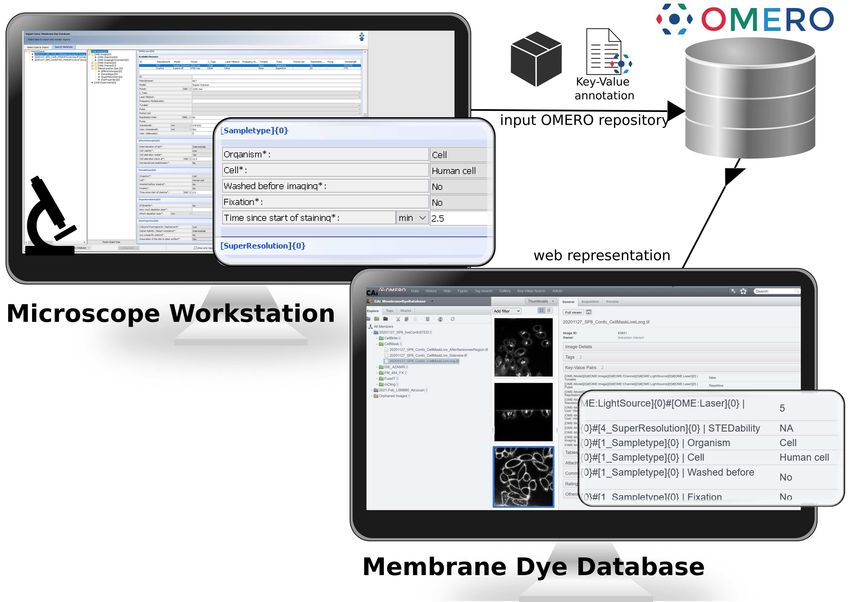
experimental setups and file formats. Therefore, the appropriate use of suitable stan-
dardized metadata and data models is a challenge [5, 10]. Accordingly, flexible tools
for capturing a complete set of metadata are in great demand by researchers applying
microscopy techniques. Moreover, it is important for imaging core facilities to be able to
provide different standards with one tool and still be flexible enough for dynamic devel-
opments. Many tools fail to strike this balance and therefore oscillate between using rigid
data models and free text input without semantic context. Similarly, the integration and
referencing of existing metadata is often lacking. Our tool, MDEmic (MetaData Editor
for microscopy), provides an easy and comfortable way for editing metadata of micro-
scopic imaging data and at the same time offers high flexibility in terms of adjustment of
metadata sets (Fig.1). As the standardization process regarding the metadata of micro-
scopic experiments is in full swing, MDEmic offers high flexibility to follow this process.
This is achieved through the dynamic configurability of both the queried or integrated
metadata and predefined values and the reference to ontology databases. MDEmic reads
the technical metadata stored in the image file using Bio-Formats [5], a software library
for reading proprietary microscopy image (meta)data and presents this metadata in the
form of the OME (Open Microscopy Environment) Data Model [4, 6, 8]. Visualizing this
model as input forms allows the researcher to adjust or correct the technical metadata.
In addition, based on the default in a configuration file associated with MDEmic, input
forms for further metadata can be generated dynamically, integrating, or extending the
OME Data Model for technical metadata. The specification of metadata can include: i)
type and category of metadata, ii) fixed terms as selectable input values loading from
subclasses by specifying ontology class identifiers, iii) definition of relation to other meta-
data categories.      For all metadata, different sets of predefinitions can be integrated
via the configuration file and selected by the user according to the respective scientific
application or image technology (Fig.2). MDEmic is part of the standard installation
package of the image database OMERO [1] and is integrated in the OMERO.importer as
OMERO.mde. The OMERO.importer can be used without a local existing OMERO in-
stallation. All metadata descriptions created in OMERO.mde can be saved and reloaded
by the user for later reuse and adaptation or can be exported to different textual for-
mats. This functionality allows the output to be easily integrated with other needs. For
example, this increases interoperability with other research data management tools to
support integration with other data types or preparation of image data for publications
or upload to public repositories [7] like the Image Data Repository, IDR [10, 2] (IDR
workflow Fig.4). In the following we describe a use case scenario where image data from
samples treated with various membrane dyes are made available in a Membrane Dye
                                                                               ”
Database“ hosted in the institutional OMERO instance and shared between the mem-
bers of a collaborative research center (CRC) with a focus in membrane research. Here,
we utilized the OMERO.mde for customized metadata annotation. To this purpose we
have defined a metadata object Membrane Dye” in OMERO.mde. This object is avail-
                                 ”
able with different sub-objects describing the membrane dyes in more detail like Effects
                                                                                       ”
On Sample” and Internalization“ of the dye. This object is provided together with the
                  ”
technical description and essential OME Data Model objects as input forms. All input
forms are summarized in the setup Membrane Dye Database” (Fig.3). This adjustment
                                     ”
for the specific object Membrane Dye” can be done by editing the configuration file
                        ”
of OMERO.mde e.g., by a data steward of the CRC or a scientist of the imaging core
facility. The use case described shows the direct benefit of a metadata annotation tool
like MDEmic (in particular here: OMERO.mde) for researchers, as it illustrates a clearly

                                              2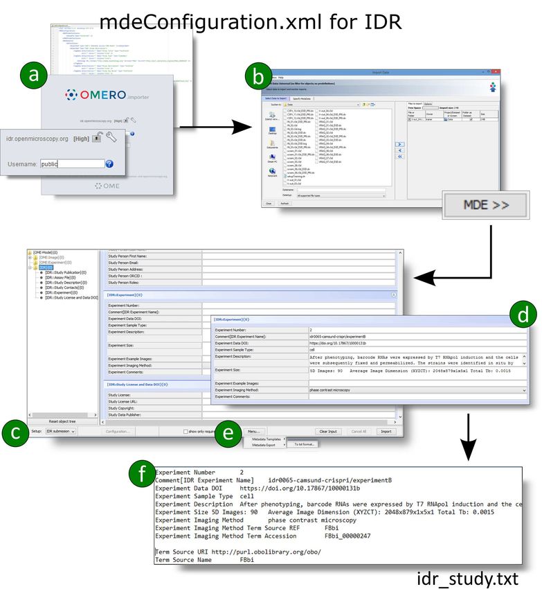
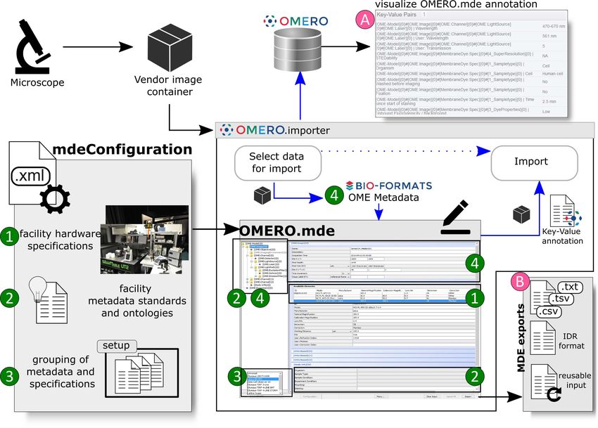
Figure 1: OMERO.importer with integrated MDEmic as OMERO.mde. In the
OMERO.importer the MDEmic is integrated as an intermediate step for the selection of data
for the import and the import itself . Metadata can now be added, which is then transferred to
the repository together with the image data (A), or the annotations can be exported in differ-
ent formats in this step (B). The MDEmic can be customised via a configuration file and loads
the specifications from this file dynamically when the OMERO.importer is started (1,2,3). All
technical metadata of the images marked in the previous step of data selection are read out by
Bio-Format(4) and provided as values in the MDEmic respectively.

defined purpose. It will help to increase the overall awareness for the importance of
metadata annotation and consistent research data management in general.

Acknowledgement
Funding: Supported by Deutsche Forschungsgemeinschaft grants – SFB 1208 (Pro-
jektnummer 267205415 INF/Z02; SFB 944 INF (S.K.); Wellcome Trust grant (Ref:
212962/Z/18/Z) and the BBSRC (Ref: BB/R015384/1) (F.W.); NIH grant (Ref:
5U01CA200059-03), and by Chan Zuckerberg Initiative DAF grant (Ref: 2019-198155
(5022)), an advised fund of Silicon Valley Community Foundation, as part of their Imag-
ing Scientist Program (C.S.D.C).

Author contributions [3]: S.K.: Software, Writing - Original Draft, Visualization;
S.W-P.: Conzeptualization, Writing - Original Draft; S.H.: Visualization, Investigation;
F.W.: Writing - Review and Editing; C.S: Writing - Review and Editing; C.S.D.C:
Writing - Review and Editing.

                                              3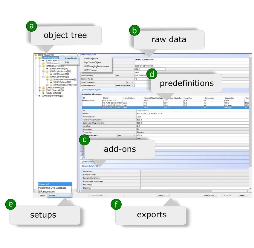
Competing Interests: The authors declare no competing interests.

Data and materials availabilty: OMERO.mde is implemented in Java,
configuration as input and templates as outputs are designed in XML. OMERO as well
as OMERO.mde are open source and available under the GNU General public license at
https://github.com/ome/omero-insight. Instructions can be found at
https://omero-guides.readthedocs.io/projects/omero-guide-mde/en/latest/.
The mdeConfiguration.xml for example workflows (”Membran Dye Database” and IDR)
are available at https://doi.org/10.5281/zenodo.5138039.

References
 [1] Chris Allan et al. “OMERO: flexible, model-driven data management for
     experimental biology”. In: Nature Methods 9.3 (2012), pp. 245–253. issn:
     1548-7105. doi: 10.1038/nmeth.1896.
 [2] Jan Ellenberg et al. “A call for public archives for biological image data”. In: Nat.
     Methods 15 (2018), pp. 849–854. issn: 1548-7105. doi:
     10.1038/s41592-018-0195-8.
 [3] Elsevier. CRediT author statement. url: https://www.elsevier.com/authors/
     policies-and-guidelines/credit-author-statement. 2021.
 [4] Ilya G. Goldberg et al. “The Open Microscopy Environment (OME) Data Model
     and XML file: open tools for informatics and quantitative analysis in biological
     imaging”. In: Genome Biol. 6.5 (2005), pp. 1–13. issn: 1474-760X. doi:
     10.1186/gb-2005-6-5-r47.
 [5] Melissa Linkert et al. “Metadata matters: access to image data in the real world”.
     In: J. Cell Biol. 189.5 (2010), pp. 777–782. issn: 0021-9525. doi:
     10.1083/jcb.201004104.
 [6] OME Data Model and File Formats 6.2.2 Documentation — OME Data Model
     and File Formats 6.2.2 documentation. url: https://docs.openmicroscopy.org/
     ome-model/6.2.2. 2020.
 [7] Jason R. Swedlow et al. “A Global View of Standards for Open Image Data
     Formats and Repositories”. In: arXiv (2020). url:
     https://arxiv.org/abs/2010.10107v1.
 [8] Jason R. Swedlow et al. “Informatics and Quantitative Analysis in Biological
     Imaging”. In: Science 300.5616 (2003), pp. 100–102. issn: 0036-8075. doi:
     10.1126/science.1082602.
 [9] Mark D. Wilkinson et al. “The FAIR Guiding Principles for scientific data
     management and stewardship”. In: Scientific Data 3.1 (2016), p. 160018. issn:
     2052-4463. doi: 10.1038/sdata.2016.18.
[10] Eleanor Williams et al. “Image Data Resource: a bioimage data integration and
     publication platform”. In: Nature Methods 14.8 (2017), pp. 775–781. issn:
     1548-7105. doi: 10.1038/nmeth.4326.

                                            4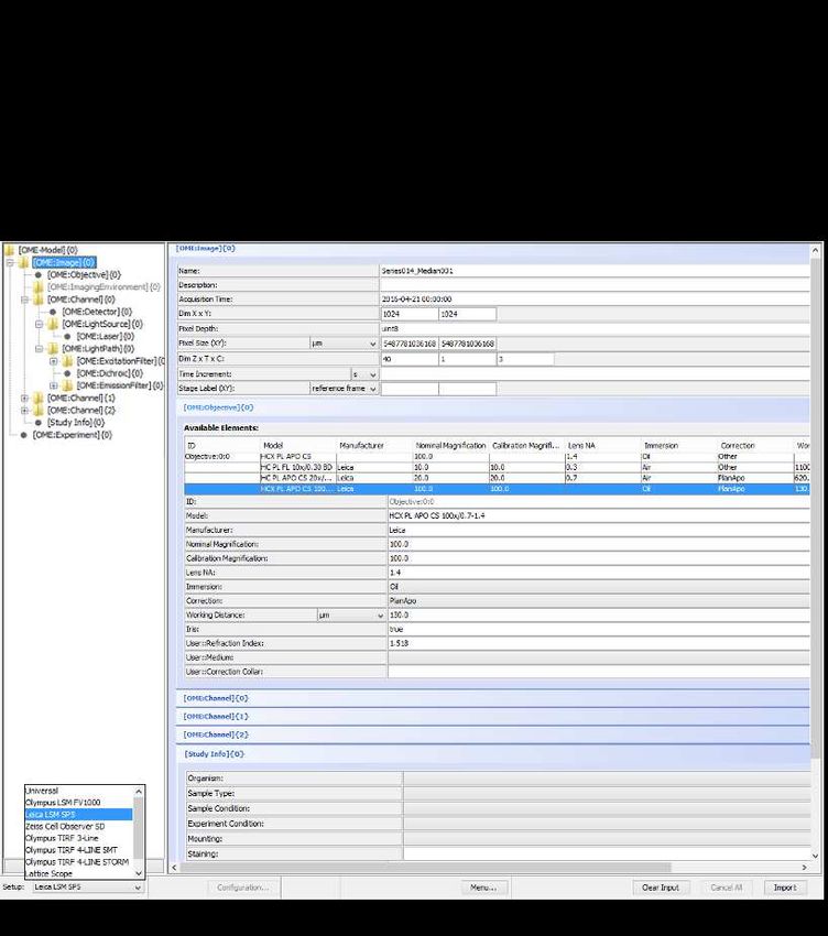
Figure 2: Graphical user interface of MDEmic. a)The object tree is based on the struc-
ture of the OME data model and can be extended by objects of this data model or self-defined
objects. Objects are a collection of related metadata. The extension can be done manually
at runtime by using the context menu or by selecting the appropriate setup from the configu-
ration file. b) The technical metadata stored in the image file is read using Bio-Formats and
provided in the input forms as editable values. c) The user can define new objects with defined
metadata keys and a selection of predefined values by specifying them in the configuration file.
These values can also be loaded automatically by reference to an ontology class. d)Predefined
metadata can be specified for all objects in the configuration, which the user can choose from.
e)A setup is a bundle of data model modifications, input form configurations and/or various
associated predefinitions (such as hardware definitions of a microscope setup or an experiment
protocol). f )All metadata can be exported directly to a text file or as a reusable template for
later annotation using MDEmic.

                                               5
Figure 3: The workflow from microscopes to (tailored) OMERO repositories. After
data acquisition on the microscope, start the software tool (OMERO.importer) for data transfer
to the Membrane Dye Database. For the selected data, the metadata contained in the data
container are read out using Bio-Formats. Now the required metadata for a membrane dye
dataset can be added using the given input mask. Finally, the data and the added metadata
are transferred to the Membrane Dye Database.
6Figure 4: Capture required metadata for IDR submission with MDEmic
integrated in OMERO. Workflow for collecting the required metadata for an
IDR submission using OMERO.mde. a) After downloading the mdeConfiguration.xml
(https://doi.org/10.5281/zenodo.5138039) for the IDR submission , you can adapt the used
ontology classes to your needs. OMERO.mde loads all subclass terms from the linked ontology
class and makes them available as a selection list. Start the OMERO.importer. By referencing
the publicly accessible IDR server, anyone can use the OMERO.importer even without a local
OMERO server. b) Select your data and switch to the mde input field. c) Select the IDR
submission setup to load the IDR-specific input masks. d) Fill in the fields. e) Exporting the
input to an IDR-formatted file automatically creates an f ) idr study.txt file with the input as
key-value pairs and the references to the selected ontology class.
7You can also read