Lung Cancer Detection Using Image Processing - Journal of ...
←
→
Page content transcription
If your browser does not render page correctly, please read the page content below

© 2021 JETIR March 2021, Volume 8, Issue 3                                                             www.jetir.org (ISSN-2349-5162)

       Lung Cancer Detection Using Image Processing
                                                            Mr. Vishal Patil
                             Assistant professor in Instrumentation department at VPM's MPCOE, Ratnagiri

                                                            Dr. Aditya Gupta
                           Assistant professor in Electronics & telecommunication department at COEP, Pune

                                                           Mr. Avinash Pawar
                                                  Principal at VPM's MPCOE, Ratnagiri

Abstract- The objective of this paper is to explore an expedient image segmentation algorithm for medical images to curtail the
physicians’ interpretation of computer tomography (CT) scan images. Modern medical imaging modalities generate large images that
are extremely grim to analyze manually. The consequences of segmentation algorithms rely on the exactitude and convergence time. At
this moment, there is a compelling necessity to explore and implement new evolutionary algorithms to solve the problems associated
with medical image segmentation. Nowadays, Image processing methods are commonly used in many medical areas for improvement
of image for earlier detection and treatment stages. Early prediction of lung cancer can increase the survival rate of patient by using
imaging tests such as computer tomography (CT) which gives better image quality and results. In Image processing procedures,
methods like pre-processing, segmentation methods like thresholding or K-means clustering and feature extraction are discussed. It is
the target to get more accurate results by using image enhancement and segmentation techniques on MATLAB software.

                                                            1.   INTRODUCTION

Lung cancer is one of the most horrible maladies in creating nations and its death rate is 19.4% [1]. Early recognition of lung tumors is
finished by utilizing many imaging strategies, for example, Computed Tomography, Sputum Cytology, Chest X-beam, and Magnetic
Resonance Imaging. Location implies grouping tumor two classes (i)non-carcinogenic tumor and (ii)cancerous tumor (malignant)[2].
The possibility of endurance at the propelled stage is less when contrasted with the treatment and way of life to endure malignant
growth treatment when analyzed at the beginning time of the disease. Manual examination and determination framework can be
extraordinarily improved with the execution of picture handling procedures.

1.1. Lungs and Lung Cancer
Lung consists of two parts as right lung and left lung within the chest. The right lung consists of 3 sections and they are called lobes.
The left lung has 2 sections [6-8]. The right lung is bigger and the left lung is smaller in order to give space for the heart. Trachea is the
wind pipe which takes air in to the lungs during breathing through nose or mouth. The trachea gets separates into tubes known as
bronchi. The bronchi which go into the lungs is further separated into smaller bronchi. These smaller branches are called as bronchioles.
At the edge of the bronchioles, minute air sacs are present called as alveoli. During breathe in the alveoli absorb oxygen into the blood
and removes carbon dioxide from the blood while breathe out. Since the primary function of the lungs is to mix the oxygen in to the
blood and removes carbon dioxide from it. Lung cancers usually start from the cells backing the bronchi and from the bronchioles or
alveoli of the lungs.

A protecting sheath surrounds the lungs known as the pleuron which protects the lungs. This pleura assists the lungs to move backward
and forward towards the chest wall during breathing. A slim, dome-shaped muscle present under the lungs, which separates the chest
and the abdomen is called diaphragm. During breathing, the air is forced in and out of the lungs by the up and down movement of the
diaphragm. Most cancers begin, when there is an abnormal growth of cells in the body. A form of cancer that is found in the lungs are

   JETIR2103137          Journal of Emerging Technologies and Innovative Research (JETIR) www.jetir.org                             1005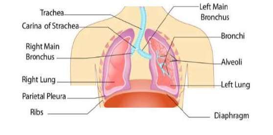
© 2021 JETIR March 2021, Volume 8, Issue 3                                                          www.jetir.org (ISSN-2349-5162)
called Lung cancers. The two major types of lung cancer are small cell lung cancers and Non-small cell lung cancers (NSCLC).It is
found that 85% of the lung cancers are NSCLC [9-10]. The NSCLC are further classified as large cell carcinoma, adenocarcinoma and
squamous cell carcinomas. Adenocarcinomas start in the cells that secrete typical substance along with mucus [11].
Lung cancers are diagnosed mostly with people who smoke [12]. Lung cancers are also visible in people who do not smoke [13] which
are due to second hand smoking. It is more common in ladies whose husbands smoke. Adenocarcinoma occurs in the exterior of the
lung and it may be detected before it spreads. Squamous cells are flat cells that rim the inner way of the airways within the lungs. The
cancer occur in it is called as Squamous cell carcinomas. This cancer is mostly related to the his-tory of smoking. Any part of the lung
may be affected by large cell carcinoma. It is a dangerous cancer which may develop and spread quickly, making the treatment tougher.
Large cell neuroendocrine carcinoma is a subtype of large cell carcinoma, which is a quick growing lung cancer. The cancer that start
form the lung only is called as lung cancer .The cancers which spread from different organs like breast, kidney and pancreas to the
lungs are not lung cancers [14].

1.2 Motivation:
Various investigate on the picture handling methods to recognize the beginning time disease location is accessible in the writing. Be
that as it may, the hit proportion of beginning time recognition of malignant growth isn't extraordinarily improved. With the progression
in the AI procedures, the early determination of the malignant growth is endeavored by a parcel of scientists. The neural system
assumes a key job in the acknowledgment of the malignancy cells among the typical tissues, which thus gives a powerful device to
building an assistive AI-based disease identification. The disease treatment will be successful just when the tumor cells are precisely
isolated from the typical cells Classification of the tumor cells and preparing of the neural system frames the reason for the AI-based
disease analysis [3]. This paper presents a CNN based technique to order the lung tumors as harmful or amiable.

1.3 Background
Lung cancer is increase in the cancer cells of the lungs. Lungs have primary work to provide oxygen to the body. But these abnormal
cells do not help lungs to carry out it’s function. Lung cancer can be spread throughout our body with the help of lymph nodes. These
stages are called metastases. In lung cancer there are two types basically and that are small cell cancer and non-small cell cancer. Non-
small cell cancer is mainly identified nowadays in many persons. There are three types of non-small cell cancer and that are
adenocarcinoma, squamous cell cancer and large cell carcinoma. In these three types, adenocarcinoma is found out in many persons [1].

This research is made to be useful for estimating survival of a lung cancer patient, but it can teach doctors in decision making about the
condition of patient and improve inform patient consent by giving a good understanding of risk which are present in treatment
procedure, based on condition of patient. We can save some costly resources also which are not necessary for patient by taking
information of condition of patient. Lung cancer is fourth in various type of cancers, in males and 6th in females. Overall in males and
females it ranked third after breast and cervical cancer. Despite many advances nowadays in terms of diagnostic methods, some minor
changes and theory interventions, the results of lung cancer patients remains very bad; hence, good understanding in risk factors may
give better impact on preventive measures to be set in community level[1]. The main causes of lung cancer is cigarette and tobacco
usually. Nowadays the causes are increased to smoke taken from any other person, exposure of certain toxins and history of family too.

1.4. Causes of Lung Cancer
The main cause of present days deaths are due to Lung cancers in both women and men. Today, nine out of 10 lung cancer deaths are
caused by smoking. The various causes of lung cancers are discussed below
1.4.1. Smoking
Cigarettes [15] are packed with cancer-inflicting chemical substances. In addition they spoil the lungs' natural protection system. To
protect the lungs, the airways of the lungs are lined with tiny hairs known as cilia. They sweep out pollution, bacteria, and viruses from
the air while breathing. The cilia is affected by the tobacco smoke and stops working in doing its job. This is the main reason for the
cancer-causing chemicals to build up in the lungs and. The cancers starts quietly. During the early stages, no symptoms or warning are
found due to cancer .But when it get worse, the following symptoms may be noticed
     1. Chest pain, during deep breathing
     2. Cough that won't go away
     3. Bloody phlegm when Coughing
     4. Wheezing or shortness of breath,
     5. Fatigue
1.4.2. Second hand Smoke
Even though the main cause of lung cancers is smoking, breathing in the second hand smoke released by a smoker is also danger. This
second hand smoke which occur at domestic or at work place also may increase the risk of danger [16- 17]. It is found that woman’s
who are married to a smoking person are 20% to 30% more likely to get lung cancer when compared the woman’s of non-smokers.
1.4.3. Risky work
Some of the working environments may also be the cause of lung cancers. People who are working with the metals such as uranium,
arsenic, and other chemical substances have more chances for cancer and they should try to limit their exposure with such metals.
People working with asbestos, for insulation, may also prone to lung cancer.

  JETIR2103137          Journal of Emerging Technologies and Innovative Research (JETIR) www.jetir.org                          1006
© 2021 JETIR March 2021, Volume 8, Issue 3 www.jetir.org (ISSN-2349-5162)
1.4.4. Radon Gas
This natural radioactive gas [18-20] which can build up inside the houses may increase the risk of lung cancers. It is found to be the
second leading cause of lung cancer in some countries. This gas can‟t be smelt or seen, but the presence of it can be identified using test
kit.
1.4.5. Air Pollution
Air pollution is also a cause for cancer, but it less prone when compared to smoking, but for good health it is better to avoid air
pollutants [21-23]. It is found that pollutants from vehicles, power plants and factories also affect the lungs like second hand smoke
does. Also it is cited that each and every car corporation is producing minimum of two hundred Cars per day in India which adds air
pollution.
1.5 Objective:
The aim of the proposed system is to detect the tumor in lung by using image segmentation technique and feature extraction.
2. REVIEW OF LITERATURE
2.1 Research Gap
Lung cancer detection using CT scan images of the chest. According to this paper, a computer aided lung cancer detection system
involves three main processing stages: enhancement, segmentation and feature extraction. Further, these stages are explained in detail
with all possible methods that can be used in each of the stages. Methods used with enhancement are: Median filter, Auto enhancement,
Fast Fourier transform. Methods used with segmentation are: Thresholding approach, Watershed Segmentation, Region Growing
Segmentation. Feature extraction includes parameters such as Area, Perimeter, Mean, Standard deviation. In the end it compares the
efficacy of methods like Neuro-Fuzzy model and Region Growing Method against CT image analysis [1].
Fuzzy C-Means (FCM) and different extensions of FCM algorithm are discussed. The exclusive FCM algorithm yields better results for
segmenting noise free images, but it fails to segment images downgraded by noise, outliers and other imaging artifacts. This paper
presents an image segmentation approach using Modified Fuzzy Possibility C-Means algorithm (MFPCM), which is a generalized
adaptation of the standard Fuzzy C-Means Clustering algorithm and Fuzzy Possibility C-Means algorithm [2].
The flaw of the conventional FCM technique is eliminated by modifying the standard technique. the Modified FCM algorithm is
defined by modifying the distance measurement of the standard FCM algorithm to permit the labeling of a pixel to be influenced by
other pixels and to restrain the noise effect during segmentation. Rather than having one term in the objective function, a second term is
included, forcing the membership to be as high as possible without a maximum frontier restraint of one. Experiments are carried out on
real images to observe the performance of the proposed modified Fuzzy Possibility FCM technique in segmenting the medical images.
Standard FCM, Modified FCM, Possibilistic C-Means algorithm, Fuzzy Possibilistic C-Means algorithm are compared with Modified
FPCM to explore the accuracy. The three most important parameters used to determine the accuracy of the Modified FPCM are
similarity, false positive and the false negative ratio [3].
SVM classifier is used for classifying the tumor as Benign or Malignant once the CAD (Computer Aided Diagnosis) system for finding
the lung tumor using the lung CT images is in place. Support vector machines are supervised learning models with corresponding
learning algorithms that perform data analysis and pattern recognition, used for classification. The basic SVM takes a collection of input
data and for each input given, predicts which of the two classes forms the input, making it a non-probabilistic binary linear classifier.
From the given set of training example data, each noted as belonging to one of two categories, an SVM algorithm builds a model that
assigns new examples into one category or the other. In the proposed system, we choose the linear classifier technique. Best hyper plane
is the one that exhibits the largest separation or margin between the two classes. So we choose the hyper plane such that its distance
from the nearest data point on each side is maximized. Such a hyper plane is known as the maximum margin hyper plane and the linear
classifier it defines is known as a maximum classifier. In case of SVM classifier out of 9 features at a time only two features are
selected for classification, which produces result as either benign or malignant [4].
Variation in nodule size, variation in the density of the nodule and difficulty of appearance of nodule anywhere in the lung field, where
they are likely to be obscured by ribs, the mediastinum and similar structures beneath the diaphragm, causing a large variation of
contrast to the background. To overcome these issues, the paper proposes a Computer Aided Diagnosis (CAD) system for detection of
lung nodules. This paper initially applies different image processing techniques for lung region extraction. Further, the Fuzzy
Possibilistic C Means (FPCM) algorithm is used for segmentation. For learning and classification, Extreme Learning Machine (ELM) Is
used. The experimentation has been performed on 1000 images obtained from reputed hospitals [5].
2.2 Literature Review
1. Title: Lung cancer detection using image processing techniques
Authors: Mokhled s. Altarwaneh
In this paper, the authors first explain the theory part of the image segmentation then he explains image segmentation into three parts:
JETIR2103137 Journal of Emerging Technologies and Innovative Research (JETIR) www.jetir.org 1007© 2021 JETIR March 2021, Volume 8, Issue 3 www.jetir.org (ISSN-2349-5162)
1. Thresholding Approach
2. Watershed Segmentation
3. Feature Extraction
4. Tumor Extraction
Here, the authors explain the Image enhancement and binarization approach techniques. But here tumor part extraction is not accurate
due to watershed segmentation.
2. Title: Medical Image Fusion via an Effective Wavelet-Based Approach.
Authors: Yong Yang, Dong Sun Park, Shuying Huang, Nini Rao.
In this paper, the Authors design wavelet transform technique for CT and MRI images. Here the author takes skull images to fusion.
After fusion compares the results with other techniques like Pixel average DWT, Gradient pyramid. here author shows that wavelet
transform is the best technique for image fusion using the following parameters Standard deviation, Average gradient, Information
entropy, Cross entropy.
3. Title: Lung Cancer detection by means evolutionary detections.
Authors: K. Senthil kumar, K. Venkatlaxami
The objective of this paper is to find out an image segmentation algorithm for various medical images. In this paper, K-means
clustering, K-median clustering and swarm optimization of particle & inertia techniques are used. It is also proved that adaptive median
filter gives better result than median or average filters. By using MATLAB software it is observed that 95.89% accuracy is obtained by
verifying sample of 20 images.
4. Identifying lung cancer using Image Processing Techniques.
Authors: Disha Sharma, Gagandeep Jindal
The automatic CAD system is explianed in this paper for the analysis of lung cancer from CT images. The main objevtive of the project
is to built a CAD system for finding the early lung cancer nodules by taking the lung CT images and classify them in two types such as
benign and malignant.
5. Lung Cancer Detection By using Fuzzy Clustering Techniques
This paper includes the steps like: Preprocesing, Thresholding, Fuzzy Clustering. Fuzzy clustering is carried out by dividing data into
homogenous region. The advancement is done by using the Hopfield nueral network for the segmentation. It is used for increase in
accuracy.
6. Lung Cancer Cell identification based on artificial neural network
Authors: Zhi hua zhou
This paper includes an artificial neural network ensemble which is a grasping paradigm where many artificial neural networks are
jointly used to answer a problem. In this paper, diagnosis procedure named as NED is proposed, which uses an ANN to detect lung
cancer cells in images of the specimens of needle which is obtained from the bodies of the patients which are diagnosed.
7. Lung Tumor detection and classification using K-means clustering
Authors: P.B Sangamitraa, S. Govindaraju
In this paper, image processing techniques are used widely in many medical areas for improvement prior detection and treatment stages,
in which the time or elapse is very essential factor to detect the disease in the patient as soon as possible.
8. Segmentation and classification of lung tumor from 3D CT image using K-means Clustering
Authors: Prionjit Sarkar, Zakir Hussain
In this paper, for detection of exact location of tumor and size of tumor, 3D CT images are used. This paper shows the utilization of
neighborhood and connectivity properties seen in CT image pixels to control this problems.
9. Lung Cancer detection system using lung CT image Processing
Authors: Moffy Vyas, Amita Dessai
In this paper, CT scan is used by radiologists to find cancer in the body and track the increase of cancer. In this paper a lung cancer
detection algorithm is presented using mathematical morphological operations for partition of the lung region of interest, in which
haralick features have extracted and utilized for categorization of cancer by ANN.
JETIR2103137 Journal of Emerging Technologies and Innovative Research (JETIR) www.jetir.org 1008© 2021 JETIR March 2021, Volume 8, Issue 3 www.jetir.org (ISSN-2349-5162)
10. Comparison of Lung Cancer detection Algorithms
Authors: Ozge Gunaydin, Melike gunay
In this paper, various machine learning methods are learnt and find out which is more efficient during detection of lung cancer.
Principal component analysis, K-nearest neighbours, SVM, Naïve Bayes, Decision trees and ANN learning methods are used in this
papers for comparison of lung cancer detection in which accuracy of decision tree is maximum as 93.24%.
11. Lung Cancer Detection on CT images by using Image Processing
Authors: Anita Chaudhary, Sonit Sukhraj Singh
In CT , problems is observed to mix in time constraints in predicting the present of lung cancer regarding the diagnosing procedure
which are used. In this paper, Image processing, Segmentation and feature extraction methods are discuss in detail.
12. Lung Cancer Detection Using image Processing and Machine Learning
Authors: Wasudeo Rahane, Himali Dalvi
In this paper it is shown that SVM is a efficient method for lung cancer detection by any other methods. In this paper, only abnormal
images are given for segmentation and tumor is find out by using segmentation.
13. A Comparative Study of Lung Cancer Detection Using Machine Learning Algorithms
Authors: Radhika P.R, Veena G.
In this Paper, lung cancer detection was done using categorization algorithms such as Naïve Bayes, SVM, Decision tree and logistic
Regression and shown that decision tree gives the highest accuracy among all algorithms.
14. Lung tumor detection and diagnosis in CT scan images
Authors: A.Amutha, R.S.D Wahidabanu
In this paper, segmentation is carried out by using histogram based feature extraction and non local denoising for pre-processing.
Experiments shows that the procedure can segment the lung field with pathology of different forms more shortly.
15. Detection of Lung Cancer in CT images using Image processing
Authors: Nidhi S. Nadkarni, Sangam Borkar
In this paper, median filtering is used for image preprocessing and by using mathematical morphological operations image
segmentation is carried out. Then features are find out from the region of interest.
16. A comparative study of Lung Cancer Detection using supervised neural network
Authors: Kyamella Roy, Madhurima Burman
In this paper, salient features like energy, entropy and variation are extracted from lung images by the use of SVM classifier. This
classification detects the image is healthy or not and 94.5% efficiency is obtained from SVM classification.
3. RESEARCH METHODOLOGY
3.1 Operational procedure contain following Steps:
1. Convert RGB to Gray image
2. Preprocessing
3. Thresholding
4. Image Segmentation
5. Feature Extraction
6. Tumor Detection
JETIR2103137 Journal of Emerging Technologies and Innovative Research (JETIR) www.jetir.org 1009© 2021 JETIR March 2021, Volume 8, Issue 3 www.jetir.org (ISSN-2349-5162)
Fig 1 . Lung Cancer Detection Steps
3.2 Image Enhancement Introduction:
The Image Pre-processing stage basically starts from image enhancement stage; Image enhancement technique is used to upgrade the
perception of image for human being who are viewers. Image enhancement techniques are also usedto give good input for automated
image processing techniques. Spatial domain methods and frequency domain methods are the types of image enhancement technique.
There are many methods for image enhancement in image processing such as median filtering, Unsharp mask filtering, histogram
equalization and linear contrast adjustment. Nowadays, binarization, intensity adjustment and morphological operations are also used
for image enhancement technique.
3.3 Image Segmentation:
Image Segmentation is the technique to divide the image into multiple parts. The aim of segmentation is to change interpretation of the
image and do it easy to inspect. Here, color based segmentation is used. In the segmentation first image acquired. Then, find out the
sample colours in L*a*b space for region of lungs. Then nearest neighbor rule is used for finding out each pixel. Finally nearest
neighbor classification is done and values of a* and b* are displayed. Image segmentation is used to separate region of interest and
other parts of the lung. In color based segmentation, similar colors in the image belongs to one cluster and other colors belongs to their
corresponding cluster in an image. Simply every cluster is a class of pixel with the same color and the properties of color. This color
based segmentation partitions the lung image into constituents regions. This segmentation has many applications like visualization and
volume estimation of ROI which can detect abnormalities in lung.
More shortly, image segmentation is the technique of giving a label to each pixel in an image so that pixels with the same label show
some visual characteristics. Tissue quantification and classification can also be done by using color image segmentation. The result of
image segmentation is set of same ROI of lung region in which all regions are dissimilar from each other. In color based segmentation
K-means clustering is used for segmentation.
3.4 Thresholding Approach:
In image segmentation, thresholding is the most useful technique. There are many uses of thresholding such as smaller space for storage
and ease in manipulation. The processing speed of thresholding is very fast as compared to other segmentation techniques in medical
images. From last 10-15 years there is lot of attention on thresholding technique[10]. In thresholding, two levels are given to the pixels
because it converts gray image to binary image. In thresholding, threshold value is find out for segmentation. In this research, Otsu’s
thresholding is used to find the global image threshold for the segmentation of lung image.
3.5 K-means Clustering:
It is the simple algorithm for dividing given samples into required number of clusters so as to reduce the sum of squared distances to the
cluster centre. This algorithm reduces the errors since no squared operation is there.
JETIR2103137 Journal of Emerging Technologies and Innovative Research (JETIR) www.jetir.org 1010© 2021 JETIR March 2021, Volume 8, Issue 3 www.jetir.org (ISSN-2349-5162)
Weaknesses :
1) Randomly choose K samples
2) Try for number of different starting points as they produce suboptimal partitions.
3) It can happens that set of samples must be empty but we can ignore it.
4) Result depends on measure ||x-m|| solution is to normalize each variable.
5) Result depends on value of K.
When the number of data is not much then initial grouping can detect or determine the cluster significantly.
Algorithm(Using Centroid):
K-means clustering is an step by step algorithm that helps us to partition the dataset into predefined different non-overlapping
subgroups where each data point belongs to one group only. Keep iterating until there is no further change in the centroids.
Begin with k number of clusters,
Get centroid updates.
Compute distance of each sample and cluster.
Again perform step 3 untill convergence.
Algorithm(Using Euclidean Distance):
1. Select any number of random cluster centers named as “C.”
2. Find out the Euclidean distance.
3. Construct the cluster having similar pixels into it if the Euclidean distance is less between constructed cluster and pixel.
4. Find out new cluster center after separation of all pixels. Here, median value is used to find out the new cluster.
5. Repeat steps 2 to 4 till any number of iterations. After required iteration, stop when some condition is encountered.
Here, We are using the first algorithm which is done by centroid calculation, because image segmentation by centroid calculation is
easy.
3.6 Feature Extraction:
Image features Extraction stage is an essential stage that utilizes algorithm to predict and isolate the different portion of the image.
Dimensions are also reduced by use of feature extraction. Binarization and masking are two methods which are used to find out
probability of lung cancer.
Features:
1. Area: It gives number of pixels.
2. Convex Area: It gives how many pixels are there in convex images.
3. Equidistant Parameter: The diameter is same as ROI
4. Eccentricity: Focci/ Major Axis Length
5. Skewness: It is a measure of symmetry
6. Energy: It is sum the of squared elements in GLCM
7. Contrast: It is measure of intensity contrast between pixel and neighbor of pixel.
8. Correlation: It is related to, how the pixel is related to its neighbor over ROI.
9. Homogenity: It is measure of nearness of elements in the GLCM.
10. Kurtosis: Shape of random variables probability distribution.
4. RESULTS AND DISCUSSION
The standard database images of lungs are taken from the available Database from IMBA Home (VIA-ELCAP Public Access)[5]. Fig2
contains the standard database image of lung and fig3 contains the image that we get after thresholding. But after thresholding we
cannot get exact tumor area in some cases.
JETIR2103137 Journal of Emerging Technologies and Innovative Research (JETIR) www.jetir.org 1011© 2021 JETIR March 2021, Volume 8, Issue 3 www.jetir.org (ISSN-2349-5162)
Fig 2. Lung Image
Fig 3. Image obtained after thresholding
Since, we cant get perfect result after thresholding for all images, K-means clustering is used to reduce this error. Because sometimes
bone defect or other abnormalities in lung can also be shown as lung tumor. Fig 4 and Fig 5 shows lung image and image after
preprocessing. Fig 6 shows the extracted tumor region from lung image.
Fig 4. Lung image
JETIR2103137 Journal of Emerging Technologies and Innovative Research (JETIR) www.jetir.org 1012© 2021 JETIR March 2021, Volume 8, Issue 3 www.jetir.org (ISSN-2349-5162)
Fig 5. Image After Thresholding
Fig 6. Tumor is Extracted
Features(Values):
For classification of lung cancer stages information of feature extraction is used. Here feature extraction of 6 lung images is carried out.
In which Lung3 and Lung5 is the image of lung having tumor because the values of features are changing due to abnormalities in the
image. Feature extraction is also used for dimensionality reduction in image processing. This values can also distinguish between lung
image and image having tumor because features of lung image having tumor should be different than normal lung image.
JETIR2103137 Journal of Emerging Technologies and Innovative Research (JETIR) www.jetir.org 1013© 2021 JETIR March 2021, Volume 8, Issue 3 www.jetir.org (ISSN-2349-5162)
Table 1. Table of values of features
Lung 1 Lung 2 Lung 3 Lung 4 Lung 5 Lung 6
Contrast 0 0 1.75 0 3.500 0
Energy 1 1 0.9305 1 0.8648 1
Kurtosis 33.8630 23.3982 24.7197 24.6175 30.1891 22.94
Entropy 0 0 0.1161 0 0.226 0
Mean 4080 4080 4080 4080 4080 4080
Smoothness 1 1 1 1 1 1
Standard 1.7946e+04 1.550e+04 1.4563e+04 1.5625e+04 1.455e+04 1.5769e+04
deviation
Skewness 5.452 4.5643 4.5720 4.6498 4.9855 4.5744
Homogenity 1 1 0.9688 1 0.9375 1
Variance 3.2785e+08 2.4646e+08 2.1535e+08 2.4913e+08 2.1577e+08 2.5146e+08
CONCLUSION
In this research, we conclude that K-means clustering and thresholding are the efficient techniques for lung cancer detection because K-
means clustering is utilized to discover groups in the image efficiently, and in the tumor of lung there is a group of cells. Similarly, in
thresholding it is observed that by setting a threshold, image processing is carried out successfully. Comparing to all algorithms, the
accuracy of the tumor extraction is better in given proposed technique. From this, it can be conclude that CT images are most useful for
lung cancer detection than MRI or X-ray because of better image quality.
FUTURE SCOPE
In future studies, many algorithms can be used to increase the accuracy. In this, thresholding is used to get edges and tumor but other
techniques such as Sobel, Canny edge detector are present to edge detection. Using this technique in the place of the thresholding will
improve the performance of edge detection. Some advanced concepts of Machine learning such as CNN(Convolution Neural Network)
and PNN(Probabilistic Neural Network) can be used for accurate detection of size and location of tumor in lung.
REFERENCES
1. Mokhled S-Altarawaneh,” Lung cancer Detection Using Image Processing Techniques”, Leonardo Electronic Journal of
Practice and Technologies, Issue 20, January-June 2012
2. K. Venkatalakshmi and S. M. Shalinie, “Classification of multispectral images using support vector machines based on
PSO and K-means clustering,” in Proceedings of IEEE In- ternational Conference on Intelligent Sensing and Information
Processing, pp. 127–133, Melbourne, VIC, Australia, December 2005.
3. K.Madheswari', N.Venkateswaran, and V.Sowmiya “Visible and Thermal image fusion using Curve let Transform and
Brain Storm Optimization” 2016 IEEE Conference.
4. Navneet kaur, Madhu Bahl,Harsimran Kaur “Review On: Image Fusion Using Wavelet and Curvelet Transform”
International Journal of Computer Science and Information Technologies, Vol. 5 (2) , 2014, 2467-2470
5. Lung Cancer Database, Available at: https://eddie.via.cornell.edu/cgi- bin/datac/signon.cgi, (accessed July 2011).
6. Non-Small Cell Lung Cancer, Available at: http://www.katemacintyrefoundation.org/ pdf/non-small-cell.pdf, Adapted from
National Cancer Institute (NCI) and Patients Living with Cancer (PLWC), 2007, (accessed July 2011).
Levner I., Zhang H.,” Classification-Driven Watershed Segmentation”, IEEE Transactions on Image Processing, 2007,
7.
16(5), 1437-45.
8. D. Lin and C. Yan, “Lung nodules identification rules extraction with neural fuzzy network”, IEEE, Neural Information
Processing, vol. 4,(2002).
JETIR2103137 Journal of Emerging Technologies and Innovative Research (JETIR) www.jetir.org 1014© 2021 JETIR March 2021, Volume 8, Issue 3 www.jetir.org (ISSN-2349-5162)
9. D. Sharma and G. Jindal, "Identifying Lung Cancer Using Image Processing Techniques," International Conference on
Computational Techniques and ArtifiCial Intelligence (ICCTAf'2011), 2011, pp. 115-120
10. Md. Badrul Alam, Miah Mohammad Abu Yousuf, “Detection of Lung Cancer from CT Image Using Image Processing
and Neural Network” 2nd Int'l Conf on Electrical Engineering and Information & Communication Technology (ICEEICT)
20 IS Jahangirnagar University, Dhaka-1342, Bangladesh, 21-23 May 2015
11. S.K. Vijai Anand, “Segmentation coupled Textural Feature Classification for Lung Tumor Prediction” ICCCC’10,
Department of Computer Science & Engineering College of Engineering Guindy, Anna University Chennai Chennai-600
025, India.
12. Levner I., Zhang H., Classification-Driven Watershed Segmentation, IEEE Transactions on Image Processing, 2007, 16(5),
1437-45.
13. Rachid Sammouda, Jamal Abu Hassan, Mohamed Sammouda, Abdulridha Al-Zuhairy, Hatem abou ElAbbas, “Computer
Aided Diagnosis System for Early Detection of Lung Cancer Using Chest Computer Tomography Images”, GVIP 05
Conference, 19-21 December 2005, CICC, Cairo, Egypt.
14. Penedo. M. G, Carreira. M. J, Mosquera. A and Cabello. D, “Computer-aided diagnosis: a neuralnetwork- based approach
to lung nodule detection”, IEEE Transactions on Medical Imaging, vol: 17, pp: 872 – 880, 1998.
15. Kanazawa. K, Kubo. M and Niki. N, “Computer aided diagnosis system for lung cancer based on helical CT images”,
Proceedings of the 13th International Conference on Pattern Recognition, pp: 381 - 385 vol.3, 1996.
16. Yamamoto. T, Ukai. Y, Kubo. M, Niki. N, Satou. H and Eguchi. K, “Computer aided diagnosis system with functions to
assist comparative reading for lung cancer based on helical CT image”, Image Processing, 2000 International Conference
on Proceedings, pp: 180 – 183, vol.1, 2000.
17. R. Wiemker, P. Rogalla, T. Blaffert, D. Sifri, O. Hay, Y. Srinivas and R. Truyen “Computer-aided detection (CAD) and
volumetry of pulmonary nodules on high-resolution CT data“, 2003.
18. Radhika P.R, Rakhi A.S Nair, Veena G “ A Comparative study of lung cancer detection using machine learning
algorithms”, 2019.
19. S. G. Armato, M. L. Giger and H. MacMahon, “Automated detection of lung nodules in CT scans: Preliminary results”,
Med. Phys., Vol.28,2001
20. A.Sindhu1 , S.Meera2 “A Survey on Detecting Brain Tumor in MRI Images Using Image Processing Techniques
”International Journal of Innovative Research in Computer and Communication EngineeringVol. 3, Issue 1, January 2015
21. Zeynel Cebeci1 , Figen Yildiz 2 “Comparison between K-Means and Fuzzy C-Means Algorithms on Different Cluster
Structures”Journal of Agricultural Informatics (ISSN 2061-862X) 2015 Vol. 6, No. 3:13-23
22. Soumi Ghosh , Sanjay Kumar Dubey “Comparative Analysis of K-Means and Fuzzy CMeans Algorithms” ((IJACSA)
International Journal of Advanced Computer Science and Applications, Vol. 4, No.4, 2013
23. Russell C. Eberhart and Yuhui Shi “Comparison between Genetic Algorithms and Particle Swarm Optimization ” Indiana
University Purdue University Indianapolis 723 W.Michigan St., SL160 Indianapolis, IN 46202
JETIR2103137 Journal of Emerging Technologies and Innovative Research (JETIR) www.jetir.org 1015You can also read