Lung hemorrhage in a green iguana (Iguana iguana) with chronic metabolic failure: a case report
←
→
Page content transcription
If your browser does not render page correctly, please read the page content below
Case Report                                                        Veterinarni Medicina, 51, 2006 (6): 356–363

Lung hemorrhage in a green iguana (Iguana iguana)
with chronic metabolic failure: a case report
V. JEKL1, R. HALOUZKA2, Z. KNOTKOVA1, G.M. DORRESTEIN3, Z. KNOTEK1
1
  Avian and Exotic Animal Clinic, 2Institute of Pathological Morphology, Faculty of Veterinary
  Medicine, University of Veterinary and Pharmaceutical Sciences, Brno, Czech Republic
3
  Diagnostic Laboratory of the NOIVBD, Veldhoven, The Netherlands

ABSTRACT: A six year old male iguana (Iguana iguana) was brought to our clinic after a fall from a height of
1.5 meters. The patient showed apathy, dehydration, and paresis of all the limbs. Postural reflexes, palpebral reflex,
and response to painful stimuli were minimal. The patient had extremely high levels of uric acid (1 734.0 µmol/l)
and phosphorus (9.80 mmol/l), ratio of calcium to phosphorus (0.18), leucocytosis (23.3×10⁹/l) with heterophilia
(17.10×10⁹/l), and high activity of ALT, AST and CK (2.09, 6.59, and 260.0 µkat/l). Tracheoscopy and pneumoscopy
revealed presence of blood clots in the trachea and lungs with haemorrhage within the parenchyma. Based on
the results of the clinical examination, endoscopy and laboratory diagnostics, the clinical diagnosis was chronic
kidney failure, liver lipidosis and lung haemorrhage. Because of a very poor prognosis, the patient was eutha-
nised. At necropsy, the signs of visceral gout and hepatomegaly with diffuse yellowish white discoloration and
with dotted brown pigmentation were also observed. The kidneys were enlarged, and stained brown grayish. The
gross and histopathological examination confirmed the clinical diagnosis of visceral gout, chronic kidney failure,
liver steatosis and granulomatous pneumonia, with lung haemorrhage associated with urate deposits in lung tissue
and posttraumatic status.

Keywords: renal disease; endoscopy; gout; trauma; liver lipidosis

  Traumatic injuries of different extensity and             (Rubel et al., 1991; Schumacher and Toal, 2001).
intensity are common in green iguanas kept by               Another cause of lung haemorrhage could be the
private owners. The lesions are usually caused by           calcification of soft tissues, urate depositions in
inappropriate manipulation and poor terrarium               lung tissue or lung vessels due to kidney failure.
construction and facilities, as well as by fights be-         This paper describes lung haemorrhage in a green
tween animals (Knotek et al., 1999). Due to the             iguana (I. iguana) associated with fall from a height,
anatomy and behaviour, superficial skin injuries            and urate deposition in lung tissue and vessels.
and fractures of limbs and tail are frequently seen
(Hernandez-Divers, 2001). However, traumatic in-
juries of internal organs are rather rare, and severe       MATERIAL AND METHODS
lung injuries are exceptional. Traumatic lung inju-
ries and possible haemorrhage in reptiles are gen-          History
erally caused by inappropriate fixation or by other
animals (Schumacher, 2003; Wellehan and Gunkel,               A six year old male green iguana (I. iguana)
2004). The diagnosis of trauma can be evident from          weighing 2.10 kg was brought to the clinic. This
physical examination, radiography or endoscopy              iguana was kept as a single animal in a terrarium

Partially supported by the Ministry of Education, Youth and Sports of the Czech Republic (Project IGA
No. 34/2004/FVL).

356
Veterinarni Medicina, 51, 2006 (6): 356–363                                                     Case Report

(2.5 × 2 × 2 m, w × h × l) equipped with a source of    Endoscopy
UV light, and heated to the maximum temperature
of 45°C underneath a bulb-heater. The photoperiod         The patient underwent an endoscopic examina-
was 12 hours of light a day. Branches were placed       tion of the trachea and lungs. Prior to the surgical
in the terrarium and coconut fibre was used as sub-     intervention, the patient was administered antibi-
strate. A waterpool was positioned at the bottom        otics (enrofloxacine, Baytril 2.5% inj., Bayer AG,
of the terrarium and the water was changed once         Germany, 15 mg/kg i.m.), rehydration solution
every two days.                                         (saline with 3% glucose, 20 ml/kg s.c.), vitamin
  The animal was fed once or twice a day, and the       K (Kanavit inj., Hoechst-Biotika, SR, 0.5 mg/kg s.c.)
diet consisted of peas, green beans, alfalfa, dan-      and B-vitamins with amino acids (Duphalyte inj.
delion leaves, and vitamin and mineral supple-          ad us.vet., Fort Dodge Veterinaria, Spain, 5 ml/kg
ments (Vitamix-REP, Biofaktory, Czech Republic).        s.c.). This examination was carried out in sternal
The iguana had never been treated before. The           position without sedation as the patient showed
only problem observed in the last 14 days was a         significant apathy. The tracheoscopy was followed
low feed consumption. The reason for the visit          by a laparoscopy with left-side access.
to the clinic was the apathy and an unwillingness         The patient was intubated and supplied with a
to move after a fall from a branch at a height of       gas mixture of oxygen with isoflurane (Isoflurane
1.5 meter.                                              Rhodia, Torrexpharma GmbH, Vienna, Austria)
                                                        and at a concentration of 1.5% and a flow rate of
                                                        100 ml/kg per minute. A half-open system with
Haematology and plasma biochemistry                     assisted patient ventilation was chosen. After ten
                                                        minutes the patient was moved to a heat blanket
  A heparinized blood sample (2 ml) was col-            set to 39°C. The isoflurane concentration was re-
lected from the ventral coccygeal vein. A differ-       duced to 1%. Rigid endoscopes were used for the
entiation of the cells was done in a blood smear        patient examination (Hopkins Documentation
stained with May-Grunwald/Giemsa-Romanowski.            Forward-Oblique Telescope 64018 BS, Ø 2.7 mm,
The total numbers of red and white blood cells          18 cm, Karl Storz Tuttlingen; Oblique Telescope
were counted manually using Natt-Herrick solu-          Ø 4.0 mm, 30 cm, Wolf Tuttlingen) with protective
tion in Burker chamber, and a dilution of 1:100.        canal (Examination Sheath 67065 CC) and xenon
Haematocrit was assessed using the microhae-            source of light (Xenon Nova 20131520, Karl Storz
matocrit method by centrifugation of the sam-           Tuttlingen). Electronic Endoflator (26 430520 Karl
ple for 6 minutes at 3 000 rpm. The haemoglobin         Storz Tuttlingen) was employed for coelom insuf-
concentration was evaluated by the classical cya-       flation.
nide-haemoglobin (haemoglobin-cyanide) method             After the endoscopic examination, the patient
using a spectrophotometer (Unicam Helios Epsilon,       was washed with oxygen for 15 minutes. Heart beat
Thermospectronic, USA). Biochemical analysis in-        and blood oxygen saturation (SpO 2) were moni-
cluded the assessment of plasmatic concentration        tored with pulse oxymeter (V3301 Pulse Oximeter,
of total protein (TP), uric acid, glucose, activity     SurgiVet, USA).
of aspartataminotranspherase (AST), alaninami-
notranspherase (ALT), alkalic phosphatase (AP),
creatinkinase (CK), and the level of calcium            Post-mortem examination
and phosphorus using CobasMira (Roche) and
Atomspec (Hilger 1550) automatic analysers.               Because of the very poor prognosis, and after con-
                                                        sultation of the owner, the animal was euthanised
                                                        (T61, Intervet, The Netherlands) and a necropsy
Radiography                                             was performed. Cytological slides stained with May-
                                                        Grunwald/Giemsa-Romanowski were prepared
  For radiological examination of the patient, a dor-   from liver, spleen and lungs and examined. Tissue
soventral projection by vertical beam and laterola-     samples from lungs, liver, spleen, kidneys, heart
teral view in sternal position by horizontal beam       and testicle were fixed in 10% buffered formalin and
using Durolux (typ 397 6142011-C 211, Chirana,          routinely embedded in paraffin. Tissue sections 4–6 µ
Czech Republic) was made.                               thick were stained with haematoxylin and eosin
                                                                                                         357
Case Report Veterinarni Medicina, 51, 2006 (6): 356–363
(HE). Evaluation was performed in the Institute of solid excrements and the urinary bladder was not
Pathological Morphology, University of Veterinary distended. Two symmetrical oval masses protrud-
and Pharmaceutical Sciences, Brno, Czech Republic ing 2 cm over the pelvis were palpated in the cau-
and in the Department of Pathobiology, Utrecht dal part of the pleuroperitoneal cavity. Suspected
University, The Netherlands. nephromegaly was confirmed by cloacal examina-
tion.
Results of the haematology and biochemistry are
RESULTS summarized in Table 1. The patient had severe leu-
cocytosis with heterophilia, high activity of ALT,
Clinical examination AST, CK, extremely high levels of uric acid and
phosphorus, and low ratio of calcium to phospho-
The patient showed significant apathy and tetra- rus (0.18).
paresis of the limbs. Postural reflexes, palpebral The radiography revealed presence of a hemiver-
reflex, and response to painful stimuli were mini- tebra between third and fourth lumbar vertebrae.
mal. Mild scoliosis and kyphosis were notified in Lung field exhibited no radioopaque shading. The
the lumbar part of the backbone. The iguana was liver was slightly enlarged, and the colon was filled
in a good nutritional state, and no external abnor- with large amount of radioopaque material with
malities were visible. The mucosa of the oral cav- small amount of gas. The enlargement of the kid-
ity and tongue were pale without erosions. A large neys was not recognized on the radiographes.
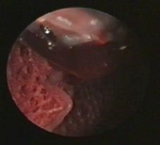
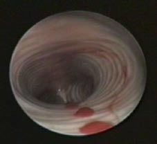
blood clot was observed in the trachea, but airflow Tracheoscopy, pneumoscopy and laparosco-
through the lumen was maintained, and there was py showed blood clots in the trachea and lungs
no dyspnoea. Palpation of the abdominal region (Figures 1 and 2). The laparoscopy demonstrated
revealed that caudal part of the intestine contained a lung trauma with small areas of haemorrhages
Table 1. Blood profile values for the green iguana
Units This case Divers et al., 1996 Knotek et al., 1999; Pejrilova et al., 2004 Harr et al., 2001
Haemoglobin g/l 72 67–122 92–94 67–122
PCV l/l 0.44 0.25–0.35 0.32–0.36 0.29–0.47
12
RBC 10 /l 1.3 1.0–1.9 0.88–1.02 1.0–1.8
9
WBC 10 /l 23.3 3.0–10.0 11.8–15.5 8.0–25.2
9
Heterophils 10 /l 17.21 0.35–5.2 2.3–5.12 0.6–6.4
9
Eosinophils 10 /l 0 0.0–0.3 0.03–0.25 0.0–0.4
9
Basophils 10 /l 0.5 0.0–0.5 0.33–0.76 0.1–1.2
9
Lymphocytes 10 /l 3.7 0.5–5.5 8.4–9.0 5.0–17.2
Monocytes 109/l 1.9 0.0–1.8 0.2–0.6 0.2–2.7
Azurophils 109/l 0 – 0.39–0.93 –
TP g/l 75.1 50–78 22.0–78.2 42–76
Glucose mmol/l 5.7 – 8.3–16.5 –
AP µkat/l 0.5 – 0.7–4.9 –
ALT µkat/l 2.09 – 0–1.2 –
AST µkat/l 6.59 0.08–0.87 0–1.6 0.12–1.70
CK µkat/l 260 – – –
Uric acid µmol/l 1 734 70–140 70.4–145.3 40–390
Ca mmol/l 1.8 2.2–3.5 2.2–3.5 2.1–5.8
P mmol/l 9.8 1.5–3 1.4–3.1 0.9–3
358Veterinarni Medicina, 51, 2006 (6): 356–363 Case Report
Figure 1. Tracheoscopy. A view of the bifurcation and Figure 2. Pneumoscopy. Blood drop in the lungs
blood drops
within the parenchyma. The pleuroperitoneal cav- ed the owner, we opted for intravenous euthanasia
ity was free of exudates. A large amount of adi- (T61, Intervet, The Netherlands) and carried out a
pose tissue was found on the pleuroperitoneum pathomorphological examination.
and pericardium; fat bodies were very large. The At necropsy, the presence of blood clots in the
enlarged liver was yellowish-white in colour, with trachea and lumen of the cranial lobes of both lungs
brown dotted pigmentation. Under the capsule was confirmed (Figures 3 and 4). Almost 30% of
of the brown-red spleen were dark spots of 1 mm the ventral lung parenchyma was affected by he-
in diameter. The kidneys were enlarged, greyish- morrhagic effusion. The pericardium was covered
brown in colour, and without subcapsular urates. with a chalky coating. The oesophagus contained
Based on the above-described examinations, the a small amount of blood clots. The cranial part of
diagnosis was made as a combination of metabolic the stomach was filled with clotted blood, which
failure and posttraumatic shock with lung haemor- was sharply separated from yellow-brown contents.
rhage. With regard to the patient’s condition, an There was no content in the small intestine and the
adverse prognosis was anticipated. Having consult- colon was stuffed with a large amount of material.
Figure 3. View of the caudal part of the heart and lungs in Figure 4. Intraluminal view the right lung (necropsy).
situ. See blood clots in cranial parts of lungs (necropsy) See blood clots in cranial parts of lungs
359Case Report Veterinarni Medicina, 51, 2006 (6): 356–363 Figure 5. View of the symmetric nephromegaly greyish- Figure 6. Lungs – hyperaemia, haemorrhage and lung brown in colour (necropsy) oedema. (histology, 100×) There were no erosions or other changes in the parts of the lung parenchyma, especially in areas of gastrointestinal mucosa. squamous metaplasia of the epithelium (Figure 6). The enlarged liver was yellow-white of colour, There was also a granulomatous pneumonia with a had rounded edges, and was of fragile consistency. number of rod-shaped bacteria inside the lesions. The cytological examination of the liver confirmed In the heart there were some small subepicardial presence of large vacuoles in the hepatocyte cy- round-nuclear infiltrates. Liver steatosis with “fatty toplasm, and the nuclei were displaced to the cell necrosis” and dispersed melanocyte granulomas membrane. The gall bladder was distended with were seen (Figure 7). The main cell types in the green-yellow liquid. Petechial haemorrhages were spleen were a diffuse mixture of histiocytes, granu- found underneath the spleen capsule (spleen size locytes, and erythrocytes with almost no lymphoid 4.5 × 1.2 cm). The kidneys were bilaterally sym- cells. Tubuli with a high epithelium and dilated col- metrically enlarged, of amber colour, and of a solid lecting tubules containing amorphous eosinophilic consistency (Figure 5). Homogenous parenchymal material and fluid in the lumen were observed in structure with no urate content in the parenchyma the kidneys (Figure 8). was visible in the tissue section. The final diagnosis was a metabolic failure – On histopathological examination, urate deposits chronic kidney failure with visceral gout, extensive and extensive haemorrhages were found in some liver steatosis and lung haemorrhage. Figure 7. Liver steatosis, fatty necrosis and melanomac- Figure 8. Mineralization of kidney tubules (histology, rophage proliferation (histology, 200×) 200×) 360
Veterinarni Medicina, 51, 2006 (6): 356–363                                                      Case Report

DISCUSSION                                               of kidney function by scintigraphy and glomerular
                                                         filtration rate (Greer et al., 2004; Hernandez-Divers
  Apathy, lack of appetite, and an overall muscular      et al., 2005).
weakness are common non-specific clinical symp-             In this case report, the endoscopic examination
toms of many diseases in lizards (Barten, 1996;          revealed extensive haemorrhages in the lungs. No
Blahak, 2000). These symptoms are mostly a result        dyspnoeic signs were recorded by this examina-
of metabolic diseases (Zwart, 2001). Haematological      tion. It was an acute state, since the haematocrit
and biochemical blood parameters give indirect           and red blood cell count was still in normal levels.
diagnostics information in reptiles (Stein, 1996;        The treatment of an acute lung haemorrhage in-
Knotek et al. 2002; Wilkinson, 2004). In the green       cludes localization and compression of bleeding
iguana, leucocytosis with heterophilia was found. A      vessels, together with enabling patency of the air-
high number of heterophils in the blood indicates        ways (Briscoe and Syring, 2004). As a supportive
an acute inflammatory reaction, traumatic inju-          treatment, an infusion of solutions with glucose and
ries of tissues, stress, bacterial or parasitic infec-   the administration of B vitamins and vitamin K are
tions, neoplastic processes or acute kidney failure      indicated (Wellehan and Gunkel, 2004).
(Campbell, 1996; Redrobe and MacDonald, 1999;               At the time of the examination of the patient,
Hernandez-Divers, 2003). In this case, the hetero-       the blood in the respiratory tract was already clot-
philia is a response to granulomatous pneumonia          ted. The expired blood was partially swallowed and
and kidney failure. The high plasma activity of AST      found in the stomach. No haemorrhages into the
and CK could be caused, in this case, by muscular        peritoneal cavity were observed. The endoscopic
tissue damage, which would be consistent with the        examination of the pleuroperitoneal cavity demon-
anamnesis mentioning the fall from a height. AST         strated a bilateral nephromegaly and a severe liver
alteration in reptiles was also observed in connec-      steatosis. Liver and kidney diseases are frequently
tion with kidney failure, due to presence of this en-    diagnosed in iguanas (Zwart, 1992; Mader, 1996).
zyme in renal tubules (Hernandez-Divers, 2003).          These diseases are often associated with both pri-
  Alterations of transaminase activity in reptil-        mary and secondary damage resulting in metabolic
ian blood can provide indirect information about         and systemic disorders (Knotek et al., 2002, 2003).
acute processes in liver, bones, kidneys, or intes-      Long-term inappropriate feeding and insufficient
tines. Interpretation of laboratory results, when        hygienic conditions are among most frequent caus-
dealing with chronic organ diseases, is complicated      es mentioned by many authors.
because a significant rise of enzyme activities were        The final diagnosis and assessment of the extent
absent because depletion of the cells. Furthermore,      of the pathological changes were based on the post-
elevation of phosphorus and uric acid, and altered       mortem examination. The fall of the iguana in asso-
ratio of calcium to phosphorus were reported in          ciation with granulomatous pneumonia and urate
chronic nephropathies in reptiles (Campbell, 1996;       deposits in lung tissue resulted in severe haemor-
Knotek et al., 2002). In our case the hyperuricae-       rhages in the lungs. The visceral gout was diag-
mia, a hyperphosphatemia as well as the low ratio        nosed along with the liver steatosis and nephrosis.
of calcium to phosphorus (0.18) indicate failure of      In this case overfeeding with protein and purine
kidney function. Zwart (1992) argued that urates         rich food, with a high content of phosphorus was
precipitate and visceral and articular gout develops     the most likely primary cause of the chronic kidney
when the uric acid level exceeds 1 457 µmol/l.           failure and liver damage. Hemivertebra between
  For anaesthesia, isoflurane was used as it is con-     third and fourth lumbar vertebrae was most prob-
sidered to be the best choice for the patients with      ably a congenital problem.
chronic metabolic disease (Bennet, 1991; Read,              Traumatic injury, based on owner information,
2004). Because of the intrapulmonary haemor-             was not the real cause of the disease. The clinical
rhage, we used positive pressure ventilation, as         examination and overall health establishment of the
described in dogs and cats (Paddleford, 1996).           patient based on blood analyses and particularly
  A valuable diagnostic contribution is a direct         by direct visualization of affected organs with the
endoscopic visualization of organs supplemented          endoscope enabled a prompt conclusion and an
with biopsy of sample tissue for laboratory analyses     objective prognosis. The chronic kidney failure and
(Divers, 1999; Hernandez-Divers, 2003; Wilkinson         the extensive liver damage resulted in the eutha-
et al., 2004). Recent literature describes evaluation    nasia of the patient.

                                                                                                          361
Case Report Veterinarni Medicina, 51, 2006 (6): 356–363
Acknowledgements Hernandez-Divers S.J., Stahl S.C., Stednam N.L., Hern-
andez-Divers S.M., Schumacher J., Hanley C.S, Wilson
The authors would like to thank to Karl Storz H., Vidyashankar A.N., Zhao Y., Rumbeiha W.K.
Veterinary Endoscopy for their technical support, and (2005): Renal evaluation in the healthy green iguana
to the technical staff of the Avian and Exotic Animal (Iguana iguana): Assesment of plasma biochemistry,
Clinic, University of Veterinary and Pharmaceutical glomerular filtration rate, and endoscopic biopsy. Jour-
Sciences, Brno, for their skilful assistance. nal of Zoo and Wildlife Medicine, 36, 155–168.
Knotek Z., Knotkova Z., Halouzka R., Modry D., Hajkova
P. (1999): Diseases of Reptiles (in Czech). CSAVA,
REFERENCES Brno. 276 pp.
Knotek Z., Hauptman K., Knotkova Z., Hajkova P., Tichy
Barten S.L. (1996): Lizards. In: Mader D.R. (ed.): Reptile F. (2002): Renal disease hemogram and plasma bio-
Medicine and Surgery. W.B. Saunders Company, Phil- chemistry in green iguanas. Acta Veterinaria Brno, 71,
adelphia. 324–332. 333–340.
Bennet R.A. (1991): A review of anesthesia and chemical Knotek Z., Knotkova Z., Doubek J., Pejrilova S., Haupt-
restraint in reptiles. Journal of Zoo and Wildlife Med- man K. (2003): Plasma biochemistry in female green
icine, 22, 282–303. iguanas (Iguana iguana) with calcium metabolism
Blahak S. (2000): Infectious diseases in reptiles with spe- disorders. Acta Veterinaria Brno, 72, 183–189.
cial references to zoonoses – an overview for practice. Mader D.R. (1996): Gout. In: Mader D.R. (ed.): Reptile
Praktischer Tierarzt, 81, 113–126. Medicine and Surgery. W.B. Saunders Company, Phil-
Briscoe J.A., Syring R. (2004): Techniques for emergency adelphia. 374–379.
airway and vascular access in special species. Seminars Paddleford R.R. (1996): Pulmonary dysfunction. In:
in Avian and Exotic Pet Medicine, 13, 118–132. Thurmon J.C., Tranquilli W.J., Benson G.J. (eds.):
Campbell T.W. (1996): Clinical pathology. In: Mader D.R. Lumb & Jones Veterinary Anesthesia. 3rd ed. Lippin-
(ed.): Reptile Medicine and Surgery. W.B. Saunders cott Williams & Wilkins, Philadelphia. 771–775.
Company, Philadelphia. 248–257. Pejrilova S., Knotkova Z., Knotek Z., Vrbas J. (2004):
Divers S.J. (1999) Lizard endoscopic techniques with par- Age-related changes of the haematological profile in
ticular reference to the green iguana (Iguana iguana). green iguana (Iguana iguana rhinolopha). Acta Vet-
Seminars in Avian and Exotic Pet Medicine, 8, 122– erinaria Brno, 73, 305–312.
129. Read M.R. (2004): Evaluation of the use of anesthesia
Divers S.J., Redmayne G., Aves E.K. (1996): Haemato- and analgesia in reptiles. Journal of American Veteri-
logical and biochemical values of 10 green iguanas nary Association, 224, 547–552.
(Iguana iguana). Veterinary Record, 138, 203–205. Redrobe S., MacDonald J. (1999): Sample collection and
Greer L.L., Daniel G.B., Shearn-Bochsler V.I., Ramsay clinical pathology of reptiles. Veterinary Clinics of
E.C. (2004): Evaluation of the use of technetium Tc99m North America: Exotic Animal Practice: Clinical Pa-
diethylenetriamine pentaacetic acid and technetium thology and Sample Collection, 2, 709–730.
Tc99m dimercaptosuccinic acid for scintigraphic im- Rubel G.A., Isenbugel E., Wolvekamp P. (1991): Atlas of
aging of the kidneys in green iguanas (Iguana iguana). Diagnostic Radiology of Exotic Pets. Small Mammals,
American Journal of Veterinary Research, 65, 87–92. Birds, Reptiles and Small Mammals. W.B. Saunders
Harr K.E., Alleman A.R., Dennis P.M., Maxwell L.K., Company, Philadelphia. 228 pp.
Lock B.A., Bennett R.A., Jacobson E.R. (2001): Mor- Schumacher J. (2003): Reptile respiratory medicine. Vet-
phologic and cytochemical characteristics of blood erinary Clinics of North America: Exotic Animal Prac-
cells and hematological and plasma biochemical refer- tice: Internal medicine, 6, 213–231.
ence ranges in green iguanas, Journal of American Schumacher J., Toal R.L. (2001): Advanced radiography
Veterinary Medical Association, 218, 915–921. and ultrasonography in reptiles. Seminars in Avian
Hernandez-Divers S.J. (2001): Clinical aspects or reptile and Exotic Pet Medicine, 10, 162–168.
behaviour. Veterinary Clinics of North America: Exotic Stein G. (1996): Hematologic and blood chemistry values in
Animal Practice: Behaviour, 4, 599–612. reptiles. In: Mader D.R. (ed.): Reptile Medicine and Sur-
Hernandez-Divers S. J. (2003): Green iguana nephrology: gery. W.B. Saunders Company, Philadelphia. 473–483.
A review of diagnostic techniques. Veterinary Clinics Wellehan J.F.X., Gunkel C.I. (2004): Emergent diseases
of North America: Exotic Animal Practice: Internal in reptiles. Seminars in Avian and Exotic Pet Medicine,
Medicine, 6, 233–250. 13, 154–159.
362Veterinarni Medicina, 51, 2006 (6): 356–363 Case Report
Wilkinson R. (2004): Clinical patology. In: McArthur S., Zwart P. (1992): Urogenital system. In: Beynon P.H.,
Wilkinson R., Meyer J. (eds.): Medicine and Surgery Lawton M.P.C., Cooper J.E. (eds.): Manual of Reptiles.
of Tortoises and Turtles. Blackwell Publishing, Oxford. BSAVA, Cheltenham. 117–127.
141–186. Zwart P. (2001): Assesment of the husbandry problems
Wilkinson R., Hernandez-Divers S., Lafortune M., Cal- of reptiles on the basis of pathophysiological findings:
vert I., Gumpenberger M., McArthur S. (2004): Diag- A review. Veterinary Quarterly, 23, 140–147.
nostic imaging techniques. In: McArthur S., Wilkinson
R., Meyer J. (eds.): Medicine and Surgery of Tortoises Received: 2006–01–18
and Turtles. Blackwell Publishing, Oxford. 187–238. Accepted after corrections: 2006–04–11
Corresponding Author:
MVDr. Vladimír Jekl, Ph.D., University of Veterinary and Pharmaceutical Sciences Brno, Faculty of Veterinary
Medicine, Avian and Exotic Animal Clinic, Palackeho 1–3, 612 42 Brno, Czech Republic
Tel. +420 541 562 368, +420 732 615 647, e-mail: jeklv@vfu.cz
363You can also read