Medical Imaging with Deep Learning - Montréal, 6 - 9 July 2020 - Medical Imaging with Deep ...
←
→
Page content transcription
If your browser does not render page correctly, please read the page content below

Medical Imaging with Deep Learning 1
 Montréal, 6 - 9 July 2020
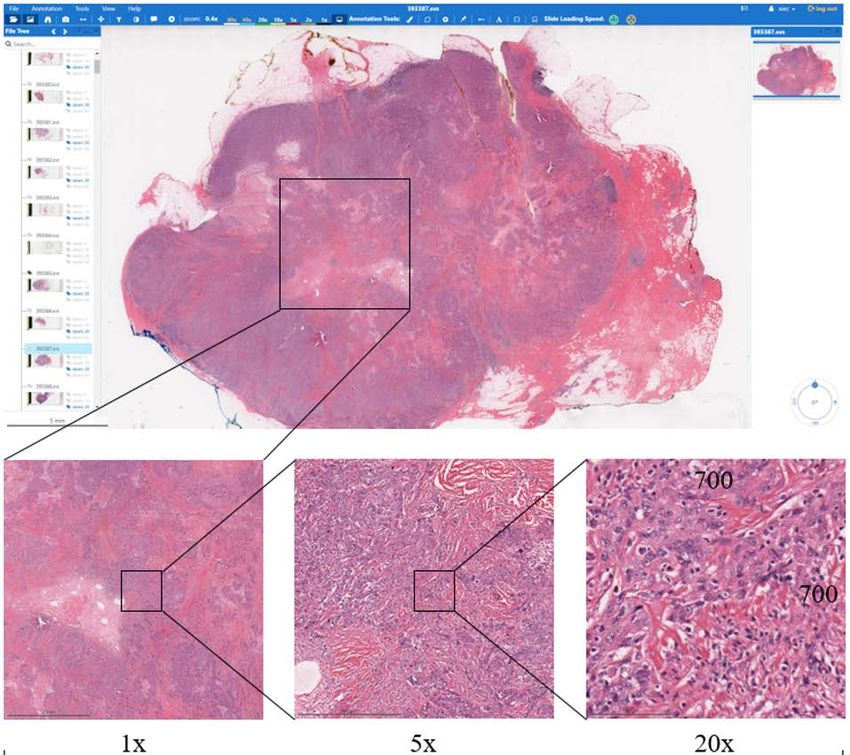
Computational Pathology

 Tissue slide cut,
 staining, and
 examination
 40,000

 65,000

 Whole Slide Image (WSI)

 Diagnosis Treatment Prognosis
 2
Challenges of Computational Pathology

 40,000
 Giga-pixel images
 Information at cellular level Slide label
 • Cancer classification
 Lack of local annotation • Treatment response
 Only slide-level labels • Survival
 65,000

 3
Two-stage approaches for WSI classification

 X: image tiles

 : latent variable Y: Slide label
 • Tile score • Cancer classification
 • Tile feature • Treatment response
 • Survival
 …

 4
Two-stage approaches for WSI classification

 X: image tiles : latent variable Y: Slide label

 With local : tile scores
 X: labeled tiles Cancer classification
 annotation • Tile label

 Multiple instance X: : tile scores
Weakly-supervised

 learning (MIL) Negative slides: all tiles • Tile “pseudo” label Cancer classification
 Positive slides: tiles with score Z > t • Assumption: Z = Y

 : tile features
 Unsupervised X: all tiles Cancer classification
 • Reconstruction
 training Survival regression
 • Self-supervision

 5

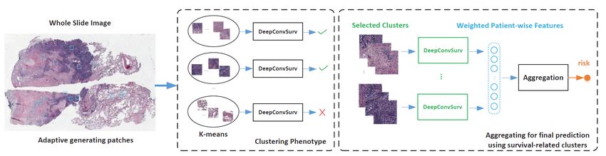
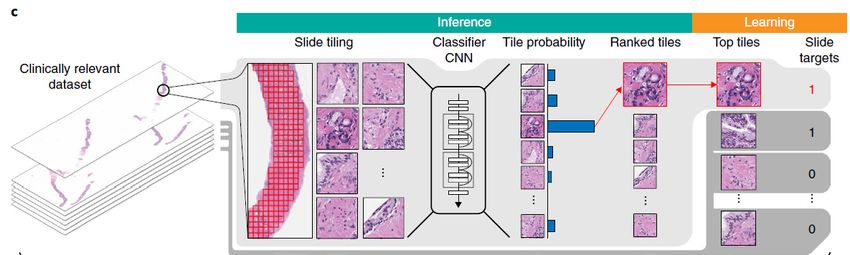
Multiple instance
learning (MIL)

 Campanella, G., Hanna, M.G., Geneslaw, L. et al. Clinical-grade computational pathology using weakly supervised deep learning on whole slide
 images. Nat Med 25, 1301–1309 (2019) doi:10.1038/s41591-019-0508-1

Unsupervised
 training

 Zhu, Xinliang, et al. "Wsisa: Making survival prediction from whole slide histopathological images." Proceedings of the IEEE 6
 Conference on Computer Vision and Pattern Recognition. 2017.

Two-stage approaches for WSI classification

 Unsupervised training:
 • Combining diverse information over whole slides
 • Decoupled training of and
 • Theoretically applicable to any learnable tasks, e.g. survival prediction
 X: image tiles

 : latent variable Y: Slide label
 • Tile score • Cancer classification
 • Tile feature • Treatment response
 • Survival
 …

 Multiple instance learning (MIL):
 • Selecting most predictive tiles
 • Assumption: Z=Y
 • Only applicable to cancer classification
 • is usually voting

 7
How do we combine diverse information of all tiles and learn slide label end-to-end?

Ideal end-to-end learning :

Represent the whole slides as K tile clusters in feature space so that
 only needs to learn aggregation over K centroids rather than all tiles:
 1,1
 1,2 1
 1,3 2 Y
 …

 …

 2,1
 2,2 Relaxation: calculating centroids -> approximating each centroid by the nearest tile
 2,3
 …

 Relaxation

 1 ,1
 2 Y + ,3
 ,8 • Approximating each centroid by the nearest tile in feature space and minimizing the Euclidean
 …

 Sampled tiles
 distance between tiles and their centroids.
 • The whole model consisting of tile encoders , which share parameters, and 1 aggregation
 8
 module can be optimized from end-to-end w.r.t any learnable target .
9
Xie, C., Muhammad, H., Vanderbilt, C. M., Caso, R., Yarlagadda, D. V. K., Campanella, G., & Fuchs, T. J. (2020, January). Beyond10
Classification: Whole Slide Tissue Histopathology Analysis By End-To-End Part Learning. In Medical Imaging with Deep Learning.
Xie, C., Muhammad, H., Vanderbilt, C. M., Caso, R., Yarlagadda, D. V. K., Campanella, G., & Fuchs, T. J. (2020, January). Beyond11
Classification: Whole Slide Tissue Histopathology Analysis By End-To-End Part Learning. In Medical Imaging with Deep Learning.
Xie, C., Muhammad, H., Vanderbilt, C. M., Caso, R., Yarlagadda, D. V. K., Campanella, G., & Fuchs, T. J. (2020, January). Beyond12
Classification: Whole Slide Tissue Histopathology Analysis By End-To-End Part Learning. In Medical Imaging with Deep Learning.
Xie, C., Muhammad, H., Vanderbilt, C. M., Caso, R., Yarlagadda, D. V. K., Campanella, G., & Fuchs, T. J. (2020, January). Beyond13
Classification: Whole Slide Tissue Histopathology Analysis By End-To-End Part Learning. In Medical Imaging with Deep Learning.
Xie, C., Muhammad, H., Vanderbilt, C. M., Caso, R., Yarlagadda, D. V. K., Campanella, G., & Fuchs, T. J. (2020, January). Beyond14
Classification: Whole Slide Tissue Histopathology Analysis By End-To-End Part Learning. In Medical Imaging with Deep Learning.
Part reassignment
 :Centroid
 approximation tile
 Iteration : Global centroids

 −1 −1
 −1 , , ,
 , , ,

 modified during training Reassign tiles to new centroids

 −1 −1 −1
 , , , , , ,

 Calculate new global centroids 1 , 2 , … , by averaging the new
 feature of each part of tiles assigned in the previous epoch − 1:
 = 1/ ∑ ( ,
 −1
 ) 15
Benchmark against traditional task
[1]
[1]
• Traditional cancer classification: S {0,1}
• Clinical-grade classification: only 4 and 6 false negative slides (undetected cancer cases) out of the 1500+ test
slides respectively
• Multi-label lung cancer architectural subtyping: S
• MIL is not applicable
1. Campanella, G., Hanna, M.G., Geneslaw, L. et al. Clinical-grade computational pathology using weakly supervised deep learning on whole slide images. Nat Med 25, 1301–1309
16
(2019) doi:10.1038/s41591-019-0508-1Cancer classification 17
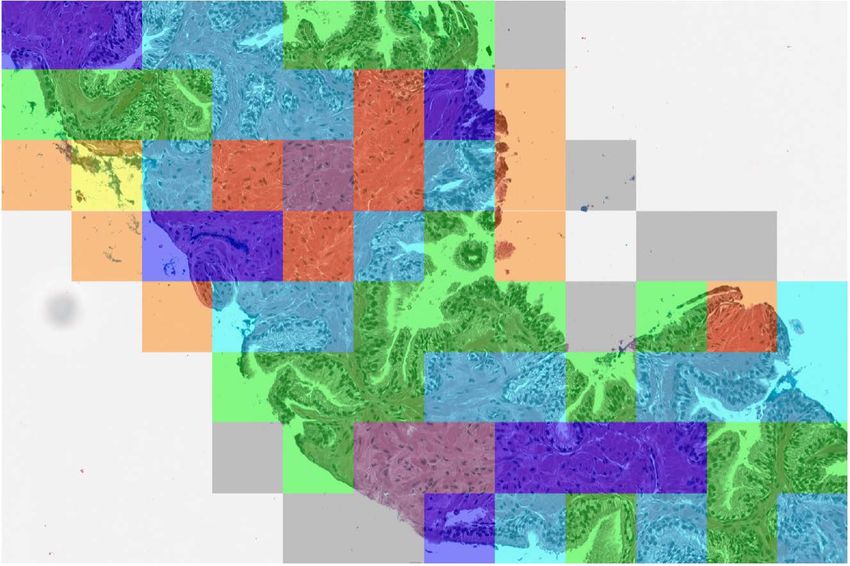
Multi-label lung cancer subtypes prediction
1. Green ink.

2. Red blood cells in blood vessels near alveolar spaces.

3. Macrophages in alveolar spaces, often with emosiderin in the macrophages.

4. Normal alveolar wall.

5. Cancer enriched for micropapillary subtype.

6. Cancer enriched for acinar subtype.

7. Black ink.

8. Cancer enriched for lepidic subtype.

9. Cancer enriched for high grade morphology, solid like.

10. Blood vessel and alveolar wall with sparse cells in spaces.

11. Cancer enrichedfor papillary subtype.

12. Stroma. 18
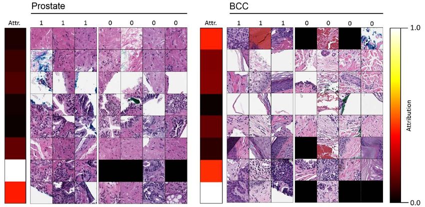
Tissue type localization and region importance scoring

 19
EPL: a general framework for the future of end-to-end WSI assessment

 • A general weakly-supervised WSI prediction algorithm; theoretically applicable to
 any learnable target .
 • Ongoing projects in the lab (with promising results):
 • EPL for survival regression
 • EPL prediction of lung cancer patient response to immunotherapy

 • Easy to be combined with tile-level proxy tasks.
 • Simply adding concurrently trained loss
 • E.g. tile labels, self-supervision targets etc.

 • Various tile encoder
 • as graph neural network (GNN) for WSI classification based on cell graph built from nuclei detection
 results of VOCA 1

 1. Xie, C., Vanderbilt, C.M., Grabenstetter, A. & Fuchs, T.J.. (2019). VOCA: Cell Nuclei Detection In Histopathology Images By Vector Oriented20
 Confidence Accumulation. Proceedings of The 2nd International Conference on Medical Imaging with Deep Learning, in PMLR 102:527-539
Thanks to Fuchs’ lab!

 21
You can also read