Micro-structuring of gold coated plates with LIPSS for localized plasmonic sensors - Laser4surf
←
→
Page content transcription
If your browser does not render page correctly, please read the page content below
Proceedings of LPM2020 – the 21st International Symposium on Laser Precision Microfabrication

Micro-structuring of gold coated plates with LIPSS for localized
                      plasmonic sensors
                       Julien Dupuy, Marc Lamblin, Fabian Dortu, Damien Bernier, Yves Hernandez

                                        Multitel a.s.b.l, Parc Initialis, Mons, BE 7000

                  Localized surface plasmon resonance (LSPR) or particle plasmon resonance (PPR) consists in
            a specific arrangement of metallic nanostructures in order to enhance their plasmonic response. This
            technique can lead to the development of high sensitivity bio-sensors but faces various difficulties
            on the fabrication side.
                  We investigate Laser Induced Periodic Surface Structuring (LIPSS) in order to produce sub
            micron periodic patterns on gold layer for localized plasmonic sensors fabrication. LIPSS are peri-
            odic structures that are generally created on a material surface by accumulation of ultra-short pulses
            exhibiting energies close to the material ablation threshold. Although LIPSS physical mechanisms
            are still under investigation, they have been induced in a wide variety of materials (metals, semicon-
            ductors, polymers, etc) for numerous applications such as realization of hydrophobic/hydrophilic
            surfaces, control of surface reflection or realization of low friction / high adhesion surfaces. Here we
            used ultrashort pulses (500 fs) at 515 nm to induce LIPSS structures on thin gold layers for LSPR
            sensors.

            Keywords: LIPSS, Plasmonic, Gold, LSPR

1. Introduction                                                     energy up to 40µJ at a repetition rate in the range of 10 kHz
    Localized Surface Plasmon Resonance (LSPR) is a                 to 300 KHz. The optical beam generated by this laser will
powerful technique for sensitive bio-sensing. LSPR in met-          be modified through three different modules before reach-
als strongly depends on the size and shape of the sensor            ing the sample.
and can be enhanced with a nanoscale pattern. LSPR sen-                 The first module permits to change the wavelength of
sors can be applied in many applications like chemical,             the laser. A second harmonic generation set-up, based on a
biological sensing… In our case we are targeting the de-            LBO crystal, is employed in order convert the optical
velopment of a label-free biosensor for plant diseases eval-        wavelength down to 515nm. This wavelength is more suit-
uation at points-of-care [1]. In this work, our team has de-        able than infrared because of the absorption spectra of Gold
veloped an original microfabrication method involving               and it will permit achieving smaller LIPSS structures.
gold gratings synthesis by pulsed laser writing.                        The second module permits to control the power/energy
Different other methods have been explored in literature for        of the laser beam and its profile. We use a halfwave plate
generating micro and nano periodic structures on gold [2,           (HWP) and a polarization beamsplitter cube for power con-
3,11] like UV lithography, interferometry, nanosphere li-           trol and a diffractive optical element (DOE) for beam shap-
thography, nanoimprint [4,5]. Another solution we are in-           ing. The DOE converts the input beam into a square top-hat
vestigating is the use of Laser Induced Periodic Surface            beam shape (4x4mm) with a homogeneous energy distribu-
Structuring (LIPSS) in order to produce sub-micron period-          tion. A second HWP is placed to control the linear laser
ic patterns on gold for localized plasmonic sensors fabrica-        polarization orientation.
tion.

LIPSS are periodic structures that are generally created on
a material surface by accumulation of ultra-short pulses
exhibiting energies close to the material ablation threshold.
High resolution structures proportional to the laser operat-
ing wavelength, typically ripples of hundreds of nanome-
ters, can be generated [6].
                                                                             Fig. 1 Laser experiment for LIPSS structuring
2. Description of the experiment

2.1 Laser and micro-machining setup                                     The third module consists of the scanning and focusing
    The micromachining set-up used for the experiments is           elements. We used here a 2D scanning galvanometric head
described in Figure 1. As we can see, the system is made of         equipped with a 160 mm f-theta lens, and coupled with a
a laser system followed by three optical modules.                   X-Y-Z translation stages platform to position precisely the
    The femtosecond laser used in this experiment operates          sample under laser beam. With this configuration, two
at a wavelength of 1030nm and delivers optical pulses of            techniques are possible for dynamic irradiation of the sam-

                                                                1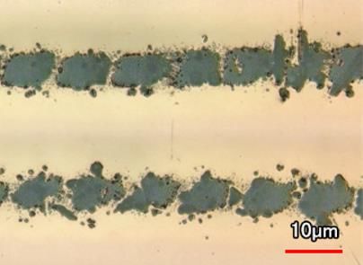
Proceedings of LPM2020 – the 21st International Symposium on Laser Precision Microfabrication
ples: move the sample using the translation stages, keeping This condition makes it possible to separate the laser im-
a fixed point of incidence for the focalized laser, or fix the pacts on the surface and to evaluate an ablation threshold
sample position and scan the surface with the galvanomet- for a single pulse. We then defined the threshold fluence as
ric head. The focused spot size (square shape) is estimated the minimum value for which an ablation profile is meas-
around 25µm. urable. The ablation threshold was estimated to 0.072
J/cm², which corresponds to an average power of 50mW,
2.2 Samples description which is in-line with literature [6,7]. The main difficulties
The samples are made of a thin layer of gold (35 nm) to evaluate this ablation threshold were that we often have
deposited on glass by vapor phase evaporation, with an a “peeling off effect” of the gold layer even at very low
intermediate 1 nm adhesion layer of Chromium. The Glass laser fluence. Experiences conducted us to use the thin
is a 1mm thick standard microscope slide. The LIPSS pro- Chromium layer as a mandatory adhesive layer.
cess permitted to generate regular patterns with a periodici-
ty of about 500 nm. Due to the thin layer of gold, careful 3.2 Fluence effect
manipulations and a precise control of the laser energy de- Dynamic studies were performed by adjusting the la-
posited on the material are needed. Initial surface rough- ser polarization with the HWP so that the ripples produced
ness of a sample is Sa=2nm. by LIPSS are oriented perpendicularly to the machining
scan lines, and by fixing the laser frequency at 100kHz for
three different fluences for a single pulse energy of 0.072
J/cm², 0.058 J/cm² and 0.042 J/cm². This corresponds to
average powers of 50, 40 and 30 mW respectively.
This time, we used a scanning speed in the range 10-
100 mm/s, giving us an overlap for each laser pulse corre-
Fig. 2 Samples scheme
sponding to 99-95%
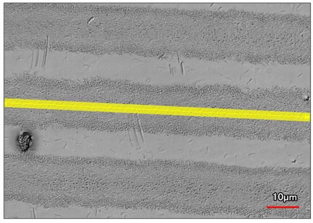
To evaluate the impact of the three fluences on the
2.3 Characterization generated LIPSS structures, we measured the surface
The analyses of the irradiated samples were performed roughness Sa on the center of the scanned line (yellow
with a 3D confocal laser microscope equipped with 20X mark on Fig.7)
and 150X objectives, and also with a SEM microscope.
The first microscope is useful to have rapid views of the
surface and precise estimation of the profile and roughness
of the LIPSS generated structures; and the second one is
mandatory to see details within the nanometer scale.
3. Main results
3.1 Ablation threshold
The first step of the work was to define the minimum
deposited laser energy density on the sample required for
removing the thin gold layer, i.e. the ablation threshold.
Fig.4 Analysis of LIPPS surface roughness as a function
of the scanning speed, for 3 different laser powers
@100kHz
Depending on the scanning speed, the overlap is more
or less important and the number of pulses varies between
1000 and 10000 pulses/mm. In all presented cases the
LIPPS patterns are very regular. The best parameter found
is 25mm/s with 30mW average power (0,042J/cm² - 100
pulses). Even if a 10X factor of deposited energy is tested
(related to the speed process) and the average value of the
surface roughness slightly decreases with the speed, we can
see that the highest speed of 100mm/s give us a good pat-
tern structure and also a faster process to generated LIPSS
on cm² surface area.
Fig. 3 Ablation of the thin gold layer obtained with a flu- 3.3 Laser Frequency effect
ence of 0.09 J / cm² We also tried different laser frequencies with different
scanning speeds in order to improve the quality and the
depth of the LIPSS structure. We compared for the same
Therefore, we studied the ablation behavior of the material
fluence of a single laser shot of 0.042J/cm² (we adapted the
as a function of the laser fluence for a high scanning speed
fixed at 1000 mm/s, and for a pulse frequency of 100kHz.
2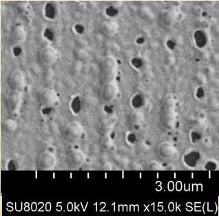
Proceedings of LPM2020 – the 21st International Symposium on Laser Precision Microfabrication

average power of the laser) the repetition rates of 100, 200
and 300kHz.

      Fig.5 Analysis of LIPPS surface roughness as a function
             of scanning speed, for 3 different laser repetition
                                   rates                                        Fig.7 Analysis for LIPSS with 0,042J/cm² with a laser
                                                                                            scanning speed of 80mm/s, 300kHz
    The trend observed in the previous paragraph is also                   The light intensity distribution on the focused laser spot,
valid if the frequency is doubled or tripled (Fig.4) with a            although having certain homogeneity thanks to the DOE, is
slight decrease of the surface roughness, i.e the depth of the         not so clean particularly at low fluence. You can see some-
LIPSS pattern, with the increase of the speed.                         times different ripples and patterns from LIPSS at the bor-
                                                                       ders of the scanning lines (Fig 7.)

     Fig.6 Analysis of LIPPS surface roughness as a function
                         of cumulative energies

    This study highlights two different behaviors of the
gold thin film under femtosecond irradiation. Within the
limits of the parameters studied, the most important param-
eter is the single pulse fluence since it is this one which
determines the evolution that matter will undergo. In a sec-
                                                                         Fig.8 SEM analysis for LIPSS with 0,042J/cm², 80mm/s
ond step, it is the number of overlapped pulses or cumula-
                                                                                                   300kHz
tive energy that allows these micro-structures to evolve
(Fig 6).                                                                 The SEM analysis reveals that the periodic structures of
                                                                       the LIPSS are made of an arranged structure of bubbles and
4. Discussion                                                          holes.
    We present the analysis of the two best results obtained
within this work. We present a case of low spatial period              4.2 Periodic ablation
LIPSS (ripples on the surface) but also one LIPSS parame-                 Here we present the laser and scanning parameter for
ter allowing a periodic ablation of the thin gold layer.               which we observed a complete ablation of the layers of
                                                                       gold and chromium. Compared with the previous experi-
4.1 Best Low Frequency LIPSS obtained                                  ments, we worked this time above the ablation threshold,
    We identified different parameters giving the best rip-            and with high speed, trying to limit the effects of total abla-
ples for the generated LIPSS, i.e the most periodic, regular           tion on LIPSS generated structure.
and contrasting ones. We measured a periodicity in the or-
der of 520nm, same range than the laser wavelength. These
experiments were done with the same single pulse laser
fluence around 0.042J/cm², and only the scanning speed
and laser frequency change (Fig 5.)

                                                                   3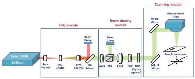
Proceedings of LPM2020 – the 21st International Symposium on Laser Precision Microfabrication

                                                                    formation of LIPSS oriented perpendicular to the radiation
                                                                    lines was carried out.
                                                                         The analysis of these samples with a scanning electron
                                                                    microscope reveals that the reliefs of the wavelets are
                                                                    peaks and aligned holes. The explanation advanced by the
                                                                    literature to explain the weak clarity of LIPSS on gold is
                                                                    the too weak coupling electron-phonon which results in the
                                                                    diffusion of the electrons before they transferred their ener-
                                                                    gy to the matrix. [6,7]
                                                                         The main obstacles encountered during this work were
                                                                    the extreme sensitivities of the parameters. Indeed, a small
                                                                    variation of the thickness of the gold layer, or of the laser
                                                                    pulses energy, leads to a large variation in the morphology
      Fig. 9 Linear Roughness Ra vs scanning speed for 6 dif-       of the observed structures. We would next try to carry out
                ferent parameters showing periodic ablation         specific periodic structures with points spaced from each
                                                                    other by the same distance. For these realizations, circular
We observe that the gold streaks, perpendicular to the laser        polarization or the creation of cross patterns with perpen-
scanning direction, are homogeneous. By analyzing their             dicular wavelet directions, are promising lines of research.
profiles (Fig 9), we observed that their depth is very close             Another technique will be Laser-induced reorganization
to the thickness of the gold and chromium layers, i.e. a            [11] using our LIPSS process, but in this case our samples
depth of about 35nm.                                                need to be adapted to this technique.

                                                                    Acknowledgments

                                                                    The samples were provided by the research center
                                                                    MATERIA NOVA in the frame of the FWVL INTERREG
                                                                    project BIOSENS (SMARTBIOCONTROL).
                                                                    This work was carried out within the frame of the project
                                                                    LASER4SURF, funding from the European Union's Hori-
                                                                    zon 2020 research and innovation program under grant
                                                                    agreement No768636.

                                                                    References
        Fig. 10 Periodic ablation for LIPSS with 0,086J/cm²,        [1] J. Hastanin & Al., "Compact multichannel spectro-
                             300kHz, 820mm/s.                           scopic label-free biosensor platform for plant diseases
                                                                        point-of-care testing (POCT)," Proc. SPIE 11361, Bio-
    We measured also a periodicity in the order of 520nm.
                                                                        photonics in Point-of-Care, 113610P (April 2020)
Nevertheless, a SEM analysis of the sample reveals that the
                                                                    [2] X. Zhang et Al., "Sensors Based on Plasmonic-
lines are not very homogeneous, which affects the periodic-
                                                                        Photonic Coupling in Metallic Photonic Crystals" -
ity analysis.
                                                                        Sensors          2012,        pp.        12082-12097;
                                                                        doi:10.3390/s120912082. (2012).
                                                                    [3] E. Petryayeva & Al.,"Localized surface plasmon reso-
                                                                        nance: Nanostructures, bioassays and biosensing—A
                                                                        review", Analytica Chimica Acta, Volume 706, Issue 1,
                                                                        7 November 2011, Pages 8-24. (2011)
                                                                    [4] Masui & Al., “Laser fabrication of Au nanorod aggre-
                                                                        gates microstructures assisted by two-photon polymer-
                                                                        ization” - Optics Express pp. 22786-96.
                                                                    [5] Wen-Shuo Kuo & Al., “Multiphoton fabrication of
                                                                        freeform polymer microstructures with gold nanorods”,
                                                                        - Optics Express pp. 27551-59,
                                                                    [6] J. Güdde & Al., "Damage threshold dependence on
                                                                        electron–phonon coupling in Au and Ni films" - Ap-
 Fig. 11 SEM analysis for Periodic ablation for LIPSS with
                                                                        plied Surface Science. (1998)
                           0,086J/cm².
                                                                    [7] J.Krüger & Al., "Femtosecond laser-induced damage
                                                                        of gold films" - Applied Surface Science.(2007)
5. Conclusion                                                       [8] A. San-Blas & Al., "Polarization conversion on
    We have studied the different steps of the self-                    nanostructured metallic surfaces fabricated by LIPSS"
organization of matter resulting from dynamic irradiation               - SPIE LASE 2019 San Francisco, Proceedings Vol-
by a femtosecond laser with Yb doped fiber doubled along                ume 10906, Laser-based Micro- and Nanoprocessing
a line on the surface of a thin layer of gold of 35nm. The              XIII; 109061H (2019)

                                                                4
Proceedings of LPM2020 – the 21st International Symposium on Laser Precision Microfabrication
[9] A. Takami & Al., "Formation of gold grating struc-
tures on fused silica substrates by femto-second laser
irradiation" – Journal of applied physics (2017)
[10] U.Hermens & Al., "Automated polarization control for
the precise alignment of laser-induced self organized
nanostructures" – Optics and lasers in engineering-
(2017)
[11] Hendrik M. Reinhardt & Al., "Directed assembly of
gold nanowires on silicon via reorganization and sim-
ultaneous fusion of randomly distributed gold nanopar-
ticles" – Optics Express 11973 (2015)
5You can also read