Study of adhesivity of surfaces using rotational optical tweezers
←
→
Page content transcription
If your browser does not render page correctly, please read the page content below
Letter                                                                                                                          Optics Letters   1

                                                  Study of adhesivity of surfaces using rotational optical
                                                  tweezers
                                                  R AHUL VAIPPULLY1 , D HANUSH B HATT1 , A NAND D EV R ANJAN1 ,                                   AND    BASUDEV R OY1
                                                  1 Physics   Department, Indian Institute of Technology Madras, Chennai, 600036
arXiv:1811.04259v1 [physics.optics] 10 Nov 2018

                                                  * Corresponding   author: basudev@iitm.ac.in

                                                  Compiled November 13, 2018

                                                  Optical tweezers are powerful tools for high resolution                          do not apply a large normal force on the surface, there is no
                                                  study of surface properties. Such experiments are tradi-                         possiblity of rupturing membranes of live cells either.
                                                  tionally performed by studying the active or the brow-                              Rotational motion of a birefringent microsphere using an cir-
                                                  nian fluctuation of trapped particles in the X, Y, Z di-                         cularly polarized optical tweezers apparatus has been described
                                                  rection. Here we find that employing the fourth dimen-                           with the following eq. 1 ([3])
                                                  sion, rotation, allows for sensitive and fast probing of                                                      dθ
                                                                                                                                                             γ      = τ = ηI                          (1)
                                                  the surface. Optical tweezers are capable of rotating                                                         dt
                                                  trapped birefringent microparticles when applied with                                where, γ is the drag coefficient, τ the rotational torque due to
                                                  circularly polarized light, thus called the Rotational                           circularly polarized light, η the efficiency of torque transfer and
                                                  Optical Tweezers. When the trapped birefringent mi-                              I is the intensity of the tweezers light. Here, we have assumed
                                                  croparticle is far enough away from the surface, the ro-                         that the gaussian varying random noise due to Brownian motion
                                                  tation rate is dependent only on the laser power. How-                           is negligible compared to the rotational torque.
                                                  ever, we find that if one traps close to a surface, the rota-                        We can generally conclude that the rotational rate would go
                                                  tion rate goes to zero even at finite tweezers laser pow-                        to zero only when the laser intensity would be brought to zero.
                                                                                                                                   An interesting case appears when we bring this rotating micro-
                                                  ers for some specific type of substrates. We suspect this
                                                                                                                                   sphere close to a surface. The rotational faxen correction to the
                                                  to be due to interaction between the substrate and the                           drag is bounded to a value (ζ(3) which is about 1.20, where ζ
                                                  birefringent particle, keeping in mind that the hydro-                           indicates the Riemann zeta function) very close to the surface
                                                  dynamic drag for this mode of rotation cannot increase                           while being compared to the value away from surface [5, 6],
                                                  beyond 1.2 times the drag away from the surface. We                              thereby indicating that the rotation rate can never become zero
                                                  use this to probe some surfaces and find that there is                           at a finite value of torque. We perform our measurements close
                                                  no binding for hydrophobic ones but hydrophilic ones                             to some mildly hydrophilic surfaces using birefringent liquid
                                                  particularly tend to show a power threshold beyond                               crystalline RM257 microspheres [7] and find that in surface prox-
                                                  which the birefringent particle starts rotating. We calcu-                       imities, the particle stops rotating even at a finite value of torque.
                                                  late that the threshold energy of the tweezers is consis-                        We suspect that this is an effect of adhesion to the surface, which
                                                                                                                                   must be overcome for rotation. A similar study was carried out
                                                  tent with the Van der Waals potential energy, when the
                                                                                                                                   for translational mode of motion of a particle moving parallel to
                                                  mode of interaction with the surface is purely physical.
                                                                                                                                   a surface when the rheology of sticking transition was studied
                                                  We also find that for chitosan, the mode of interaction                          [8]. However, this mode of translation faces much larger Faxen
                                                  is possibly different from Van der Waals. We place the                           corrections than the yaw rotational one [9], not to mention the
                                                  particle on the threshold and observe ”stick-slip” kind                          time scales of interaction inviting slower effects.
                                                  of rotational behaviour. © 2018 Optical Society of America                           In order to perform the measurement, we optically trap
                                                                                                                                   and rotate a birefringent microparticle (liquid crystalline col-
                                                  http://dx.doi.org/10.1364/ao.XX.XXXXXX                                           loid of RM257 material (Merck), chemical name 2-Methyl-1,4-
                                                                                                                                   phenylene bis(4-(3-(allyloxy)propoxy)benzoate) [7, 10], of diam-
                                                     The study of adhesivity at the microscopic scales has gener-                  eter 1 ± 0.2 µm, typical birefringence of 0.01) close to the top
                                                  ally been performed using Atomic Force Microscopes (AFM) [1].                    surface in an inverted microscopy configuration using the op-
                                                  This applies a nanonewton force and can also detect interactions                 tical Tweezers Kit (Thorlabs USA) as shown in Fig. 1. Colloids
                                                  to a minimum of about 100 pN nm due to limitations in the                        of RM257 are stable in water. The illumination objective is 1.25
                                                  cantilever size [2]. Further, hard probing of live biological cells              NA, E Plan 100x objective from Nikon at the bottom with the
                                                  using AFM probes can rupture the membrane thereby killing                        illumination aperture being overfilled. The collection objective is
                                                  them. Here we present an alternative technique using birefrin-                   E Plan 10x, 0.25 NA also from Nikon. The laser used for optical
                                                  gent probes trapped in optical tweezers [3] which can probe                      trapping is 976 nm Butterfly laser (Thorlabs, USA). The parti-
                                                  softly [4] and sense interactions at higher resolution. Since we                 cles are trapped close to the top surface of the sample chamber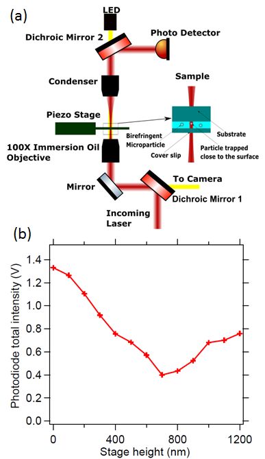
Letter                                                                                                                Optics Letters   2

and all the measurements performed in this configuration. We
collect all the forward scattered light incident on a photodiode
and move the particle close to the surface. The scatter intensity
keeps on reducing till it reaches a minimum, whereafter it keeps
increasing. The position of the surface when the inflection point
of forward scattered light is attained is known to be about 100
nm from the surface, given in Fig. 5(a) of [11], and also shown
in Fig. 1. The slopes of the lines before and after the surface is
reached is noticably different, from where the surface position
can be inferred. As the surface is reached, where the inflection
point is attained, the particle starts to get pushed away from the
equilibrium of the trap upon the effect of the surface, thereby
showing a different slope.
    When we trap the particle close to the glass surface and grad-
ually reduce the laser power, we find a threshold beyond which
the particle stops rotating. It has been shown in Fig. 2. There is
however no such threshold if a PDMS substrate is used. We in-
vestigate this further by considering that PDMS is hydrophobic
in nature while the glass surface that we have used is hydrophilic.
We put droplets of water onto the glass slide and estimate the
contact angle which we find to be about 45 degrees. We also
consider a quartz slide that shows a contact angle of 75 degrees,
indicating that it is less hydrophilic. The respective rotation rate
as a function of laser power curve has been shown on Fig. 2. In
all these measurements for rotation rate as a function of laser
power, we reduce the power till the rotation rate is about 1 Hz
under which the trapping becomes so weak that the vertical ex-
cursions from equilbrium are comparable to the rotation events
what we are trying to detect. Thus reducing laser power under
such values do not give trustworthy results. Further, consider-
ing that the suspensions of RM257 form stable colloids in water
and that these tend to stick to the hydrophilic subtrates tends to
indicate that this particle itself is hydrophilic in nature.
    The hydrophobic surface hardly has a threshold, as the
straight line passes through the origin within the error bar of the
fit. However, there are noticable thresholds for the quartz and
the regular glass slide, with the quartz having a lower threshold
than the glass slide. This can be explained by the following
equation

                            dθ
                         γ     = η ( I − I0 )                      (2)
                            dt
    where, I0 is the threshold that needs to be overcome to initiate
rotation. This can be understood as bonds being formed between
the probe and the surface, which requires η I0 amount of torque
                                        η
to be overcome. The slope of line is γ which for similar types of
particles would be same since η only depends upon the particle
size and birefringence of the particle, while the γ depends upon         Fig. 1. (a) Schematic diagram showing the optical tweezer set
the viscosity of the medium and the size of the particle. We do          up. The trapping is made close to the top surface of the sample
indeed find the slopes to be same for the three different surfaces       chamber. (b) Change in the total intensity of the scattered light
since the sizes of all of them are about 1 micron.                       as the particle is brought closer to the surface using a transla-
    We compute the corresponding threshold energy that these             tion stage in steps of 100 nm. The position of the minimum
particles must overcome to initiate rotation. The threshold power        of the scattered light signal (the point of inflection) indicates
for rotation on glass is about 4.25 mW and the corresponding             an axial distance of of 100 nm from the surface. Here 0 nm
threshold a = -2.12 Hz. The drag coefficient γ = 8 π η1 r3 , where       indicates a position futher away from the surface than 1200
η1 is the viscosty of water and r is the radius of the particle. Then    nm.
the coefficient η/γ = 0.52 ×(2π)) Hz/mW, such that the value
of η = 8.05 × 10⁻²¹ pN nm Hz/mW. Then, the threshold torque
is η × I0 which is 34.2 pN nm and the subsequent energy is to
rotate it by 360 degrees which is 34.2 pN nm × 2π = 215 pN nm.
We find them to be of the order of 50-220 pN nm, as shown in
Fig. 3. These have been plotted as a function of contact angle
of a water droplet on the surface and exhibit a straight line fit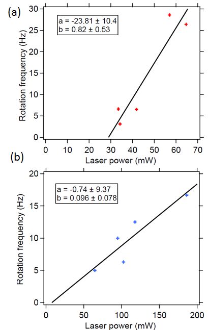
Letter Optics Letters 3
25 PDMS substrate
a = -0.53 ± 0.89
b = 0.53 ± 0.06
20 Quartz substrate
Rotation frequency (Hz)
a = -0.82 ± 0.67
b = 0.51 ± 0.04
15 Glass substrate
a = -2.12 ± 0.62
b = 0.52 ± 0.03
10
5
0
0 10 20 30 40 50
Laser power (mW)
Fig. 2. Rotation rate as a function of laser power for PDMS,
Quartz & Glass Surfaces. Quartz and glass seem to have a
threshold while PDMS shows no threshold. The data have
been fitted to an equation of the form y = b x + a.
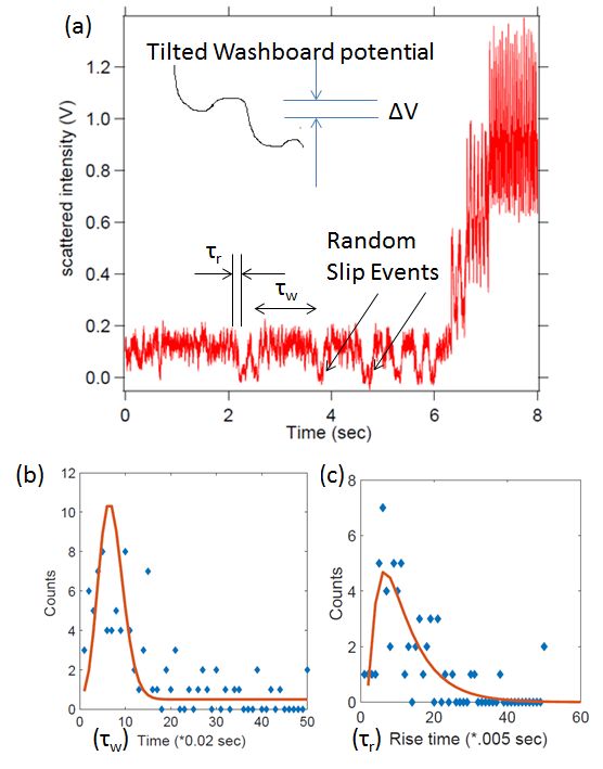
Fig. 4. (a) Time series for rotation at four different laser pow-
ers on a glass substrate. At the minimum laser power used
here, the rotational events become random and can be de-
scribed as slip events. (b) Time distribution of slip events fitted
to a Poisson distribution. (c) Distribution of rise times for the
slip events fitted to a curve indicated in [13], expected for tran-
sitions in a biased double well potential. This gives the height
250 of the bistable potential well.
Threshold energy (pN nm)
200
within error bars. The threshold energy corresponds very well
to the Van der Waals interaction [12] with a Hamaker constant of
150 about 2 × 10⁻²⁷ J m separated from the particle to the surface by
less than 100 nm. Thus, the rotation threshold is a good measure
100 of the hydrophilicity of the substrate given that the mechanism
of interaction is Van der Waals interaction.
50 We show a typical curve for rotational motion of the micro-
sphere close to the surface in Fig. 4 (a). At high values of laser
power, the particle continues to rotate periodically. However, as
0 the power is reduced slowly, the rotation rate also reduces till a
point where the rotational events start to become random. When
-50 we study waiting time distribution for the delay between two
0 20 40 60 80 100 120 slip events, we find a distribution of the form Fig. 4(b) which
Contact Angle (degrees) can be fitted well to a Poisson distribution. We also study the
time it takes for the rotation of the particle by 180 deg during
Fig. 3. Threshold laser energy to initiate rotation as function the slip events which follows a distribution given in [13], shown
of a water drop contact angle on the substrate. The energy in Fig. 4(c) and expected for a biased double well potential.
required to initiate rotation seems to indicate van der waals A tilted washboard potential can be also referred to a biased
potentials as the binding mechanism to the surface. double well potential due to rotational symmetry. We estimate
that the bistable potential has a barrier (∆V) of the height 0.8
k B T, quite consistent with other types of such jumps in a tilted
washboard potential [10, 13–15].
We go on to study a different type of substrate where theLetter Optics Letters 4
motion close to the surface upon the influence of specific binding
to the surface [17]. We suspect that our increase in drag is due
to partial nonspecific binding to the surface of the cell.
Thus, to conclude, we have demonstrated a system using
birefringent microspheres which can sense the surface adhesiv-
ity. If surfaces without chemical adhesivity are used, the Van der
Waals force ensures that only hydrophilic ones show rotational
threshold. This can subsequently be used to determine the hy-
drophilicity of the surface using a hydrophilic probe. Further,
even if other mechanisms of binding are present, the rotational
threshold indicates the binding energy. We also show that at
the point of threshold, particularly on the glass surfaces which
are mildly hydrophilic, the rotational motion follows a stick-slip
behaviour where the slip events happen randomly. This kind
of technique can eventually be used to study adhesivity at the
nanometric scales and is possibly a more sensitive probe than
Atomic Force Microscopes.
We thank the Indian Institute of Technology Madras for the
seed grant. We also thank Amal Kanti Bera for providing us the
CHO cells required for the experiment, Dillip Satapathy for the
PDMS and Chitosan sample and Erik Schaffer for the RM257
liquid crystal powder.
A. Materials and Methods
Fig. 5. Rotation rate as a function of laser power for (a) Chi-
tosan substrate and (b) CHO cell surface. The chitosan sample The chitosan (Sigma-Aldrich 50000) substrate was prepared by
seems to exhibit a proper threshold while the CHO cell seems dissolving in 5% solution of formic acid in double distilled water.
have no threshold with a much lower slope than the harder We took 3% (W/V) concentration of chitosan in the formic acid
surfaces. The data have been fitted to a form y = b x + a. solution for making the film. This solution is stirred at 75◦ C for
one hour. The prepared solution is filtered and spin coated on
glass substrate.
mechanism of interaction is not expected to be of Van der Waals The PDMS (Dow Corning’s Sylgard 184 elastromer kit) substrate
type, namely that of chitosan. We find that even though the is prepared by curing the Silicone elastomer in 10% (W/W) with
contact angle for the chitosan substrate is about 75 degrees, the the curing agent. The viscous solution is spin coated on glass
threshold is much larger than that of glass, at 1400 pN nm. This slides and baked at 150◦ C.
can be explained by the fact that chitosan is a well known bio-
adhesive with stickiness extending to a pH of 7 [16]. We believe REFERENCES
to have recorded this in the threshold of rotation. We show this
1. E. L. Florin, V. T. Moy, and H. E. Gaub, Science 264, 415 (1994).
Fig. 5(a).
2. M. B. Viani, T. E. Schaffer, A. Chand, M. Rief, H. E. Gaub, and P. K.
We also tried to perform the rotation close to a Chinese Ham-
Hansma, J. App. Phys. 86, 2258 (1999).
ster Ovary (CHO) cell which was prepared to be adhered to a 3. M. E. J. Freise, T. A. Nieminen, N. R. Heckenberg, and H. Rubinsztein-
glass substrate and find a very different behavior, as shown in Dunlop, Nature. 394, 348 (1998).
Fig. 5(b). The slope of the curve seems to be much smaller (0.09 4. L. Friedrich and A. Rohrbach, Nat. Nano 10, 1064 (2015).
Hz/mW compared to about 0.51 Hz/mW in Fig. 2) while the 5. Q. Liu and A. Prosperetti, J. Fluid Mech. 657, 1 (2010).
threshold seems to be absent. We can explain this by consid- 6. J. Leach, H. Mushfique, S. Keen, R. D. Leonardo, G. Ruocco, J. M.
ering that the threshold η I0 indicated in eq. 2 is dependent on Cooper, and M. J. Padgett, Phys. Rev. E 79, 026301 (2009).
frequency as γ0 ω. 7. A. Ramaiya, B. Roy, and E. Schaffer, Proc. Nat. Acad. Sci. (USA) 114,
10894 (2017).
8. P. Sharma, S. Ghosh, and S. Bhattacharya, Nat. Phys. 4, 960 (2008).
γω = η I − γ0 ω (3)
9. B. Roy, A. Ramaiya, and E. Schaffer, J. Opt. 20, 035603 (2018).
Then we get the following equation where the slope can be 10. B. Roy and E. Schaffer, Curr. Sci. 111, 2005 (2016).
much lower. 11. E. Schaffer, S. F. Norrelykke, and J. Howard, Langmuir. 23, 3654
(2007).
(γ + γ0 )ω = η I (4) 12. R. H. French, K. I. Winey, M. K. Yang, and W. Qiu, Aust. J. Chem. 60,
251 (2007).
Although the viscous drag coefficient depends upon the particle 13. L. Shao, D. Andren, S. Jones, P. Johansson, and M. Kall, Phys. Rev. B
size, the RM257 sample diameter is monodisperse to within 1000 98, 085404 (2018).
± 200 nm [7], also confirmed with video imaging. This does not 14. F. Pedaci, Z. Huang, M. van Oene, S. Barland, and N. H. Dekker, Nat.
explain the low slope. This indicates that the CHO cell surface Phys. 7, 259 (2011).
appears like a viscous medium for the rotating particle with a 15. F. Pedaci, Z. Huang, M. van Oene, and N. H. Dekker, Opt. Exp. 20,
viscosity which is 5 times that of a particle in proximity to glass. 3787 (2012).
16. N. Mati-Baouche, P.-H. Elchinger, H. de Baynast, G. Pierre, C. Delattre,
Since the trapping light enters the sample chamber from the
and P. Michaud, Euro. Poly. J. 60, 198 (2014).
bottom while the CHO has been attached to the substrate at the
17. A. Pralle, E.-L. Florin, E. H. K. Stelzer, and J. K. H. Horber, Single Mol.
top, the cell itself has no effect on the trapping light. This kind of 1, 129 (2000).
effect has also be reported in a similar work using translationalLetter Optics Letters 5
FULL REFERENCES
1. E. L. Florin, V. T. Moy, and H. E. Gaub, “Adhesion forces between
individual ligand-receptor pairs,” Science. 264, 415 (1994).
2. M. B. Viani, T. E. Schaffer, A. Chand, M. Rief, H. E. Gaub, and P. K.
Hansma, “Small cantilevers for force spectroscopy of single molecules,”
J. App. Phys. 86, 2258 (1999).
3. M. E. J. Freise, T. A. Nieminen, N. R. Heckenberg, and H. Rubinsztein-
Dunlop, “Optical alignment and spinning of laser-trapped microscopic
particles,” Nature. 394, 348 (1998).
4. L. Friedrich and A. Rohrbach, “Surface imaging beyond the diffraction
limit with optically trapped spheres,” Nat. Nano 10, 1064 (2015).
5. Q. Liu and A. Prosperetti, “Wall effects on a rotating sphere,” J. Fluid
Mech. 657, 1 (2010).
6. J. Leach, H. Mushfique, S. Keen, R. D. Leonardo, G. Ruocco, J. M.
Cooper, and M. J. Padgett, “Comparison of faxen’s correction for a
microsphere translating or rotating near a surface,” Phys. Rev. E 79,
026301 (2009).
7. A. Ramaiya, B. Roy, and E. Schaffer, “Kinesin rotates unidirectionally
and generates torque while walking on microtubules,” Proc. Nat. Acad.
Sci. (USA) 114, 10894 (2017).
8. P. Sharma, S. Ghosh, and S. Bhattacharya, “Microrheology of a sticking
transition,” Nat. Phys. 4, 960 (2008).
9. B. Roy, A. Ramaiya, and E. Schaffer, “Determination of pitch rotation in
a spherical birefringent microparticle,” J. Opt. 20, 035603 (2018).
10. B. Roy and E. Schaffer, “Directed rotational motion of birefringent
particles by randomly changing the barrier height at the threshold in a
washboard potential,” Curr. Sci. 111, 2005 (2016).
11. E. Schaffer, S. F. Norrelykke, and J. Howard, “Surface forces and
drag coefficients of microspheres near a plane surface measured with
optical tweezers,” Langmuir. 23, 3654 (2007).
12. R. H. French, K. I. Winey, M. K. Yang, and W. Qiu, “Optical proper-
ties and van der waals–london dispersion interactions of polystyrene
determined by vacuum ultraviolet spectroscopy and spectroscopic el-
lipsometry,” Aust. J. Chem. 60, 251 (2007).
13. L. Shao, D. Andren, S. Jones, P. Johansson, and M. Kall, “Optically
controlled stochastic jumps of individual gold nanorod rotary motors,”
Phys. Rev. B 98, 085404 (2018).
14. F. Pedaci, Z. Huang, M. van Oene, S. Barland, and N. H. Dekker,
“Excitable particles in an optical torque wrench,” Nat. Phys. 7, 259
(2011).
15. F. Pedaci, Z. Huang, M. van Oene, and N. H. Dekker, “Calibration of
the optical torque wrench,” Opt. Exp. 20, 3787 (2012).
16. N. Mati-Baouche, P.-H. Elchinger, H. de Baynast, G. Pierre, C. Delattre,
and P. Michaud, “Chitosan as an adhesive,” Euro. Poly. J. 60, 198
(2014).
17. A. Pralle, E.-L. Florin, E. H. K. Stelzer, and J. K. H. Horber, “Pho-
tonic force microscopy: A new tool providing new methods to study
membranes at the molecular level,” Single Mol. 1, 129 (2000).You can also read