Modern dolomite formation caused by seasonal cycling of oxygenic phototrophs and anoxygenic phototrophs in a hypersaline sabkha - Nature
←
→
Page content transcription
If your browser does not render page correctly, please read the page content below
www.nature.com/scientificreports
OPEN Modern dolomite formation caused
by seasonal cycling of oxygenic
phototrophs and anoxygenic
phototrophs in a hypersaline
sabkha
1*
Zach A. Diloreto , Sanchit Garg1, Tomaso R. R. Bontognali2,3 & Maria Dittrich1
The “Dolomite Problem” has been a controversy for over a century, owing to massive assemblages
of low-temperature dolomite in ancient rocks with little dolomite forming today despite favorable
geochemical conditions. Experiments show that microbes and their exopolymeric substances
(EPS) nucleate dolomite. However, factors controlling ancient abundances of dolomite can still
not be explained. To decode the enigma of ancient dolomite, we examined a modern dolomite
forming environment, and found that a cyclic shift in microbial community between cyanobacteria
and anoxygenic phototrophs creates EPS suited to dolomite precipitation. Specifically, EPS show
an increased concentration of carboxylic functional groups as microbial composition cycles from
cyanobacterial to anoxygenic phototroph driven communities at low-and high- salinity, respectively.
Comparing these results to other low-T forming environments suggests that large turnover of organic
material under anoxic conditions is an important driver of the process. Consequently, the shift in
atmospheric oxygen throughout Earth’s history may explain important aspects of “The Dolomite
Problem”. Our results provide new context for the interpretation of dolomite throughout Earth’s
history.
Low-temperature (low-t) dolomite has been an enigmatic mineral for over a century and the focus of a long-
lasting dispute known as a “the dolomite problem”. The puzzle of low-t dolomite stems from its considerable
abundance in ancient sedimentary rocks, while its presence inexplicably decreases in younger sequences1–3. Find-
ings on synthesis of low-t dolomite have shown that abiotic formation at low temperature is impossible without
any heterogeneous surfaces, e.g., seeds2,4–7. This includes biological surfaces such as EPS2,4–6 and abiotic surfaces
such as clays which have been shown to facilitate the formation of proto-dolomite7. The lack of precipitation was
thought to be caused by inhibitors of reaction kinetics such as cation desolvation8, an absence of nucleation sites9,
inhibition by sulfate ion, and formation of neutral MgSO4 over dolomite4. The question about kinetic inhibition
seemed to be addressed with the observation that low-t dolomite could form in the presence of sulfate-reducing
bacteria10,11. Later on, multiple laboratory studies demonstrated that low-t dolomite precipitation could occur
in presence of microbes under multiple metabolic pathways including aerobic r espiration12, methanogenesis13,
methane oxidation14, and even sulfide oxidation15. More recent findings have suggested that organic material of
microbial origin was responsible for catalyzing low-t dolomite p recipitation16–18. Specifically, low-t Mg-dolomite
precipitation under surficial conditions could be catalyzed by the presence of E PS18–21. The pathway of this process
was later proposed as a possible dehydration of M g2+ facilitated by carboxylic groups which act as a nucleation
sites for overcoming kinetic barriers6,13. Additionally, laboratory experiments have shown that salinity also plays
a role in low-t dolomite precipitation by increasing Mg ions available in addition to increasing the amount of
EPS produced and available for n ucleation23–27.
Although these findings reinforced the view that microbial life, including EPS, represent an important com-
mon denominator for the mechanism of low-t dolomite formation, fundamental aspects of this mineralization
1
Department of Physical and Environmental Sciences, Biogeochemistry Group, University of Toronto Scarborough,
Toronto, Canada. 2Space Exploration Institute, Fbg de l’Hopital 68, 2002 Neuchâtel, Switzerland. 3Department of
Environmental Sciences, University of Basel, Klingelbergstrasse 27, Basel, Switzerland. *email: zach.diloreto@
utoronto.ca
Scientific Reports | (2021) 11:4170 | https://doi.org/10.1038/s41598-021-83676-1 1
Vol.:(0123456789)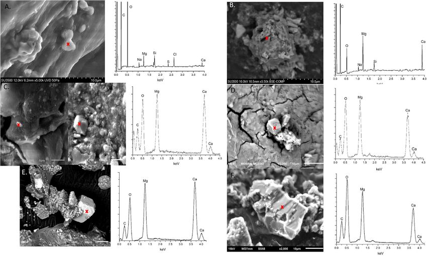
www.nature.com/scientificreports/
process throughout Earth’s history remain unclear. In particular, although several environmentally ubiquitous
microbial species have been reported to mediate dolomite during laboratory experiments, dolomite formation is
not a common process in modern times. Even more intriguing is that while the studied microorganisms represent
different metabolic pathways and thus, are present in a wide range of geochemical regimes, dolomite can be found
only under very specific environmental conditions. Example of such specific environments include hypersaline
settings such as coastal l agoons10,11, evaporative l akes28, alkaline l akes29 and sabkhas18,19,30,31, as well as hemipe-
lagic or “Deep-Sea” environments that occur on continental m argins32–34 and “cold-seeps”35,36. Consequently,
laboratory culture experiments, although being a generally valuable approach to discover the mechanisms, are
not capable of capturing the complexity of natural dolomite-forming environments, in which several microbial
metabolisms simultaneously interact with environmental factors changing dynamically (e.g., hourly, diurnal,
monthly, seasonal, long-term climatic fluctuations). By integrating these observations under natural conditions
light can be shed on these interactions and their influence on the mechanisms of low-t dolomite formation.
Thus, we have conducted an in-situ field study of dolomite-forming microbial mats that colonize the inter-
tidal zone of the Khor Al Adaid sabkha in Qatar. This type of coastal sabkha, where low-t dolomite forms in
association with other common evaporitic sedimentary facies, is considered as an excellent modern analogue
for many ancient, carbonate-rich sedimentary sequences37,38. Instead of using a “snap-shot-type” approach based
on a single field campaign, the same microbial mats were monitored and sampled in different seasons during a
period of three years. This holistic approach provided new insight that, as discussed in the paper, shed new light
on the factors that may have controlled the uneven distribution of dolomite throughout the geological record.
Results
The Khor Al-Adaid sabkha is located in the South-Eastern of Qatar. This area has been previously described
from an environmental as well as geological perspective37,38. Within the sabkha two locations were selected as
sampling sites. One within the lower intertidal zone, referred to as KAAS-1, and one within the upper intertidal
zone, referred to as KAAS-2. The microbial mats at these two locations were sampled over multiple seasons
in March and October of 2016, and February of 2018 (Fig. 1). Some data collected in March of 2016 has been
previously presented in37.
The geochemistry of water columns and mats at both sites follows a typical progression from photo-oxic to
anoxic with depth, oxygen and redox potential follow a typical progression from oxidized to reduced conditions
for both mats during all seasons, excluding KAAS-1 in March of 2016 (Fig. 2). pH values in March 2016 and
February 2018 are circum-neutral (7.8–8.9) within the water column but show increased pH (8.6–9.2) at the first
5 mm of mat, referred to as the sediment water interface (SWI). Deeper in the mats, in first three centimeters
we observed the decreases in pH to values between 6.5 and 7.5. The large increase of pH at depths below 4 cm
in KAAS-2 can be linked to a process generating alkalinity or a large accumulation of brine. In our previous
study37 we reported the presence of halophiles that can only survive at extreme salt concentrations. KAAS-1
during October 2016 shows a different trend with values of 7.6–7.65 in the water column leading up to the SWI
interface. Below the SWI within the mat pH values become more basic increasing to 7.95. The trend of pH depths
profiles in February 2018 are comparable with those measured in March of 2016.
In October 2016, the mat KAAS-2 has dried out (Fig. 1H,K). Concentrations of major ions as well as salinity,
pH and temperature are shown for both sampling sites in (Table S1) and (Table S2). Salinity changes occur in
conjunction with temperature increases. In March of 2016 KAAS-1 had consistent salinity values throughout the
surface water and mat of ≈ 48‰. In October 2016 salinity of KAAS-1 triples compered to March values within the
surface water and almost doubles compered to March values at the SWI and within the mat itself with values of
137.79‰ and 72.57–74.00‰ respectively. February 2018 measurements are similar to values measured in March
of 2016, however, salinity in the surface water is much higher, 77.03‰, while at the SWI and within the mat is
much lower, 39.46‰ and 25.93‰ respectively. KAAS-2 shows differences in salinity throughout March 2016
and February 2018. March values remain consistent throughout the mat profile ranging from 78.63–81.04‰,
while measurements in February are the highest with salinity in the surface waters of 140.12‰, 92.30‰ at the
SWI, and 102.76‰ within the mat.
Mineralogy of the mats reveals that dolomite is present within each mat, at every depth, during all sampling
season, with the exception of the top layer (or Crust) of KAAS-2 in October 2016. XRD spectra for each sea-
son show a distinguish main reflection peak at two theta (2θ) 30.91 which reflects Miller indices hkl = 104 for
dolomite as well as ordering peaks at 101 (22.0 for 2θ), 015 (35.19 for 2θ) and, rather less distinguished in some
spectra, 021 (43.78 2θ) (Figure S1). The presence of ordering peaks is key in delineating dolomite from disordered
dolomite, proto-dolomite, and high Mg c alcite39. Some of the main reflection peaks exhibit a slight right shift
suggesting enrichment in Mg40. SEM data displays representative particles with elemental composition similar to
dolomite from each of the mats during each season (Fig. 3). The presence of such particles with similar elemental
composition to dolomite from KAAS-1 during each season suggests a mix of both proto-dolomite and rhombo-
hedral stoichiometric dolomite and is observed within all layers during each of the measuring seasons. Although
it may be present as a precursor mineral, SEM investigations in KAAS-2 revealed no proto-dolomite, instead
mostly stacked poly-crystalline aggregates with a composition similar to dolomite, which remain consistent in
morphology and crystallinity throughout each sampling season. Large amounts of gypsum and silica (sand from
the desert) were detected visually, as well as through XRD and SEM. Generally, more detailed information on
these phases was collected during the course of SEM investigations but were not pertinent to this study. There
are likely many sulfosalts as well, but confirmation of any specific phases is difficult and speculative due to the
high diversity of such phases and complex XRD patterns.
Microbial community composition at the phylum level for KAAS-1 is shown in (Fig. 4). Community com-
position at higher and lower taxonomic classifications can be found in (SI ZOTU Files). The most significant
Scientific Reports | (2021) 11:4170 | https://doi.org/10.1038/s41598-021-83676-1 2
Vol:.(1234567890)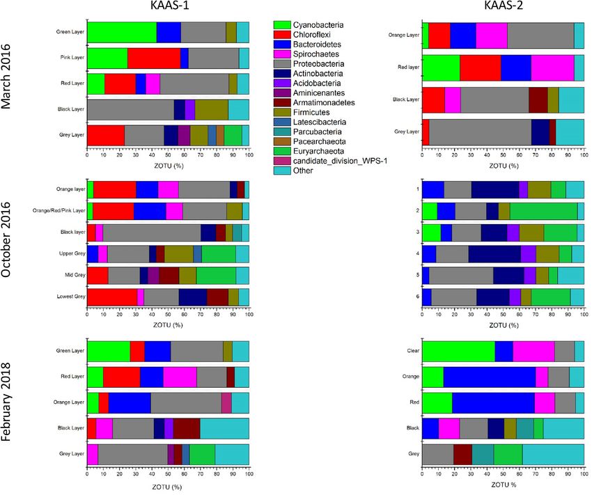
www.nature.com/scientificreports/
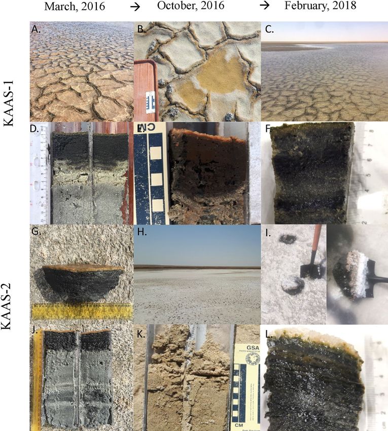
Figure 1. (A) Surface of KAAS-1 during March 2016. (B) Surface of KAAS-1October 2016. (C) Surface
of KAAS-1 February 2018. (D) Vertical profile of KAAS-1 during March 2016 consisting of the thin upper
green layer with intermixed pink and red layers (≈0.5 cm), the thicker middle black layer (≈4.5 cm), and the
bottom-most grey layers composing the rest of the profile. (E) Vertical profile of KAAS-1 October 2016 with
the uppermost orange layer and intermixed pink and orange layer (≈0.5 cm), the middle black layer (≈5 cm),
and upper (6 cm), middle (8 cm) and lower grey (> 8 cm) layers. (F) Vertical profile of KAAS-1 in February
2018 with green, red and orange layers intermixed (≈0.5 cm), middle black layer (5 cm) and bottom grey
layer (> 5 cm) (G) Surface of KAAS-2 during March 2016 with extracted portion of mat (middle). (H) Dried
out surface of KAAS-2 October 2016. (I) Salt encrusted surface of KAAS-2 February 2018 and extracted mat
portion (left). (J) Vertical profile of KAAS-2 during March 2016 showing the orange and red layers (≈0.2 cm),
middle black layer (4 cm), and bottom grey layer (> 4 cm). (K) Vertical profile of KAAS-2 in October 2016
showing a surficial crust and desiccated core. (L) Vertical profile of February 2018 showing a clear-gel
underneath the removed salt-encrustation, an intermixed orange, green and red layer (≈1 cm), followed by a
middle black layer (3 cm) and the intermixed transition between the black and bottom grey layers (> 3 cm).
Scientific Reports | (2021) 11:4170 | https://doi.org/10.1038/s41598-021-83676-1 3
Vol.:(0123456789)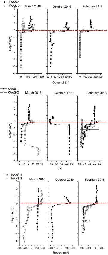
www.nature.com/scientificreports/
Figure 2. [Top]: Vertical microsensor profiles oxygen [μmol L−1] from KAAS-1 and KAAS-2 from March
and October 2016, as well as February 2018. [Middle]: Vertical microsensor profiles of pH from KAAS-1 and
KAAS-2 from March and October 2016, as well as February 2018. [Bottom]: Vertical microsensor profiles of
redox [mV] from KAAS-1 and KAAS-2 from March and October 2016, as well as February 2018.
changes occur within the uppermost photo-oxic zone of the mats. During March 2016 KAAS-1 mat is denoted
by a green uppermost layer which is dominated by cyanobacterial sequences (42.99%) with large abundances
of Proteobacteria (27.82%), and Bacteriodetes (14.92%), and minor amounts of Firmicutes (6.36%) within the
upper green layer. The composition of the following pink layer shows a significant decrease in cyanobacterial
sequences (24.89%), while Chloroflexi flourish to 32.7%, accompanied by a minor increase in Proteobacteria
(31.25%) and decline in Bacteroidetes (4.79%). In October 2016 the uppermost layer of KAAS-1 is dominated
Scientific Reports | (2021) 11:4170 | https://doi.org/10.1038/s41598-021-83676-1 4
Vol:.(1234567890)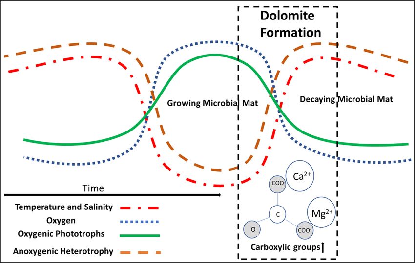
www.nature.com/scientificreports/

                                  Figure 3.  (A) Proto-dolomite phase from KAAS-1 during March of 2016 and associated EDS spectra. Area of
                                  interest indicated by cross. (B) Dolomite phase from KAAS-2 during March of 2016 and associated EDS spectra.
                                  Area of interest indicated by cross. (C) Proto-dolomite and dolomite phases from KAAS-1 during October of
                                  2016 and associated EDS spectra. Area of interest indicated by cross. (D) Dolomite phase from KAAS-2 during
                                  October of 2016 and associated EDS spectra. Dolomite only located in the lower layers of the mat at this time.
                                  Area of interest indicated by cross. (E) Dolomite phase from KAAS-1 during February of 2018 and associated
                                  EDS spectra. Area of interest indicated by cross. (F) Proto-dolomite phase from KAAS-1 during February of
                                  2018 and associated EDS spectra. Area of interest indicated by cross.

                                  by Proteobacteria (31.86%), Chloroflexi (26.38%), with almost equal amount of Bacteroidetes (13.55%), and
                                  Spirochaetes (12.57%). The following mixed red/pink layer shows similar composition with abundances of Pro-
                                  teobacteria (27.03%), Chloroflexi (25.32%), Bacteroidetes (19.79%), and Spirochaetes (10.41%). In February 2018
                                  KAAS-1 reverts to a comparable composition as seen in March of 2016. The top layer is mostly a green colora-
                                  tion and its microbial community is dominated by cyanobacterial sequences (26.49%). There are also abundant
                                  sequences of Chloroflexi (9.04%), Bacteroidetes (15.92%), Proteobacteria (32.44%), and Firmicutes (5.73%). The
                                  proceeding red layer denotes a decrease in Cyanobacteria sequences (10.01%), and Proteobacteria (18.69%), but
                                  an increase in Chloroflexi (22.78%) similar abundances of Bacteroidetes (14.10%) as the previous layer, and the
                                  appearance of Spirochaetes (20.65%). In all three seasons the microbial community of the lower most layers (red/
                                  black/grey) show compositions indicative of reduced conditions with an increased amount of Proteobacteria
                                  and Archaea with depth.
                                      KAAS-2 also undergoes the most pronounced changes in the upper photo-oxic zone of the mats. During
                                  March 2016 is denoted by a bright orange uppermost layer which has a microbial community consisting of
                                  Cyanobacteria (3.9%), Chloroflexi (13.51%), Bacteroidetes (15.81%), Spirochaetes (19.32%), and Proteobacteria
                                  (41.13%). Below the orange layer is an intermittent red layer consisting of Cyanobacteria (23.21%), Chloroflexi
                                  (25.39%), Bacteroidetes (18.62%), and Spirochaetes (26.55%). During October 2016 the community composi-
                                  tion drastically changes in KAAS-2. Within the uppermost “crust” there are high abundances of Proteobacteria
                                  (16.29%), Bacteroidetes (13.42%), Actinobacteria (29.28%), Frimicutes (14.18%), and Euryarchaeota (9.33%). This
                                  composition is similar throughout the entire profile. In February of 2018 KAAS-2 has a clear top layer directly
                                  beneath the precipitated salt crystals. The composition of this clear layer is dominated by Cyanobacteria (44.92%),
                                  Bacteroidetes (11.00%), Spriochaetes (25.86%), and Proteobacteria (12.29%). Underneath this is a layer with an
                                  intermixing of green, pink and orange. Within this layer the microbial community is composed of Cyanobacteria
                                  (13.19%), Bacteroidetes (56.77%), Spirochaetes (7.72%), and Proteobacteria (13.08%). In conjunction with this top
                                  layer is an intermittent red layer with similar composition, consisting of Cyanobacteria (18.58%), Bacteroidetes
                                  (50.68%), Spriochaetes (12.60%), and Proteobacteria (12.79%).
                                      In all of the sampling seasons the amount of proteins that comprise purifiable EPS are low in comparison to
                                  carbohydrates and uronic acids often falling near the detection limit of the employed Bradford assay (1ug/g for
                                  micro-assay)41. Uronic acid concentration is markedly higher during times of increased salinity, and a majority
                                  of all three fractions appear concentrated in the uppermost portion of the mat. Total concentrations of each

Scientific Reports |   (2021) 11:4170 |                https://doi.org/10.1038/s41598-021-83676-1                                                  5

                                                                                                                                              Vol.:(0123456789)
www.nature.com/scientificreports/
Figure 4. [Top]: Phylum level classification of 16 s rRNA ZOTU abundance (%) from KAAS-1 and KAAS-2
in March of 2016. [Middle]: Phylum level classification of 16 s rRNA ZOTU abundance (%) from KAAS-1 and
KAAS-2 in October of 2016. [Bottom]: Phylum level classification of 16 s rRNA ZOTU abundance (%) from
KAAS-1 and KAAS-2 in February of 2018.
KAAS-1 KAAS-2
KAAS-1 KAAS-2 KAAS-1 February February
March 2016 March 2016 October 2016 2018 2018
Average Average Average Average Average
Functional Group pKa Range LT (mMol/g EPS) LT (mMol/g EPS) LT (mMol/g EPS) LT (mMol/g EPS) LT (mMol/g EPS)
Carboxyl 3–5.8 0.113 ± 0.08 0.173 ± 0.04 0.461 ± 0.23 0.075 ± 0.03 0.091 ± 0.05
Phosphoryl 6–8 0.149 ± 0.01 0.103 ± 0.04 0.260 ± 0.32 0.044 ± 0.02 0.020 ± 0.01
Amine 8–9
0.845 ± 0.25 0.224 ± 0.03 0.721 ± 0.48 0.545 ± 0.01 0.223 ± 0.02
Amine (hydroxyl) 9–10
Table 1. Summary of Total functional group concentrations over their pKa ranges from the uppermost mat
layers during each sampling season. These measurements originate from EPS extracted from the green and
orange layers or top 0.5 cm of each mat given a particular sampling season.
functional group are summarized in (Table 1). The spectra analysis suggests that there are four assemblages
of functional groups based on pKa ranges carboxylic (3–5.8), phosphoryl (6–8), amine (8–9) and hydroxyl
(9–10)42–45 (Figure S4).
In March of 2016 EPS extracted from KAAS-1 contains 0.113 ± 0.08 mMol g −1 carboxylic functional groups,
0.149 ± 0.01 mMol g−1 phosphoryl functional groups, and 0.845 ± 0.25 mMol g −1 amine functional groups. EPS
Scientific Reports | (2021) 11:4170 | https://doi.org/10.1038/s41598-021-83676-1 6
Vol:.(1234567890)www.nature.com/scientificreports/
extracted from KAAS-1 during October of 2016 exhibit much more variability in composition and mark an
increase in carboxylic groups up to 0.461 ± 0.23 mMol g −1, in phosphoryl groups up to 0.260 ± 0.32 mMol g −1, and
a decrease to 0.721 ± 0.48 mMol g −1 in amine groups. In February of 2018 functional group density experiences
an overall decrease with concentrations of 0.075 ± 0.03 mMol g−1 for carboxylic groups, 0.044 ± 0.02 mMol g−1 for
phosphoryl groups, and 0.545 ± 0.01 mMol g −1 for amine groups. Similar to KAAS-1 the amount of extractable
and purifiable proteins from KAAS-2 during all sampling seasons is negligible with some values below the assay
detection limit. KAAS-2 shows increased concentrations of the uronic acid fraction during times of high salin-
ity, this trend we also observed in KAAS-1. EPS extracted from KAAS-2 in March of 2016 has concentrations
of 0.173 ± 0.04 mMol g −1 for carboxylic groups, 0.103 ± 0.04 mMol g −1 for phosphoryl groups, and 0.224 ± 0.03
mMol g−1 for amine groups. In October of 2016 there was no extractable EPS from the dried-out mat. During
February 2018 EPS sampled from KAAS-2 had functional group concentrations of 0.091 ± 0.05 mMol g−1 for
carboxylic groups, 0.020 ± 0.01 mMol g−1 for phosphoryl groups, and 0.223 ± 0.02 mMol g−1 for amine groups.
Quality control of titration data for KAAS-1 and KAAS-2 show good agreement between both measured and
modeled titrations (Figure S3).
Discussion
The Khor Al-Adaid sabkha undergoes distinct seasonal fluctuations as a result of the local c limate37. Air tem-
perature changes of 5–10 °C are the root cause of the differences between mats. There are a number of smaller
scale interlinked geographical factors that can influence the mat environment such as water recharge rate and
source, topography, and tidal range; however, the effect of these factors is probably limited. Instead, a more
appropriate frame of reference is the geomorphology of the sampling sites, or more specifically the intertidal
nature of the environment. The position in intertidal zonation strongly governs mat morphology, evaporative
effects, and water e xchange46,47. Our previous s tudy37 reveals that KAAS-1 falls within the lower intertidal zone,
while KAAS-2 is located within the mid to upper intertidal zone, subjecting KAAS-2 to more extreme changes
in environmental conditions.
Among all environmental factors, our observations show that salinity, which fluctuates drastically between
seasons, appears to be a major driver in low-t dolomite precipitation. Salinity influences geochemical parameters
by governing solution chemistry and consequently, saturation indices of the mineralogical phases48. Conditions
of supersaturation with respect to dolomite are, however, insufficient for dolomite formation, which is kineti-
cally inhibited at low t emperatures2. In light of the proposed role of microbial produced EPS in low-t dolomite
formation, it is critical to evaluate the role salinity fluctuations in the expression of the microbial community
and consequences for EPS characteristics.
Our observations in sabkha environment show that the mat microbial communities respond strongly to the
fluctuations in salinity, undergoing significant changes during lower and higher saline conditions. The changes
to microbial composition are most pronounced within SWI zone in the uppermost layers of both microbial mats.
We observed that during cool periods microbial mats at KAAS-1 are dominated by Cyanobacteria (oxygenic pho-
totrophs) within its uppermost layers, while microbial mats at KAAS-2 are dominated by Chloroflexi (anoxygenic
phototrophs). Interestingly, anoxygenic phototrophs occur where oxygen is still saturated. Under such conditions,
anoxygenic phototrophy is suppressed, which most probable cause the organisms to respire heterotrophically49.
During warm periods KAAS-1 experiences salinity resembling KAAS-2 during cool periods, which is reflected
by a comparable microbial community composition. Once the temperature becomes cooler again, the micro-
bial mat composition in KAAS-1 returns similar to that found in March, while KAAS-2 remains dominated by
heterotrophic organisms (Salinibacter, e.g., 50) reflecting an ecological succession or hysteresis effect. The cyclic
nature of the microbial community composition suggests a reproducible sequence of events that operate over
longer time periods, especially when considering the re-establishment of KAAS-2 after complete desiccation.
This hysteresis effect in microbial communities is accompanied by cyclic changes in EPS composition. The
concurrent molecular characterization of EPS and analysis of microbial compositions during three seasons reveal
that microbial mat dynamics can be closely linked to the functional groups of EPS, their densities as well as the
amount of EPS. We found that high salinity, and the shift from Cyanobacteria to Chloroflexi, or Salinibacter was
coexisting with a rise in the amount of extractable EPS, as well as an increase in the fraction carboxylic functional
groups, the proposed ligands for the formation of low-t d olomite6,13. Cumulatively, these results indicate that by
shifting a microbial community from oxygenic phototrophs to heterotrophs as a result of salinity creates EPS
favorable to dolomite nucleation.
The previous studies which focused either on microbial community analysis in the Arabian Peninsula, Pacific
atolls, Mexico, Spain, Washington (USA)51–56,58,61 or geochemical conditions in Spain and C uba57,59,60, docu-
mented similar environmental interactions between microbial communities succession and salinity in microbial
mats. This fact suggests that our observations in the Khor Al-Adiad sabkhas are not locally restricted. However,
the lack of multi-seasonal and interdisciplinary approach, as well as limited attention to mineral-microbe inter-
actions hinder the previous studies to draw the conclusions about the biogeochemical dynamics in microbial
composition, consequences for EPS quantity and quality and at long last on low-t dolomite formation.
The discovery of this truly biogeochemical interplay between bio- and geosphere in our field studies is even
more astonishing, since many investigators observed this particular pathway for low-t dolomite formation during
a transition in microbial mats driven by environmental factors, especially salinity57–60. For example, very recent
in the playa lakes, maximum appearance of dolomite was reported to occur concurrently with dominance of
anoxygenic phototrophs57,58. In another study, increased carbonate mineralization was documented as microbial
mats experienced a transition from oxic to anoxic conditions59,60. These types of dynamic fluctuations do not
appear to be unique to surficial examples of low-T dolomite forming environments. Indeed, literature examining
Scientific Reports | (2021) 11:4170 | https://doi.org/10.1038/s41598-021-83676-1 7
Vol.:(0123456789)www.nature.com/scientificreports/
the occurrence of hemipelagic or “deep-sea” dolomite describes low-T dolomite formation in environments rich
in organic material in the presence anaerobic heterotrophic organisms.
Hemipelagic or “deep-sea” low-t dolomite has been documented in a variety of locales, generally in areas of
upwelling across the globe, including, for example: the west coast of A frica32, the west coast of south America in
33,34 35,36
Peru , and cold-seeps in the Gulf of M exico . Due to the nature of these sites, samples on which these studies
are based consist of drill cores and microbial activity is mostly inferred through isotopic analysis of carbonates
and pore waters, such as δ13C, δ18O, carbonate-associated sulfate (CAS), or stable magnesium isotopes includ-
ing δ25Mg and δ26Mg. Instead, data based on direct measurements of the microbial community, which would
provide consistent and valuable information into the occurrences of low-T dolomite under these conditions,
remains limited. It has been observed that strong depletion in 13C values may suggest biogenic methane as a main
carbon source35. Similar 13C depletion results as well as δ13C and CAS ratios were also obtained from dolomite
bearing samples36. It was suggested that dolomite was formed in the shallow subseafloor, due to sulfate-driven
anaerobic methane oxidation (AOM), which is performed by a consortium of sulfate-reducing b acteria36,77. The
combination of these metabolism results in the depletion of sulfate and buildup of sulfide in a zone referred to as
the deep sulphate-methane transition zone (SMTZ). It was noted that the kinetic barrier being overcome under
these conditions was still unclear36. It is possible that the high turnover of organic material by AOM prokaryotes
results in a similar effect as the transition between oxygenic and anoxygenic organism observed in the sabkha
environment. Unfortunately, without a direct characterization of organic material from the seep environment,
this explanation remains hypothetical.
A closer examination of the process of AOM (1) shows the processes can shift equilibria toward carbonate
formation.
CH4 + SO2− − −
4 → HCO3 + HS + H2 O (1)
It has been suggested that the increase in alkalinity and HCO3 result in more CO32− to compensate for the
increased CO2 created32. Although this is a sound argument, how the precipitation process overcomes the kinetic
barriers preventing mineral formation remains unknown. Data from the sabkha study provides a possible expla-
nation as it sheds light on the biogeochemical interplay between a synchronized transition in the geochemical
conditions, microbial composition, EPS (or, more generally, organic material) and its role for low-t dolomite
formation. In the case of hemipelagic low-T dolomite, the presence of large abundances of additional abiotic
nucleation surfaces, such as c lays7, could further facilitate low-T dolomite precipitation. However, until now a
direct examination of naturally occurring organic material and its influence on low-t dolomite formation have
not been systematically investigated.
Referencing the sabkha microbial mats, a phase of dominance of Cyanobacteria can be described as a growing
microbial mat (GMM) in opposite to a period of time of decaying microbial mat (DMM) with high abundances
of anoxygenic phototrophs and heterotrophs [e.g.,57]. The cyclic transitions between these two phases of GMM
and DMM are linked to changes in salinity, a community shift to heterotrophic metabolisms, synchronized with
molecular changes in functional groups of EPS and, finally, with more crystalline morphologies of dolomite-
like phases. Within the sabkha’s microbial mats of our study, such solid phases evolve from proto-dolomite or
stacked aggregates to fully formed rhombs. It has been hypothesized that rising salinity or the transition from
oxic to anoxic conditions cause EPS degradation by heterotrophic activity resulting in the release of Ca and Mg
into solution and freeing of previously bound functional groups responsible for carbonates nucleation47–60,62 In
the case of hemipelagic low-t dolomite, this process is also characterized by an increase in alkalinity associated
with AOM. Additionally, it should be noted that, within the Khor-Al Adaid sabkhas, massive, continuous lay-
ers of low-T dolomite have not been observed but are present within the northern Dohat Faishakh sabkhas31
and sabkhas of Abu Dhabi18. These sites along with many other low-T dolomite forming environments10,11,28–35
show continuous layers of dolomite 10 s of centimeters thick formed under what can be considered similar
conditions. The sabkha environments in Abu Dhabi and northern Qatar [e.g.,18,31] are older, more extensive and
result in thicker profiles (≈40 cm) than the Khor Al-Adaid sabkha. Additionally, much of the dolomite found
in these environments is at lower depths (10–40 cm) and has been proposed to initially form at the surface as
proto-dolomite and disordered dolomite, the same as the Khor Al-Adaid mat, then progress and accumulate to
dolomite due to burial and diagenetic “ageing”. The observed lack of large continuous layers of low-T dolomite
in this study could be due to sample variability although is more likely due to discontinuous presence of active
microbial mats (for example the completely desiccated KAAS-2 mat during the October 2016 sampling), which
would result in less accumulation and decreasing burial rates. Despite the lack of continuous, thick layers of
low-T dolomite, the dolomite-like phases formed within these mats is a uthigenic37. Observations from the
sabkha highlight the mechanism of initial nucleation and indicate a more nuanced process of low-T dolomite
formation than previously proposed.
We observed that the high abundance of anoxygenic phototrophs or Bacteroidetes, Chloroflexi and Salini-
bacter, occur in zones saturated with oxygen. Under such conditions, anoxygenic phototrophy is suppressed,
which may cause Chloroflexi to respire h eterotrophically49, creating a comparable metabolic dynamic. Ca and
Mg and an augmented alkalinity can surely cause an additional increase in dolomite saturation index. But,
this is not sufficient for dolomite precipitation, which is kinetically inhibited29. The functional groups of EPS
may support overcoming the kinetic barriers that prevent Mg to be incorporated into the carbonate mineral at
low temperature. Once this kinetic barrier has been overcome the formation of proto-dolomite and dolomite
would proceed in the upper layers of the sabkha through an adsorption-displacement mechanism onto growing
Ca-Mg carbonate3,7 followed by a currently poorly defined “ageing” process to form ordered dolomite [e.g.,10].
The ageing process is a combination of diagenesis and time, which would proceed at lower depths in the mat
as it accumulates. Overall, we suggest that the presence of EPS with a specific composition, rather than cation
Scientific Reports | (2021) 11:4170 | https://doi.org/10.1038/s41598-021-83676-1 8
Vol:.(1234567890)www.nature.com/scientificreports/
Figure 5. Conceptual diagram of proposed multi-seasonal low-t dolomite formation. Shifting environmental
conditions cycle between higher and lower temperature and salinity. This results in the cycling of a growing and
decaying microbial mat with low-t dolomite formation facilitated during the transition between growing and
decaying conditions.
release during EPS degradation or alkalinity increases [e.g.,29] during AOM, is the key factor for the nucleation
of dolomite during environmental transitions, like the described SMTZ or GMM and DMM.
Our findings indicate that geochemical driven cycles of growth and decay within specific microbial com-
munities such as oxygenic and anoxygenic phototrophs or heterotrophs, promote low-t dolomite formation.
In other words, biogeochemical conditions coupled with a specific type of microbial community creates EPS
that has a higher and lower affinity for low-t dolomite formation (Fig. 5). This hypothesis is in agreement with
both sites for low-t dolomite precipitation, as well as current microbial models, which attribute low-t dolomite
precipitation to low oxygen concentrations and the activity of sulfate-reducing bacteria10,63–65. These models
suggest that low-t dolomite precipitation proceeds as the kinetic barriers caused by S O42− are removed. However,
experiments have shown that S O42− presents less of a problem than previously t hought66,67. Instead, we propose
that in environments with low oxygen and high organic matter turnover, heterotrophic microbes become more
pronounced and consume organic material creating substrates with compositions better suited to dolomite
precipitation. This is corroborated by recent laboratory studies that have suggested a similar “new-way” of pro-
ducing low-t dolomite in the presence of anoxygenic s trains26,68, which is also in agreement with our concept of
dolomite formation. Our hypothesis is soundly corroborated in modern environments and laboratory experi-
ments, but attempting to apply this model to paleo-studies regarding low-t dolomite can allow us to evaluate its
applicability over longer time scales.
From a geological perspective, a fundamental consequence of our concept is that the interplay between
production and consumption of EPS in conjunction with the GMM to DMM transition is an important factor
in low-T dolomite formation. Consonant with our hypothesis, are ancient dolomites that have been described
in several recent studies. For example, formation of massive Precambrian carbonate platforms is suggested
to be connected to the interaction between cyanobacteria and heterotrophic bacteria or anoxygenic bacteria
metabolizing heterotrophically (heterotrophic sulfate reduction) by production of H O32− caused by
2O and C
dissociation of ammonia and hydroxyl ions during buffering and may have also raised carbonate alkalinity69.
Additionally, a recently compiled dataset of ocean chemistry and carbonate strata from the late Archaean through
the Cambrian, shows similar links between the distribution of aerobic and anaerobic metabolisms and dolomite70.
This study reported higher abundances of dolomite at all ocean depths during the Archean, but a higher affinity
for dolomite in shallow sedimentary environments as time progressed. This occurred in conjunction with the
spread of aerobic organisms across the seafloor which may have restricted anaerobic microorganisms to shallow
areas70. Given that the change in ocean geochemistry and redox conditions, as well as microbial metabolism,
these secular changes may have resulted in large turnovers of organic material, a process analogous to the sea-
sonal fluctuations observed within the Khor-Al Adaid mats. Dolomite formation in the quaternary has also been
linked to changes in water and redox chemistry that led to elevated cell mortality and organic matter turnover29.
Thus, it seems that the interactions between microbial mats impacted by changing redox conditions in the
environment and consequent microbial community are imperative for dolomite formation through geological
Scientific Reports | (2021) 11:4170 | https://doi.org/10.1038/s41598-021-83676-1 9
Vol.:(0123456789)www.nature.com/scientificreports/
time. Specifically, our observations in the sabkhas as well as paleoenvironmental and laboratory studies have
indicated that occurrence anoxygenic heterotrophs may play a key role in low-T dolomite formation aside from
sulfate-reduction15,23,26,27,29,32–36,66–68.
More specific paleo-environmental also corroborate our findings and the compilation of existing data dis-
covers striking similarities between the modern environments and ancient low-t dolomite locations. The sedi-
mentological studies on ancient dolomite, namely cap dolostones approximately 635 Ma old, from the Puga
Formation in Brazil, confirm our concept even for dolomite formation over a much larger extent. A progressive
change in temperature and salinity has been isotopically identified in cap dolostones from the Puga Formation
in Brazil71. Within these dolostone caps a stromatolitic environment dominated by cyanobacteria existed and
caps may have been deposited as conditions transitioned from anoxic to suboxic as suggested by the observed
sedimentary features71. Another very recent study on the same post-glacial dolostone cap rocks discovered
enrichment of newly identified biomarker 25,28-bisnorgammacerane (25,28-BNG) which can be formed as a
result of a transition of microbial mat dominated by cyanobacteria to one dominated by heterotrophs72. Further
paleo-evidence for the proposed mechanism facilitating low-t dolomite formation comes from studies that
examined dolocretes were formed within the Hammersley Basin during the quaternary age (37 to > 45 ka) and
Late Miocene and Pliocene (5.3–3.5 ma)73,74. In both cases, dolocrete formation is suggested to be stimulated
by more saline evaporitic paleoenvironmental conditions, possibly playa lakes or saline mudflats, as evidenced
by δ18O values. Interestingly, the most recent study observed extensive fossilized EPS and organic filaments
in more shallow sections of the same c ores74 indicating a possible presence of microbial mats at the dolomite
forming locations, even though no information about microbial composition exists. The paleoenvironment in
which this occurred looks remarkably resembles the Khor Al-Adaid sabkha. Consequently, it is probable that the
higher saline conditions would have promoted similar dynamics in which a growing mat would have undergone
degradation stimulating dolomite precipitation.
Overall, our observations demonstrate that environmental fluctuations, in particular salinity, and an accom-
panying transition from oxygenic to anoxygenic heterotrophic dominance in microbial mats created an organic
substrate (EPS) particularly suited to facilitate low-t dolomite precipitation. Specifically, increased salinity leads
to a community dominated by anoxygenic phototrophs which degrade EPS produced by cyanobacteria. This
resulted in elevated concentrations of carboxylic functional groups, which are known to promote Mg dehydra-
tion and subsequent incorporation into carbonate minerals. Importantly, this study reveals the interplay between
oxygenic and anoxygenic phototrophs and the GMM to DMM is cyclical and may propagate over longer time
scales. Currently established models of dolomite precipitation, as well as several modern, and paleoenvironmental
studies purport findings that are in agreement with our interpretation of low-t dolomite precipitation and we
suggest that low-t dolomite may have decreased with time due to secular changes in the Earths’ atmosphere and
ocean chemistry.
Methods
Depth profiles of O2, pH, redox potential, and salinity. Depth profiles of geochemical characteris-
tics, including O2, pH and redox potential, were collected using needle-tip microsensors (Unisense, Denmark)
connected to a Unisense 4 channel multimeter. These profiles were measured in-situ between 11-2p.m (AST),
recorded water temperature was of 30-35ºC. Salinity of surface water at each site was measured using a Metrohm
914 pH/conductometer and conductivity probe with built in temperature sensor (Metrohm part #6.0917.080).
Measurements were then converted to salinity using a temperature-corrected calibration curve. Detailed infor-
mation on sensor configuration and calibration can be found in supplementary data.
Porewater analysis. Porewater was collected using Rhizons during March and October 2016 (Rhizosphere
research products, Netherlands) from pooled water above the mat, as well as using short cores with pre-drilled
and covered holes. Porewater from 2018 was collected using prefabricated diffusive gradient thin films or DGT
samplers (DGT Research Products, Lancashire, UK). Porewater samples were preserved for Inductively Couple
Plasma – Optical Emission Spectrometry (ICP-OES) in 5 ml centrifuge tubes with 5% HNO3 from Rhizons,
while DGT samples were sealed and both stored at 4 °C until analysis. Detailed information on preparation of
pore-water and analysis using ICP-OES can be found in the supplementary data.
Scanning electron microscopy (SEM), energy‑dispersive X‑ray spectroscopy (EDS), and X‑ray
diffraction (XRD) analysis. Morphological examination, elemental composition of samples was per-
formed through a combination of scanning electron microscopy (SEM), and energy-dispersive X-ray spectros-
copy (EDS) respectively. The mineralogical characterization was performed by X-ray diffraction (XRD). Details
on the specific machines and sample preparation and analysis can be found in the supplementary data.
16s rRNA amplicon library preparation and data analysis. Samples of up to 10 g were isolated
from short cores for metagenomic analysis following the same procedure from37. Cores were sliced under an N 2
atmosphere using aseptic techniques and preserved via freezing immediately at − 80 °C. Samples were stored at
− 80 °C until DNA extraction was performed using a MoBiopower biofilm DNA isolation kit as per the manu-
facturer’s instructions. DNA isolates were stored at − 20 °C until sequenced. 16S rRNA amplicon sequencing tar-
geted the V4 variable region using the combined bacterial and archaeal primer set of 341F (CCTACGGGNGGC
WGCAG) and 785R (GACTACHVGGGTATCTAATCC). Further details on amplicon library preparation and
bioinformatic analysis can be found in the supplementary data.
Scientific Reports | (2021) 11:4170 | https://doi.org/10.1038/s41598-021-83676-1 10
Vol:.(1234567890)www.nature.com/scientificreports/
Extraction and characterization of exopolymeric substances (EPS) from microbial mats. EPS
composition throughout each sampling season was evaluated from the uppermost, middle, and bottom layers
of each mat (Fig. S2). EPS were extracted and characterized following modified protocols outlined in75. Details
on extraction procedure and colorimetric analysis of fractions as well as quality control can be found in the sup-
plementary data.
Potentiometric titrations. Potentiometric titrations were performed to determine acidity constants (Pka)
and concentration of functional groups in EPS from microbial mats. All potentiometric titrations were per-
formed on a 905 Titrando auto-titration system (Metrohm) on dynamic endpoint titration (DET) mode in gas-
tight-vessel (Metrohm1-50 ml with thermostat jacket). A mass of 20–80 mg of purified EPS were used for each
titration with 5 ml of 0.01 M KCl as an electrolyte solution and 0.1 M HCl and 0.01 M NaOH as titrants. Each
sample was degassed for 1 h under N 2 gas prior to titration and was analyzed in triplicate. Following the titration
the data was expressed in the form of charge excess which is calculated using the first half following equation42,76:
+
−
n Kai LTi
Cbj − Caj + H j − OH j = +S
i=1 Kai + [H + ]j
bj are the concentrations of acid and base at each j addition of titrant [ H+]j and [ OH−]j are the
where Caj and C
proton concentrations at each j addition of titrant.
The second half of the equation represents the surface binding sites based on charge excess and are deter-
mined as a sum of the number (n) of monoprotic ligands (LTi) with resulting in their total concentration along
with acidity constants, as well as the constant (S) acid neutralization capacity of the EPS surface. The functional
group determination by this method was performed using a linear programming method (LPM)42,76 over a pH
range of 4–10 at 0.2 unit intervals. Prior to LPM charge excess obtained from titrations was plotted to determine
agreement between forward and reverse runs as well as sample replicates.
Data availability
All datasets generated or analyzed will be made available by request in a timely manner to any qualified
researcher. 16S rRNA sequences were submitted for individual samples to the NCBI SRA database and will be
available under accession numbers PRJNA489272 and PRJNA634701 upon publication.
Received: 2 October 2020; Accepted: 1 February 2021
References
1. Graf, D. L. & Goldsmith, J. R. Some hydrothermal syntheses of dolomite and protodolomite. J. Geol. 64, 173–186 (1956).
2. Land, L. S. Failure to precipitate dolomite at 25 C from dilute solution despite 1000-fold oversaturation after 32 years. Aquat.
Geochem. 4, 361–368 (1998).
3. Petrash, D. A. et al. Microbially catalyzed dolomite formation: from near-surface to burial. Earth Sci. Rev. 171, 558–582 (2017).
4. Baker, P. A. & Kastner, M. Constraints on the formation of sedimentary dolomite. Science 213, 214–216 (1981).
5. Land, L. S. The origin of massive dolomite. J. Geol. Educ. 33, 112–125 (1985).
6. Roberts, J. A. et al. Surface chemistry allows for abiotic precipitation of dolomite at low temperature. Proc. Natl. Acad. Sci. 110,
14540–14545 (2013).
7. Liu, D. et al. Experimental evidence for abiotic formation of low-temperature proto-dolomite facilitated by clay minerals. Geochim.
Cosmochim. Acta 247, 83–95 (2019).
8. Brady, P. V., Krumhansl, J. L. & Papenguth, H. W. Surface complexation clues to dolomite growth. Geochim. Cosmochim. Acta 60,
727–731 (1996).
9. Bosak, T. & Newman, D. K. Microbial nucleation of calcium carbonate in the Precambrian. Geology 31, 577–580 (2003).
10. Vasconcelos, C. & McKenzie, J. A. Microbial mediation of modern dolomite precipitation and diagenesis under anoxic conditions
(Lagoa Vermelha, Rio de Janeiro, Brazil). J. Sediment. Res. 67, 378–390 (1997).
11. Vasconcelos, C., McKenzie, J. A., Bernasconi, S., Grujic, D. & Tiens, A. J. Microbial mediation as a possible mechanism for natural
dolomite formation at low temperatures. Nature 377, 220 (1995).
12. Sánchez-Román, M. et al. Aerobic microbial dolomite at the nanometer scale: Implications for the geologic record. Geology 36,
879–882 (2008).
13. Kenward, P. A. et al. Ordered low-temperature dolomite mediated by carboxyl-group density of microbial cell walls. Aapg Bull.
97, 2113–2125 (2013).
14. More, T., Yadav, J. S. S., Yan, S., Tyagi, R. D. & Surampalli, R. Y. Extracellular polymeric substances of bacteria and their potential
environmental applications. J. Environ. Manage. 144, 1–25 (2014).
15. Moreira, N. F., Walter, L. M., Vasconcelos, C., McKenzie, J. A. & McCall, P. J. Role of sulfide oxidation in dolomitization: sediment
and pore-water geochemistry of a modern hypersaline lagoon system. Geology 32, 701–704 (2004).
16. Vasconcelos, C. et al. Lithifying microbial mats in Lagoa Vermelha, Brazil: modern Precambrian relics?. Sed. Geol. 185, 175–183
(2006).
17. Bontognali, T. R. et al. Microbes produce nanobacteria-like structures, avoiding cell entombment. Geology 36, 663–666 (2008).
18. Bontognali, T. R. et al. Dolomite formation within microbial mats in the coastal sabkha of Abu Dhabi (United Arab Emirates).
Sedimentology 57, 824–844 (2010).
19. Bontognali, T. R., McKenzie, J. A., Warthmann, R. J. & Vasconcelos, C. Microbially influenced formation of Mg-calcite and Ca-
dolomite in the presence of exopolymeric substances produced by sulphate-reducing bacteria. Terra Nova 26, 72–77 (2014).
20. Krause, S. et al. Microbial nucleation of Mg-rich dolomite in exopolymeric substances under anoxic modern seawater salinity:
new insight into an old enigma. Geology 40, 587–590 (2012).
21. Zhang, F., Xu, H., Konishi, H., Shelobolina, E. S. & Roden, E. E. Polysaccharide-catalyzed nucleation and growth of disordered
dolomite: a potential precursor of sedimentary dolomite. Am. Miner. 97, 556–567 (2012).
22. Kenward, P., Goldstein, R., Gonzalez, L. & Roberts, J. Precipitation of low-temperature dolomite from an anaerobic microbial
consortium: the role of methanogenic Archaea. Geobiology 7, 556–565 (2009).
Scientific Reports | (2021) 11:4170 | https://doi.org/10.1038/s41598-021-83676-1 11
Vol.:(0123456789)www.nature.com/scientificreports/
23. Qiu, X., Wang, H., Yao, Y. & Duan, Y. High salinity facilitates dolomite precipitation mediated by Haloferax volcanii DS52. Earth
Planet. Sci. Lett. 472, 197–205 (2017).
24. Qiu, X., Yao, Y., Wang, H., Shen, A. & Zhang, J. Halophilic archaea mediate the formation of proto-dolomite in solutions with
various sulfate concentrations and salinities. Front. Microbiol. 10, 480 (2019).
25. Huang, Y.-R. et al. Aerobically incubated bacterial biomass-promoted formation of disordered dolomite and implication for
dolomite formation. Chem. Geol. 523, 19–30 (2019).
26. Al Disi, Z. A. et al. Characterization of the extracellular polymeric substances (EPS) of Virgibacillus strains capable of mediating
the formation of high Mg-calcite and protodolomite. Mar. Chem. 216, 103693 (2019).
27. Al Disi, Z. A. et al. Influence of temperature, salinity and M g2+:Ca2+ ratio on microbially-mediated formation of Mg-rich carbon-
ates by Virgibacillus strains isolated from a sabkha environment. Sci. Rep. 9, 1–12 (2019).
28. Wright, D. T. The role of sulphate-reducing bacteria and cyanobacteria in dolomite formation in distal ephemeral lakes of the
Coorong region, South Australia. Sediment. Geol. 126, 147–157 (1999).
29. McCormack, J., Bontognali, T. R., Immenhauser, A. & Kwiecien, O. Controls on cyclic formation of Quaternary early diagenetic
dolomite. Geophys. Res. Lett. 45, 3625–3634 (2018).
30. Illing, L.V., Wells, A.J., and Taylor, J.C.M. Penecontemporaneous dolomite in the Persian Gulf (1965).
31. Brauchli, M. et al. The importance of microbial mats for dolomite formation in the Dohat Faishakh sabkha, Qatar. Carbonates
Evaporites 31, 339–345 (2016).
32. Moore, T., Murray, R., Kurtz, A. & Schrag, D. Anaerobic methane oxidation and the formation of dolomite. Earth Planet. Sci. Lett.
229, 141–154 (2004).
33. Meister, P. et al. Dolomite formation in the dynamic deep biosphere: results from the Peru Margin. Sedimentology 54, 1007–1032
(2007).
34. Mavromatis, V., Meister, P. & Oelkers, E. H. Using stable Mg isotopes to distinguish dolomite formation mechanisms: a case study
from the Peru Margin. Chem. Geol. 385, 84–91 (2014).
35. Roberts, H. H., Feng, D. & Joye, S. B. Cold-seep carbonates of the middle and lower continental slope, northern Gulf of Mexico.
Deep Sea Res. II 57, 2040–2054 (2010).
36. Tong, H. et al. Environments favoring dolomite formation at cold seeps: a case study from the Gulf of Mexico. Chem. Geol. 518,
9–18 (2019).
37. DiLoreto, Z. A. et al. Microbial community composition and dolomite formation in the hypersaline microbial mats of the Khor
Al-Adaid sabkhas. Qatar. Extremophiles 23, 1–18 (2019).
38. Paulo, C. & Dittrich, M. 2D Raman spectroscopy study of dolomite and cyanobacterial extracellular polymeric substances from
Khor Al-Adaid sabkha (Qatar). J. Raman Spectrosc. 44, 1563–1569 (2013).
39. Kaczmarek, S. E. et al. Dolomite, very-high magnesium calcite, and microbes: implications for the microbial model of dolomitiza-
tion. SEPM Spec. Publ. 109, 1–14 (2017).
40. Gregg, J. M., Bish, D. L., Kaczmarek, S. E. & Machel, H. G. Mineralogy, nucleation and growth of dolomite in the laboratory and
sedimentary environment: a review. Sedimentology 62, 1749–1769 (2015).
41. Bradford, M. M. A rapid and sensitive method for the quantitation of microgram quantities of protein utilizing the principle of
protein-dye binding. Anal. Biochem. 72, 248–254 (1976).
42. Cox, J. S., Smith, D. S., Warren, L. A. & Ferris, F. G. Characterizing heterogeneous bacterial surface functional groups using discrete
affinity spectra for proton binding. Environ. Sci. Technol. 33, 4514–4521 (1999).
43. Aiken, G., McKnight, D., Wershaw, R. & MacCarthy, P. Humic substances in soil, sediment, and water. 1985. Soil Sci. 142, 323
(1986).
44. Martell, A. E. & Smith, R. M. Critical stability constants Vol. 1 (Springer, New York, 1974).
45. Mozes, N. Microbial cell surface analysis. (VCH Publishers, 1991).
46. Strohmenger, C. J. et al. The sabkha sequence at Mussafah Channel (Abu Dhabi, United Arab Emirates): facies stacking patterns,
microbial-mediated dolomite and evaporite overprint. GeoArabia 15, 49–90 (2010).
47. Strohmenger, C. J. et al. Facies stacking patterns in a modern arid environment: a case study of the Abu Dhabi sabkha in the vicinity
of Al‐Qanatir Island, United Arab Emirates. Quaternary carbonate and evaporite sedimentary facies and their ancient analogues:
a tribute to Douglas James Shearman, 149–182 (2011).
48. Stumm, W. & Morgan, J. J. Aquatic chemistry: chemical equilibria and rates in natural waters Vol. 126 (Wiley, Hoboken, 2012).
49. Pierson, B. K. & Castenholz, R. W. A phototrophic gliding filamentous bacterium of hot springs, Chloroflexus aurantiacus, gen.
and sp. nov. Arch. Microbiol. 100, 5–24 (1974).
50. Oren, A. Salinibacter: an extremely halophilic bacterium with archaeal properties. FEMS Microbiol. Lett. 342, 1–9 (2013).
51. Al-Thani, R. et al. Community structure and activity of a highly dynamic and nutrient-limited hypersaline microbial mat in Um
Alhool Sabkha, Qatar. PLoS ONE 9, e92405 (2014).
52. Perri, E. et al. Carbonate and silicate biomineralization in a hypersaline microbial mat (Mesaieed sabkha, Qatar): roles of bacteria,
extracellular polymeric substances and viruses. Sedimentology 65, 1213–1245 (2018).
53. Abed, R. M., Kohls, K. & De Beer, D. Effect of salinity changes on the bacterial diversity, photosynthesis and oxygen consumption
of cyanobacterial mats from an intertidal flat of the Arabian Gulf. Environ. Microbiol. 9, 1384–1392 (2007).
54. Vogt, J. C., Abed, R. M., Albach, D. C. & Palinska, K. A. Bacterial and archaeal diversity in hypersaline cyanobacterial mats along
a transect in the intertidal flats of the Sultanate of Oman. Microb. Ecol. 75, 331–347 (2018).
55. Schneider, D., Arp, G., Reimer, A., Reitner, J. & Daniel, R. Phylogenetic analysis of a microbialite-forming microbial mat from a
hypersaline lake of the Kiritimati Atoll, Central Pacific. PLoS ONE 8, e66662 (2013).
56. Harris, J. K. et al. Phylogenetic stratigraphy in the Guerrero Negro hypersaline microbial mat. ISME J. 7, 50 (2013).
57. Cabestrero, Ó., Sanz-Montero, M., Arregui, L., Serrano, S. & Visscher, P. Seasonal variability of mineral formation in microbial
mats subjected to drying and wetting cycles in alkaline and hypersaline sedimentary environments. Aquat. Geochem. 24, 79–105
(2018).
58. Cabestrero, Ó. & Sanz-Montero, M. E. Brine evolution in two inland evaporative environments: influence of microbial mats in
mineral precipitation. J. Paleolimnol. 59, 139–157 (2018).
59. Bouton, A. et al. External controls on the distribution, fabrics and mineralization of modern microbial mats in a coastal hypersaline
lagoon, Cayo Coco (Cuba). Sedimentology 63, 972–1016 (2016).
60. Pace, A. et al. Formation of stromatolite lamina at the interface of oxygenic–anoxygenic photosynthesis. Geobiology 16, 378–398
(2018).
61. Lindemann, S. R. et al. The epsomitic phototrophic microbial mat of Hot Lake, Washington: community structural responses to
seasonal cycling. Front. Microbiol. 4, 323 (2013).
62. Dupraz, C. et al. Processes of carbonate precipitation in modern microbial mats. Earth Sci. Rev. 96, 141–162 (2009).
63. Burns, S. J., Mckenzie, J. A. & Vasconcelos, C. Dolomite formation and biogeochemical cycles in the Phanerozoic. Sedimentology
47, 49–61 (2000).
64. Gandin, A. & Wright, D. Evidence of vanished evaporites in Neoarchaean carbonates of South Africa. Geol. Soc. Lond. Spec. Publ.
285, 285–308 (2007).
65. Nutman, A. P., Friend, C. R., Bennett, V. C., Wright, D. & Norman, M. D. ≥ 3700 Ma pre-metamorphic dolomite formed by micro-
bial mediation in the Isua supracrustal belt (W. Greenland): simple evidence for early life?. Precambr. Res. 183, 725–737 (2010).
Scientific Reports | (2021) 11:4170 | https://doi.org/10.1038/s41598-021-83676-1 12
Vol:.(1234567890)You can also read