Molecular and Cellular Mechanisms Driving Cardiovascular Disease in Hutchinson-Gilford Progeria Syndrome: Lessons Learned from Animal Models - MDPI
←
→
Page content transcription
If your browser does not render page correctly, please read the page content below
cells
Review
Molecular and Cellular Mechanisms Driving Cardiovascular
Disease in Hutchinson-Gilford Progeria Syndrome: Lessons
Learned from Animal Models
Ignacio Benedicto 1, *, Beatriz Dorado 1,2 and Vicente Andrés 1,2, *

                                          1   Centro Nacional de Investigaciones Cardiovasculares (CNIC), 28029 Madrid, Spain;
                                              beatrizjulia.dorado@cnic.es
                                          2   CIBER en Enfermedades Cardiovasculares (CIBER-CV), Spain
                                          *   Correspondence: ibenedicto@cnic.es (I.B.); vandres@cnic.es (V.A.)

                                          Abstract: Hutchinson-Gilford progeria syndrome (HGPS) is a rare genetic disease that recapitulates
                                          many symptoms of physiological aging and precipitates death. Patients develop severe vascular
                                          alterations, mainly massive vascular smooth muscle cell loss, vessel stiffening, calcification, fibrosis,
                                          and generalized atherosclerosis, as well as electrical, structural, and functional anomalies in the heart.
                                          As a result, most HGPS patients die of myocardial infarction, heart failure, or stroke typically during
                                          the first or second decade of life. No cure exists for HGPS, and therefore it is of the utmost importance
                                          to define the mechanisms that control disease progression in order to develop new treatments to
                                          improve the life quality of patients and extend their lifespan. Since the discovery of the HGPS-causing
                                          mutation, several animal models have been generated to study multiple aspects of the syndrome
                                and to analyze the contribution of different cell types to the acquisition of the HGPS-associated

                                          cardiovascular phenotype. This review discusses current knowledge about cardiovascular features
Citation: Benedicto, I.; Dorado, B.;
                                          in HGPS patients and animal models and the molecular and cellular mechanisms through which
Andrés, V. Molecular and Cellular
                                          progerin causes cardiovascular disease.
Mechanisms Driving Cardiovascular
Disease in Hutchinson-Gilford
                                          Keywords: Hutchinson-Gilford progeria syndrome (HGPS); progerin; cardiovascular disease; atheroscle-
Progeria Syndrome: Lessons Learned
from Animal Models. Cells 2021, 10,
                                          rosis; endothelial dysfunction; vascular smooth muscle cell; vascular calcification; extracellular matrix;
1157. https://doi.org/10.3390/            fibrosis; vessel stiffening; progeroid animal models
cells10051157

Academic Editor: Christoph Englert
                                          1. Hutchinson-Gilford Progeria Syndrome (HGPS)
Received: 1 April 2021                          HGPS (also termed progeria) is a rare genetic disorder (estimated prevalence of 1 in
Accepted: 7 May 2021
                                          18–20 million people) caused by a mutation in the LMNA gene that provokes accelerated
Published: 11 May 2021
                                          aging and premature death [1–3]. In mammals, LMNA normally encodes two major alterna-
                                          tively spliced A-type lamin variants, lamin A and lamin C, which are main components of
Publisher’s Note: MDPI stays neutral
                                          the nuclear lamina. Post-translational modifications of prelamin A that yield mature lamin
with regard to jurisdictional claims in
                                          A include C-terminal farnesylation and carboxymethylation and subsequent cleavage of
published maps and institutional affil-
                                          the modified C-terminus by the zinc metalloprotease ZMPSTE24. “Classical” progeria
iations.
                                          is caused by a heterozygous de novo c.1824C>T (p.G608G) point mutation in the LMNA
                                          gene. This creates an aberrant splice site in exon 11 that deletes 150 nucleotides, resulting
                                          in the synthesis of a truncated prelamin A variant called progerin that lacks the cleavage
                                          site needed to remove the farnesylated and carboxymethylated C-terminus [4]. Progerin
Copyright: © 2021 by the authors.         is expressed in most differentiated cells and induces multiple cellular and physiological
Licensee MDPI, Basel, Switzerland.
                                          anomalies, including defects in nuclear morphology, chromatin disorganization, elevated
This article is an open access article
                                          DNA damage, impaired stem cell maintenance and differentiation, metabolic alterations,
distributed under the terms and
                                          autophagy deregulation, and systemic inflammation [5].
conditions of the Creative Commons
                                                HGPS patients appear normal at birth, but start to develop a premature and accelerated
Attribution (CC BY) license (https://
                                          aging phenotype between 1 and 2 years of age. Typical features include failure to thrive,
creativecommons.org/licenses/by/
                                          alopecia, loss of subcutaneous fat, skin alterations, tooth and bone abnormalities, muscle
4.0/).

Cells 2021, 10, 1157. https://doi.org/10.3390/cells10051157                                                    https://www.mdpi.com/journal/cells
Cells 2021, 10, 1157 2 of 20
weakness, and progressive joint contractures. However, the most clinically relevant feature
of HGPS is the development of generalized atherosclerosis and cardiac dysfunction, which
ultimately lead to death mainly due to myocardial infarction, heart failure, or stroke at
an average age of 14.6 years [4,6]. The following section describes the HGPS-associated
cardiovascular phenotype in detail.
2. The Cardiovascular Phenotype in HGPS Patients: A Historical Perspective
Cardiovascular disease (CVD) is the main cause of death in HPGS, and it is therefore
crucial to decipher the underlying mechanisms in order to design effective therapies.
Because HGPS patients typically lack many of the classical cardiovascular risk factors such
as hypercholesterolemia, high C-reactive protein, obesity, and smoking, the study of the
mechanisms underlying CVD in HGPS also presents an opportunity to understand CVD
in non-HGPS individuals in relative isolation from confounding risk factors. A robust
characterization of the HGPS cardiovascular phenotype is also critical for developing
clinical standards of care and objective cardiovascular readouts of therapeutic efficacy in
HGPS clinical trials.
The extreme rarity of HGPS hinders systematic cardiovascular examination of patients,
because the very few HGPS patients are spread around the world, may be diagnosed at
different ages, and have very different access to primary and specialized care. However,
information gathered over the years from individual HGPS case reports, autopsies, and a
few clinical trials has helped to define a fairly consistent cardiovascular phenotype. The
invaluable HGPS literature prior to the discovery of the genetic cause of the disease (from
1886 to 2003) included patients diagnosed by their characteristic physical appearance,
including the so called “old-mannish” look, with a disproportionally big head, baldness,
prominent eyes, thin lips, pointed nose, wrinkled and thin skin, and extreme thinness [7,8].
This allowed the differentiation of HGPS patients from patients with other progeroid-
like diseases. Since 2003, HGPS clinical reports include only individuals with confirmed
genetic tests, which have identified the classical LMNA c.1824C>T mutation in most pa-
tients (https://www.progeriaresearch.org/wp-content/uploads/2021/04/FINAL1-PRF-
By-the-Numbers_-March-2021.pdf, accessed on March 2021). These recent reports support
the previously described HGPS phenotype and have broadened our understanding of
HGPS cardiovascular pathology through the use of modern clinical techniques for as-
sessing vasculopathy, such as the measurement of vessel wall echodensity, ankle-brachial
index, and pulse wave velocity (PWV). Here, we present a chronological overview of the
cardiovascular characterization of HGPS patients that gathers together information from
the landmark literature on the clinical definition of HGPS pathology.
2.1. Atherosclerosis and Cardiovascular Calcification
The first thorough anatomical description of a child with remarkable signs of prema-
ture aging was reported in 1886 by J. Hutchinson [9]. A decade later, H. Gilford found that,
with no family relationship, Hutchinson’s patient shared a striking physical resemblance
to one of his own patients, who looked like an old man, had heart problems, and died at
the age of 17 [10]. In the detailed autopsy of this second patient, Gilford noted extensive
atheromatous degeneration of the mitral and aortic orifices, obliteration of the lumen
of the coronary arteries, soft atheromatous patches on the convex surface of the aortic
arch, patches of calcareous material on the concave surface, and extensive atheroma with
collections of organized fibrin in the aortic branches [10].
In 1972, F. L. DeBusk reported four new cases of HGPS and compiled the first thorough
list of progeria-like cases from the worldwide literature. In one of his patients, he found
enlargement of the right ventricle and a calcified heart valve, and in another he found
obstruction of the proximal left coronary artery [7]. Examination of autopsies revealed
that most patients with typical HGPS die of congestive heart failure or acute myocardial
infarction due to primary underlying coronary atherosclerosis, and that all patients had
varying degrees of generalized atherosclerosis, mainly affecting the large arteries [7].Cells 2021, 10, 1157 3 of 20
In a later review of 12 autopsies, P. B. Baker and colleagues found that cardiac com-
plications in progeria patients were rarely due to cardiomyopathy (interstitial fibrosis in
the myocardium without severe coronary artery disease); rather, most cardiac manifes-
tations were due to atherosclerosis-related coronary artery narrowing or occlusion [11].
Atherosclerosis and calcification of aortic and mitral valves were also common, and all
cases featured atherosclerotic plaques in the aorta, ranging from small fatty streaks to
complicated calcified lesions. In addition, Baker et al. reported the case of a patient with
significant thickening of the intima and media in small cardiac intramural arteries accom-
panied by diffuse interstitial fibrosis, suggesting small artery disease as a possible cause of
cardiac ischemia [11].
2.2. Vascular Smooth Muscle Cell (VSMC) Loss and Vascular Fibrosis
In 2001, W. E. Stehbens and colleagues reported an unusual histological finding upon
reviewing two new cases of HGPS patients who had died of myocardial infarction [12].
Both patients had severe depletion of VSMCs in the aortic media. The VSMCs were replaced
by collagen fibrils, and their depletion correlated with the presence of atherosclerosis and
hemodynamic stress around branch sites. The author concluded that the VSMC debris
in the fibrosed medial layer indicated muscle degeneration rather than muscle atrophy,
and that increased arterial fibrosis may reduce the viscoelastic properties of the wall and
contribute to increased blood pressure.
A further analysis in 2006 by R. C. M. Hennekam reported 10 new cases of HGPS and
reviewed 132 cases from the literature [8]. The report covered the range from individual
phenotypic features to common symptoms shared by all patients. None of the patients
showed signs of a cardiovascular phenotype until around 6 to 8 years of age, when it
began to manifest as shortness of breath with exertion and easy fatigability. Other common
cardiovascular features included an increase in heart rate and blood pressure with age,
heart enlargement and impaired coronary function attributable to an enlarged left ventricle,
hypertrophy of myocardial cells and cardiac interstitial fibrosis, angina pectoris appearing
up to five years before death, extensive VSMC loss and thickening of the coronary arteries
with atheroma plaques (with and without calcification), and thickening of the aortic leaflets,
which were often calcified. In addition, half the patients had mitral valve defects, and
the aorta had a very variable appearance, from almost normal to severely atherosclerotic.
Vascular problems in these patients often extended to the brain, with silent or symptomatic
strokes and cerebral infarction frequently reported.
The first prospective clinical characterization of HGPS patients was reported in 2008
by M. A. Merideth et al. [13]. These authors followed 15 HGPS patients aged between 1
and 17 years over 16 months. Most of the patients had elevated platelet counts, a prolonged
prothrombin time, and elevated serum phosphorus. Another frequent feature was the
development of age-dependent impaired vascular function, including elevated blood
pressure, increased arterial augmentation rate, and reduced ankle-brachial index, together
with adventitial thickening. Less frequent abnormalities were thickened aortic valves with
regurgitation, left ventricular hypertrophy, pulmonary hypertension, stenotic lesions in the
cerebral and carotid arteries, and electrocardiographic alterations that were not associated
with ischemia [13].
In 2010, M. Olive et al. published the first structural and immunohistological compari-
son of cardiovascular tissues between HGPS and non-HGPS individuals, examining two
HGPS patients who died of myocardial infarction and a small non-HGPS cohort composed
of 29 individuals with or without CVD with ages ranging from 1 month to 97 years [14].
Atherosclerosis in HGPS patients and in normally aged individuals shared several features,
such as severe stenosis, extensive arterial calcification, and the presence of a range of early-
to late-stage atherosclerotic lesions displaying calcification, inflammation, and evidence of
erosion or rupture. However, HGPS lesions tended to have smaller atheromatous cores,
very large regions of calcification, and thick fibrosis and were categorized predominantly as
fibro-calcific lesions and less frequently as fibro-atheromas. Moreover, compared with geri-Cells 2021, 10, 1157 4 of 20
atric vessels, all HGPS vessels (arteries and veins) showed prominent adventitial fibrosis.
This study also revealed that progerin is expressed not only in the vessels of HGPS patients,
but also in a small subset of cells in the coronary arteries of non-HGPS individuals, with
progerin levels increasing with age [14]. Additional studies have confirmed low progerin
expression in several human cell types and tissues [15–28], but its functional relevance
remains to be determined.
2.3. Vascular Stiffening: A Useful Readout for Clinical Trials
Researchers have also investigated whether end-stage cardiovascular events in HGPS
are related to progressive impairment of vascular compliance. In a prospective single-
center study, M. Gerhard-Herman et al. enrolled 26 HGPS patients and 62 age- and
gender-matched healthy children [29]. All the HGPS patients exhibited vascular stiffness,
evidenced by markedly elevated carotid-femoral PWV values comparable to those in adults
older than 60 years. This finding was later corroborated in another study [30]. Other HGPS
patient characteristics included higher carotid echobrightness in the intima-media, near the
adventitia and deep adventitia, indicating increased arterial wall density and suggesting
accumulation of thick collagen fibrils, as well as elevated internal carotid artery mean flow
velocity and abnormal ankle-brachial indices, indicating occlusive stenosis and peripheral
vascular disease, respectively [29]. HGPS was therefore classified as a disease of vascular
stiffening in the setting of gradual vascular stenosis, similar to CVD in normal aging.
Subsequently, PWV and arterial wall echodensity became key cardiovascular readouts of
therapeutic efficacy in HGPS clinical trials [31–33]. The first HPGS clinical trial showed im-
proved vascular stiffness upon treatment with the farnesyltransferase inhibitor lonafarnib,
from pre-therapy PWV values typical of people aged 60–69 years to end-of-therapy PWV
values corresponding to 40–49-year-old individuals [31]. Moreover, lonafarnib treatment
reduced the echobrightness of the carotid intima-media and near/deep adventitia. These
findings thus suggested that lonafarnib therapy had the potential to reduce fatal and not-
fatal HGPS cardiovascular events and strokes. A second clinical trial showed that triple
therapy with lonafarnib plus the statin pravastatin and the bisphosphonate zoledronic
acid produced benefits in bone mineral density but did not improve vascular stiffness or
structure more than lonafarnib alone [32]. Instead, the triple therapy resulted in increased
development of carotid and femoral arterial plaques and extraskeletal calcifications, thus
discouraging recommendation of its use for the clinical treatment of HGPS [32]. The
results from both clinical trials indicated that single lonafarnib therapy can significantly
lower mortality rate [33], and in light of these studies, in November 2020 lonafarnib (mar-
keted as Zokinvy) became the first U.S. Food and Drug Administration-approved drug for
HGPS [34].
2.4. Cardiac Dysfunction
Clinical evaluation of potential treatments for HGPS also requires the definition of
appropriate cardiac endpoints. Several electrocardiographic defects have been reported in
HGPS patients [13,30,35], and one defect type frequently found as patients age is cardiac
repolarization anomalies [35,36]. Regarding cardiac anatomy and function, Prakash et al.
recently reported that the most frequent echocardiographic abnormality showing increased
prevalence with age in HGPS patients is left ventricular (LV) diastolic dysfunction [30]. The
authors speculated that LV dysfunction might be a consequence of myocardial interstitial fi-
brosis and endocardial thickening, which was observed in two previous case reports [8,14].
Other cardiac alterations, such as LV hypertrophy and aortic valve calcification and dys-
function, were less common and more frequently seen during the second decade of life [30].
In line with previous observations in the carotid artery [29], 70% of HGPS patients had
unusually high echobrightness in the aortic root wall, and these patients were more likely
to present with diastolic LV dysfunction [30]. Interestingly, increased echobrightness was
not associated with age, suggesting that extracellular matrix (ECM) remodeling may be an
early alteration potentially underlying the development of the cardiac phenotype. AnotherCells 2021, 10, 1157 5 of 20
recent study [37] applied three-dimensional echocardiography (speckled tracking imaging)
to analyze seven HGPS patients (five children ≤ 8 years of age and two children in their
teens), 21 aged-matched healthy children, and 14 older healthy volunteers (mean age:
65.7 ± 7.5). Despite the fact that most HGPS patients were in their first decade of life, the
authors found one case of diastolic dysfunction and another of aortic valve calcification,
severe aortic stenosis, and LV hypertrophy, in agreement with some of the findings reported
by Prakash et al. [30].
The data accumulated over several decades indicates that the HGPS cardiovascular
phenotype is defined by generalized atherosclerosis with a wide spectrum of early- to
late-stage atherosclerotic plaques, prominent VSMC loss, vascular stiffening, calcification
and fibrosis, cardiac repolarization abnormalities, LV diastolic dysfunction, and cardiac
valve disease, all of which probably contribute to the death of HGPS patients from my-
ocardial infarction, stroke, or heart failure. However, many fundamental questions remain
unanswered, and the natural history of CVD leading to these death-causing pathologies is
ill defined. Moreover, it remains to be determined whether LV diastolic dysfunction and
the altered electrocardiographic activity commonly observed in HGPS patients are early
cardiovascular complications or are secondary to other chronic pathologies that precede
their onset, such as long-term vascular stiffening and calcification, fibrosis, and ischemia.
It is therefore essential to investigate the natural history of HGPS-associated CVD and
its underlying mechanisms in order to provide clinicians with the scientific support they
need to fight disease progression earlier and more efficiently. The discovery of the genetic
mutation causing classical HGPS in 2003 [2,3] triggered the generation of a number of
HGPS experimental models that play a pivotal role in the investigation of progerin-induced
alterations and the testing of new therapeutic approaches. In the next section we describe
the major HGPS-like animal models, with a special emphasis on those that recapitulate
aspects of HGPS-associated CVD.
3. The Cardiovascular Phenotype in Animal Models of HGPS
The use of animal models in HGPS research is particularly important given the
low number of HGPS patients worldwide, which makes it very challenging to perform
longitudinal studies and clinical trials and reduces the availability of human samples for
ex vivo analysis. Over the past two decades, several HGPS animal models have been
developed to study different aspects of the disease (Table 1). In 2002, two laboratories
developed the Zmpste24−/− mouse model, which lacks the metalloprotease ZMPSTE24
responsible for cleaving the farnesylated and carboxymethylated C-terminus of prelamin A.
Homozygous Zmpste24−/− mice accumulate irreversibly modified prelamin A, lack mature
lamin A, and develop a bona fide progeroid phenotype, including muscle and bone defects,
alopecia, lipodystrophy, cardiac alterations, and reduced lifespan [35,38,39]. However, with
the exception of perivascular fibrosis in coronary arteries, no major vascular alterations
have been reported in Zmpste24−/− mice. This situation thus left a need for mouse models
that more accurately resemble HGPS pathology. The first progerin-expressing HGPS
murine model was the LmnaHG mouse, generated in 2005 [40,41]. Heterozygous LmnaHG/+
mice express progerin, lamin A, and lamin C, whereas homozygous LmnaHG/HG mice
only express progerin. Both LmnaHG/+ and LmnaHG/HG mice develop HGPS-like disease,
including slow growth, bone abnormalities, fat loss, and premature death. However, these
mice have not been reported to develop vascular abnormalities and therefore are not
suitable for the study of HGPS-associated vascular pathology, the most clinically relevant
feature of the disease.Cells 2021, 10, 1157 6 of 20
Table 1. Cardiovascular phenotype of HGPS animal models.
Vascular Features Cardiac Features
Altered Vessel Altered Vessel
Adventitial Medial Coronary/Aortic
Progerin VSMC Vascular Contraction Relaxation Aorta/Carotid Interstitial Perivascular Electrical Diastolic
Species Animal Model Thickening/ Collagen Atherosclerosis Root References
Expression Loss Stiffening (Phenyle- (Acetyl- Calcification Fibrosis Fibrosis Anomalies Dysfunction
Fibrosis Accumulation Calcification
phrine) choline)
Zmpste24−/− N NR NR NR NR NR NR NR NR NR Yqo /N 1 Y Y N [35,38,39]
G608G BAC Y Y Y NR NR NR NR Yht NR NR NR NR NR [42–44]
LmnaG609G Y Y Y N Y Y Y Yht NR NR NR Y Y/N 2 [45–57]
Apoe−/− Ubiquitous
Y Y NR Y NR NR NR NR N Y Y Y* NR [53,58]
LmnaG609G/G609G
Ldlr−/− Y Y Yqo Y NR NR NR NR NR NR Yqo NR NR [59]
LmnaG609G/G609G
Mus
musculus LmnaLCS/LCS Yqo Yqo NR NR Y Y N NR NR NR NR NR NR [50,51,53]
SM22αCre
VSMCs,
Apoe−/− cardiomyocytes
LmnaLCS/LCS Y Y NR Y NR NR NR NR Y Y Y N* NR [53]
SM22αCre
LmnaLCS/LCS NR NR NR NR N N N NR NR NR NR NR NR [50,51]
Tie2Cre ECs, some
leukocytes
Lmnaf/f ;TC Yqo Yqo Yqo Yqo NR NR Y NR NR Yqo Yqo NR NR [57]
Prog-Tg ECs N Y NR NR NR NR N N NR Y Y NR Y [60]
LMNA c.1824C>T
Sus scrofa Yucatan Y Y Y NR NR NR NR NR NR Y Y Y Y [61]
minipig
LMNA Ubiquitous
Macaca c.1824C>T
NR Y NR NR NR NR NR NR NR NR NR NR NR [62]
fascicularis cynomolgus
monkey
Y, yes; N, no; NR, not reported; ht , reported only in heterozygous mice; qo , qualitative observation (no quantification). 1 , Yqo in [39], N in [35,38]. 2 , Y in [52] (n = 12–16 mice), N in [45] (n = 4 mice). *, results
obtained in 16-week-old mice; however, 26-week-old Apoe−/− LmnaLCS/LCS SM22αCre mice present modest but significant cardiac electrical anomalies [53].Cells 2021, 10, 1157 7 of 20
The G608G BAC mouse model, developed in 2006, expresses human lamin A, lamin
C, and progerin together with endogenous mouse lamin A and lamin C and was the
first HGPS animal model to recapitulate some aspects of the HGPS-associated vascular
phenotype [42]. Heterozygous G608G BAC animals have a normal lifespan and do not
present consistent abnormalities in bone, muscle, skin, or heart [42], whereas homozygous
mice show musculoskeletal and skin anomalies, lack subcutaneous fat, and die prema-
turely [43,63]. However, both heterozygous and homozygous G608G BAC mice develop a
marked vascular phenotype in the aorta, carotid artery, and iliac artery, including progres-
sive loss of VSMCs, thickening of the adventitia and medial layer, and vessel calcification
in older animals. Histological analysis and transmission electron microscopy studies also
revealed medial accumulation of proteoglycans and disorganized collagen fibrils, together
with alterations to elastic fibers. These structural abnormalities are accompanied by de-
fective arterial responses to the VSMC-dependent vasodilator sodium nitroprusside [42].
G608G BAC mice therefore constitute a very valuable tool for studying several features of
the HGPS-associated vascular phenotype and testing new therapeutic approaches. Indeed,
work in homozygous G608G BAC mice recently demonstrated that treatment with LMNA-
targeted antisense oligonucleotides reduces progerin levels and increases longevity [44,64],
and this was accompanied by amelioration of VSMC depletion and adventitial thickening
in one of the studies [44]. In another study, partial correction of the HGPS-associated muta-
tion LMNA c.1824C>T by in vivo systemic base editing in homozygous G608G BAC mice
led to the total prevention of VSMC loss and adventitial thickening and a striking 2.4-fold
increase in lifespan [43]. Potential limitations of the G608G BAC model are the unknown
effects of overexpressing human progerin in the context of endogenous mouse lamin A
and lamin C expression, and the presence of the human genes UBQLN4, MAPBPIP, and
RAB25 in the LMNA-containing bacterial artificial chromosome used to generate G608G
BAC mice [42].
The LmnaG609G HGPS mouse model, generated in 2011 by the López-Otín laboratory,
was the first mouse model to express progerin due to aberrant splicing of the endogenous
Lmna gene, resulting from the introduction of the 1827C>T (p.G609G) mutation, which
is equivalent to the human mutation found in HGPS patients [45]. Using this model,
these authors provided the first in vivo evidence that antisense morpholino-based ther-
apy to prevent the pathogenic Lmna splicing might be viable for HGPS. Homozygous
LmnaG609G/G609G mice express progerin, lamin C, and residual levels of lamin A and show
many HGPS features, including failure to thrive, bone defects, loss of fat deposits, brady-
cardia, prolonged QRS waves (indicating altered heart ventricular depolarization), and
premature death [45,49,52]. LmnaG609G/G609G cardiomyocytes have structural, conduction,
and excitation-contraction coupling defects, all of which can be partially corrected by
chronic treatment with the microtubule-stabilizing drug paclitaxel [49]. Vascular alter-
ations include VSMC depletion in the medial layer of the aorta and other arteries, collagen
and proteoglycan accumulation in the aortic media, reduced elastin fiber undulations, and
increased vessel stiffness assessed by wire and pressure myography [45,51,55,56,65]. Aortic
VSMC loss and adventitial fibrosis were also features of a separate LmnaG609G/G609G mouse
model developed independently in 2016 by the Fong laboratory [54]. Compared with
age-matched wild-type animals, the aortic and carotid medial layers of LmnaG609G/G609G
mice show marked accumulation of the collagen-crosslinking enzyme lysyl oxidase (LOX),
which probably contributes to progerin-induced vessel stiffening [55]. Heterozygous
LmnaG609G/+ mice also die prematurely, but their average lifespan (34 weeks) is consider-
ably longer than that of homozygous LmnaG609G/G609G mice (15 weeks) [45]. The longer
lifespan of the heterozygotes allows the study of vascular alterations that take longer to
manifest, such as vessel calcification, which is prominent in the aortic arch and thoracic
aorta of ≈30-week-old LmnaG609G/+ mice [46–48].
Although the G608G BAC and LmnaG609G mouse models recapitulate many of the
vascular alterations found in HGPS patients, they do not develop atherosclerosis, one
of the main features in HGPS patients. This is most likely due to very low levels ofCells 2021, 10, 1157 8 of 20
pro-atherogenic lipoproteins in mice, which confers natural resistance to developing
atherosclerosis [66]. To overcome this limitation, the Apoe−/− LmnaG609G/G609G mouse model
was generated [53]. These mice harbor the progerin-producing Lmna mutation and are
engineered to lack apolipoprotein E, a genetic manipulation that has been extensively
demonstrated to induce atherosclerosis in rodents [66]. Similar to LmnaG609G/G609G mice,
Apoe−/− LmnaG609G/G609G mice show postnatal growth defects, VSMC loss in the aortic
arch, cardiac electrical anomalies, and reduced lifespan compared to Apoe−/− Lmna+/+
controls; however, Apoe−/− LmnaG609G/G609G mice also show adventitial thickening, accu-
mulate fluorescently-labeled human low-density lipoproteins (LDLs) in the medial layer of
atheroma-free aortic regions, and develop aggravated aortic atherosclerosis without evi-
dencing higher serum cholesterol or LDL than Apoe−/− Lmna+/+ mice. Apoe−/− LmnaG609G/G609G
mice also showed signs of plaque vulnerability in the aortic root, accompanied by fibrosis
and inflammation in the adjacent cardiac tissue, and some showed evidence of myocardial
infarction at necropsy [53]. The Apoe−/− LmnaG609G/G609G mouse is thus the first HGPS
animal model to allow the study of HGPS-associated atherosclerosis.
The same laboratory subsequently developed another atheroprone HGPS model, the
Ldlr−/− LmnaG609G/G609G mouse [59], which harbors the progerin-causing Lmna mutation
and ablation of the LDL receptor gene (Ldlr), another well-established strategy for inducing
atherosclerosis in mice [66]. The Ldlr−/− LmnaG609G/G609G mouse phenotype is very similar to
that of Apoe−/− LmnaG609G/G609G mice, but with less pronounced adventitial thickening and
less fibrosis and inflammation in the tissue neighboring the aortic root. The similar vascular
phenotypes of these models indicates that the observed alterations are due to the effects of
progerin expression, and not to the specific deletion of Apoe or Ldlr. A potential limitation
of both the Apoe−/− LmnaG609G/G609G and Ldlr−/− LmnaG609G/G609G mouse models is that the
animals develop severe hypercholesterolemia and hypertriglyceridemia, conditions that
are generally absent in HGPS patients [13,67], who also show reduced HDL cholesterol
levels with age [67]. It is therefore possible that the altered lipid profile might to some
extent modulate the vascular anomalies induced by progerin expression.
Studies using G608G BAC mice and the LmnaG609G/G609G model and its atheroprone
variants have made a significant contribution to the assessment of the HGPS-associated
vascular phenotype. The most suitable model for analysis will depend on the vascular
alteration of interest. For example, G608G BAC and homozygous LmnaG609G/G609G mice
can be used to decipher the mechanisms of VSMC loss, medial collagen accumulation,
increased arterial stiffness, and other HGPS-related vascular anomalies in the absence of
hyperlipidemia. In contrast, heterozygous G608G BAC and LmnaG609G/+ mice are useful
tools for analyzing progerin-induced vessel calcification, whereas Apoe−/− LmnaG609G/G609G
and Ldlr−/− LmnaG609G/G609G mice are suitable models for the study of HGPS-
associated atherosclerosis.
Research with mouse models has significantly advanced knowledge about HGPS-
associated pathology, including CVD, and has paved the way to the development of
therapies with the potential to ameliorate or even cure HGPS [43,44,64,68–70]. However,
therapies that are effective in HGPS-like mouse models have yielded only modest benefit
in HGPS patients [31–33]. In this context, decision-making about scheduling candidate
treatments for clinical trials [71] would be greatly facilitated by prior validation in large
animal models more closely resembling human physiology. Large animal models also
allow the use of the same devices employed to study cardiovascular alterations in human
patients, which can help standardize surgical models or medical protocols before moving
into clinical practice. The first large animal model of HGPS was generated in 2019 in
Yucatan minipigs. The modified pigs bear a heterozygous c.1824C>T mutation in the
LMNA gene and show severe growth retardation, lipodystrophy, skin and bone alterations,
and premature death [61]. Importantly, they also show cardiovascular alterations seen
in HGPS patients, including LV diastolic dysfunction, altered cardiac electrical activity,
and VSMC depletion. Moreover, the hearts of HGPS-like minipigs develop interstitial
fibrosis and defective myocardial perfusion, together with degenerated coronary arteriesCells 2021, 10, 1157 9 of 20
and increased collagen content in the medial and adventitial layers of cardiac arterioles.
The human-like size of the HGPS minipig model allowed continuous in vivo monitoring
of cardiac electrical activity with subcutaneously implanted loop recorders, which revealed
severe pre-mortem cardiac conduction abnormalities in some animals. These included
advanced third-degree atrio-ventricular block at the moment of death and complete atrio-
ventricular block preceded either by short-duration polymorphic ventricular tachycardia or
by ST-segment elevation, which was not present 29 h before death, suggesting a coronary
ischemic event potentially related to microvascular dysfunction and vascular fibrosis. In
2020, the same c.1824C>T mutation was introduced into the LMNA gene of cynomolgus
monkeys to generate the first non-human primate HGPS model [62]. Although lifespan
studies were not reported, progeroid monkeys recapitulated typical HGPS phenotypes,
including growth retardation, bone and joint alterations, skin problems, alopecia, and loss
of subcutaneous fat. HGPS-like monkeys also show an increase in vascular wall collagen
deposition and signs of intimal hyperplasia in the aorta, although these alterations were
only reported in monkeys homozygous for the LMNA mutation.
Over almost two decades, the generation of multiple HGPS animal models has enabled
a thorough characterization of the HGPS-associated cardiovascular phenotype and the
discovery of mechanisms that mediate vascular damage. In the next section, we examine
progerin-induced alterations in VSMCs and endothelial cells (ECs), major vessel-wall
constituents that play key roles in the development and complications of atherosclerotic
CVD. We also discuss the potential role of progerin expression in these cell types in the
acquisition of HGPS-associated cardiovascular phenotype. Multiple in vitro studies have
made important contributions to the characterization of progerin-induced alterations in
VSMCs and ECs [72–82]; however, in the interest of conciseness, here we focus mainly on
in vivo and ex vivo data obtained from HGPS animal models.
4. Role of VSMCs and ECs in HGPS-Associated Cardiovascular Dysfunction
Progerin expression has been detected in VSMCs and ECs in HGPS patients [14,16].
VSMCs are a key structural and functional element of the vessel wall, indispensable for
maintaining vascular tone and regulating blood pressure thanks to their contractile proper-
ties [83,84]. In pathological scenarios such as atherosclerosis, VSMCs progressively acquire
a defective contractile phenotype and undergo transdifferentiation into a wide range of
different cell types, including macrophage-like cells, adipocyte-like cells, cells with an
osteochondrogenic phenotype, and ECM-producing myofibroblast-like cells. Endothelial
dysfunction is a major determinant of the initiation and progression of atherosclerosis [85].
Increased endothelial permeability facilitates subendothelial lipid deposition and leukocyte
extravasation into the intima, which drives atherosclerotic plaque formation [86]. Moreover,
there is strong evidence that an abnormally stiff ECM perturbs intercellular endothelial
junctions [87] and can promote paracellular leukocyte transmigration [88,89]. Interestingly,
reducing arterial ECM stiffness with the LOX inhibitor β-aminopropionitrile (BAPN) atten-
uates atherosclerosis in Apoe knockout mice [90]. ECM stiffening in HGPS could therefore
contribute to atherosclerosis development by directly altering endothelial phenotype and
function. Because the individual functions of VSMCs and ECs are dependent on proper
communication between these cell types [91,92], slight perturbations to this signaling
circuit can generate a feedback loop with detrimental effects on vessel structure and func-
tion. Understanding progerin-induced structural, functional, and molecular alterations
to the vascular ECM, VSMCs, and ECs and their interactions is therefore crucial for the
development of new vascular-targeted therapies for HGPS.
4.1. VSMCs in HGPS Mouse Models
HGPS patients and animal models both show prominent VSMC depletion, which
may be involved in the development of life-threatening cardiovascular complications
associated with the disease. To date, the most widely used experimental system for the
study of progerin-induced VSMC alterations in vivo is the LmnaG609G model. HomozygousCells 2021, 10, 1157 10 of 20
LmnaG609G/G609G mice show no obvious aortic structural alterations at 8 weeks of age [55,65],
but VSMC depletion is evident in the aortic arch at 12–14 weeks [45,51] and also in the
thoracic aorta at 17 weeks [65]. These observations reinforce the idea that HGPS-associated
VSMC loss is progressive and that the aortic arch may be especially sensitive to progerin-
induced vascular damage, due to environmental factors such as turbulent blood flow and
other mechanical cues characteristic of this aortic segment [93]. Consistent with this idea,
VSMC depletion in the aortic arch of LmnaG609G/G609G mice is more severe on the inner than
the outer curvature [65], correlating directly with exposure to more pronounced blood flow
disturbance. The thoracic aorta of ≈14-week-old LmnaG609G/G609G mice shows no evidence
of VSMC loss but does show reduced numbers of smooth muscle fibers (measured by
hematoxylin staining) and increased deposition of medial collagen [51]. Moreover, vessel
contraction in response to phenylephrine and potassium chloride is impaired in thoracic
aorta segments from these progeroid mice [50,56], and this effect is not rescued by collagen
disruption after collagenase treatment [50]. In line with this evidence of progressive dam-
age, dysfunction, and dedifferentiation of VSMCs in LmnaG609G/G609G mice, atheroprone
Apoe−/− LmnaG609G/G609G mice show VSMC depletion in the aortic arch at 16 weeks of age
but not at 8 weeks [53]. An RNA sequencing analysis to identify potential mechanisms
causing VSMC loss explored gene expression differences between aortas from 8-week-old
Apoe−/− LmnaG609G/G609G mice and age-matched control Apoe−/− Lmna+/+ animals [58]. To
enrich for VSMC-expressed genes, the adventitia was removed before RNA extraction.
The most significant differences between control and progerin-expressing aortas were in
genes related to fibrosis, oxidative stress, and, interestingly, the endoplasmic reticulum
(ER) stress response and the ER stress-related unfolded protein response. Treatment of
Apoe−/− LmnaG609G/G609G mice with tauroursodeoxycholic acid (TUDCA), a chemical chap-
erone that inhibits ER stress, significantly reduced medial VSMC loss and atherosclerosis
burden [58].
Additional genetic and pharmacological strategies have proved successful in reducing
or preventing VSMC loss in HGPS-like mice. These strategies include deletion of the
matrix metalloprotease 13 (Mmp13) gene, the use of the matrix metalloprotease inhibitor
batimastat [94], and the blockade of interleukin-6 signaling with the neutralizing antibody
tocilizumab [95] in LmnaG609G/G609G mice, suggesting that HGPS-associated VSMC deple-
tion involves ECM remodeling and inflammation. VSMC loss in LmnaG609G/G609G mice was
also prevented by inhibition of N-acetyltransferase 10 with remodelin, and this treatment
also ameliorated aortic adventitial thickening and extended lifespan [96]. In G608G BAC
mice, VSMC depletion was reduced by treatment with the farnesyltransferase inhibitor
tipifarnib [97] and by lowering progerin levels with LMNA-targeting antisense oligonu-
cleotides [44]. Another effective means of ameliorating VSMC loss in LmnaG609G/G609G mice
is the inhibition of progerin carboxymethylation by inactivating the isoprenylcysteine car-
boxylmethyltransferase (Icmt) gene, a strategy that also increased lifespan in heterozygous
LmnaG609G/+ mice [98]. These findings were complemented by in vitro experiments show-
ing that drug-based ICMT inhibition delayed senescence and stimulated proliferation of
late-passage HGPS cells and Zmpste24-deficient mouse fibroblasts but had no effect on the
proliferation of wild-type human cells or Zmpste24-deficient mouse cells lacking Icmt [98].
A number of therapeutic strategies have also shown beneficial effects on other HGPS-
associated vascular phenotypes related to VSMC dysfunction. In LmnaG609G/G609G mice,
increased vascular stiffness and defective vascular tone were partially prevented by dietary
supplementation with sodium nitrite [50,51], and vessel calcification was reduced after
pyrophosphate treatment [48]. Interestingly, LOX inhibition with BAPN prevented carotid
stiffening and diastolic dysfunction in 8-week-old LmnaG609G/G609G mice [55], strongly sug-
gesting that ECM remodeling plays a key role in the acquisition of the HGPS-associated
cardiovascular phenotype. Moreover, aortic calcification in heterozygous LmnaG609G/+ mice
was reduced by ATP-based therapy or dietary supplementation with magnesium, and this
outcome was accompanied by a concomitant increase in lifespan [46,47].Cells 2021, 10, 1157 11 of 20
VSMC depletion in HGPS patients and animal models is well established; however,
it is only recently that attempts have been made to assess the causal role of progerin-
expressing VSMCs in the acquisition of HGPS-associated vascular phenotype. In one
approach, SM22αCre transgenic mice (with Cre activity in embryonic VSMCs and cardiomy-
ocytes) [99] were crossed with LmnaLCS/LCS mice (harboring the 1827C>T mutation in the en-
dogenous Lmna gene after an intronic transcriptional stop signal flanked by LoxP sites) [45];
the resulting LmnaLCS/LCS SM22αCre mice express progerin predominantly in VSMCs and
cardiomyocytes [50,51,53]. Like LmnaG609G/G609G mice with ubiquitous progerin expres-
sion, LmnaLCS/LCS SM22αCre mice have aortic VSMC loss, increased stiffness, and impaired
contraction [50,51,53]. In the context of Apoe deletion, Apoe−/− LmnaLCS/LCS SM22αCre mice
develop similar vascular alterations to those found in Apoe−/− LmnaG609G/G609G mice, includ-
ing VSMC depletion, adventitial thickening, and increased atherosclerosis burden [53], all of
which are also prevented by treatment with the ER stress-targeting chaperone TUDCA [58].
These results strongly suggest that VSMC-intrinsic alterations play a central role in the
development of the HGPS vascular phenotype. However, it is worth noting that unlike 14–
16-week-old LmnaG609G/G609G mice, SM22αCre-driven progerin expression in age-matched
animals was not sufficient to impair acetylcholine-induced vasorelaxation [50] (see below)
or induce cardiac electric alterations in the context of Apoe deletion [53]. Therefore, it is
likely that full development of the HGPS-associated cardiovascular phenotype requires
progerin expression in other cell type(s), in addition to VSMCs and cardiomyocytes. More-
over, although LmnaLCS/LCS SM22αCre and Apoe−/− LmnaLCS/LCS SM22αCre mice both show
VSMC depletion and adventitial thickening, the former have normal lifespan [53]. In line
with the normal lifespan of VSMC-depleted non-atherogenic HGPS-like mice, heterozy-
gous G608G BAC mice show aortic VSMC loss but have a normal life expectancy [42].
These observations indicate that, at least in mice, progerin-induced VSMC loss, increased
vascular stiffness, and reduced vascular contractility in the absence of a pro-atherogenic
environment are not sufficient to shorten lifespan. Moreover, when hyperlipidemia is not
present (as is the case with HGPS patients), the development of life-threatening cardiovas-
cular complications may require progerin expression in other cell type(s) in addition to
VSMCs. Further studies are needed to fully define the direct and indirect roles of VSMCs
in the development of the HGPS vascular phenotype and to determine whether target-
ing progerin expression or downstream effectors specifically in VSMCs can prevent the
development of HGPS-associated vascular alterations.
The cytoskeleton and nuclear lamina are connected through the linker of nucleoskele-
ton and cytoskeleton (LINC) complex, which mediates physical force transmission between
these compartments. Progerin-expressing cells show altered expression and diffusional
mobility of LINC nuclear membrane proteins [100,101], and SM22αCre-driven disruption
of the LINC complex in LmnaG609G/G609G mice ameliorates aortic VSMC loss and adventitial
fibrosis [65]. In addition, progerin expression in cultured mouse VSMCs increases cell
death in response to mechanical stress in a LINC complex-dependent manner [65]. In line
with these findings, in vitro biomechanical assays have shown that progerin expression in-
duces nuclear stiffening and defective nucleocytoskeletal force transmission [77,82,102,103].
Notably, mechanical strain decreases the viability of progerin-expressing primary skin
fibroblasts and increases pro-inflammatory gene expression in progerin-expressing iPSC-
derived VSMCs, both generated from HGPS patients [104,105]. These results strongly sug-
gest a direct link between progerin-induced anomalies in LINC-mediated VSMC nuclear
mechanosensing and the development of HGPS-related vascular alterations. Nevertheless,
it has not been reported whether VSMC-specific disruption of the LINC complex increases
the lifespan of LmnaG609G/G609G mice [65], and further studies are therefore needed to assess
the efficacy of VSMC targeting to extend survival in HGPS animal models.
4.2. ECs in HGPS Mouse Models
In contrast to the progerin-induced alterations in the medial and adventitial layers—
enriched in VSMCs and fibroblasts, respectively—the aortic luminal endothelium of HGPSCells 2021, 10, 1157 12 of 20
mice appears to be histologically intact, even in areas with severe VSMC depletion [42].
Moreover, a cohort of 14 HGPS patients showed normal flow-mediated brachial artery dila-
tion, suggesting preserved endothelial reactivity [13]. However, even subtle perturbations
in ECs can induce endothelial dysfunction, considered central to the onset and develop-
ment of atherosclerosis [85]. LmnaG609G/G609G mice show defective endothelium-dependent
aortic dilation in response to acetylcholine, a clear sign of endothelial dysfunction [50,57],
prompting interest in progerin-induced endothelial alterations and their potential role in
the development of the HGPS-associated cardiovascular phenotype. Recent single-cell
RNAseq studies in native, non-cultured mouse lung ECs revealed significant enrichment in
transcriptional changes related to the inflammatory response in cells from LmnaG609G/G609G
mice (ubiquitous progerin expression) [57]. Further studies are needed to identify and
assess the role of progerin-induced transcriptional alterations in ECs from tissues more
closely related to HGPS pathology, such as heart and aorta.
Some recent studies have investigated the effects of endothelium-specific progerin
expression on the development of HGPS-associated features. Mice carrying human lamin A
minigenes with the HGPS 1824C>T mutation were crossed with transgenic mice expressing
a tetracycline-responsive transcriptional activator under the control of the EC-specific Cdh5
promoter, generating Prog-Tg mice with EC-specific expression of human progerin and
lamin A together with endogenous mouse lamin A and lamin C [60]. These mice have low
body weight, develop cardiac hypertrophy and diastolic dysfunction, and die prematurely
at a median age of ≈25 weeks. Prog-Tg mice also develop myocardial interstitial and
perivascular fibrosis, which was suggested to result from depressed cardiac levels of
endothelial nitric oxide synthase (eNOS). Although aortas from Prog-Tg mice show no
significant VSMC loss or defective acetylcholine-mediated vasorelaxation, they do display
adventitial thickening and impaired EC alignment with blood flow. In line with these
findings, iPSC-derived ECs from HGPS patients have lower expression of flow-responsive
genes than control cells [75]. Moreover, progerin-expressing ECs from Prog-Tg mice show
increased levels of SUN1 and SUN2, members of the LINC complex, and mislocalization
of emerin, a nuclear membrane protein involved in nuclear mechanoresponse [60]. The
analysis of Prog-Tg mice thus demonstrated a causal role of EC-expressed progerin in
the development of some HGPS-associated cardiovascular alterations, suggesting that
defective endothelial mechanosensing may be one of the underlying mechanisms. A
possible limitation of the Prog-Tg model is that ectopic progerin and lamin A are expressed
at ≈4 times the level of endogenous lamin A, which could affect the interpretation of the
observed alterations.
Another system developed to test the effects of EC-specific progerin expression is the
LmnaLCS/LCS Tie2Cre model [50,51]. These animals were generated by crossing LmnaLCS/LCS
mice [45] with mice expressing Cre recombinase driven by the Tek (a.k.a. Tie2) pro-
moter [106]. ECs in the progeny LmnaLCS/LCS Tie2Cre mice express mouse progerin driven
by the endogenous Lmna promoter. Unlike mice with ubiquitous and SM22αCre-driven
progerin expression, LmnaLCS/LCS Tie2Cre mice show no increase in vascular stiffness [51].
Moreover, consistent with the Prog-Tg model, LmnaLCS/LCS Tie2Cre mice lack significant
alterations in acetylcholine-mediated vasorelaxation [50]. These studies were therefore
unable to demonstrate any effect of EC-specific progerin expression; however, the sacrifice
of all animals at 13–15 weeks prevented analysis of survival or any vascular features that
may take longer to appear.
A similar Tie2-Cre-based strategy was adopted to generate the Lmnaf/f ;TC model of
EC-specific progerin expression [57]. Lmnaf/f ;TC mice share many features with the Prog-
Tg model, including modest but significant body weight reduction, cardiac hypertrophy,
fibrosis and functional alterations, signs of aortic adventitial thickening and reduced eNOS
expression, and premature death at a median age of ≈25 weeks. However, unlike Prog-
Tg and LmnaLCS/LCS Tie2Cre mice, Lmnaf/f ;TC mice also show significantly impaired aortic
vasorelaxation in response to acetylcholine, suggesting that progerin expression in ECs may
be sufficient to induce endothelial dysfunction. This observation is in line with the impairedCells 2021, 10, 1157 13 of 20
vasorelaxation of tissue-engineered blood vessels generated with induced pluripotent stem
cell-derived ECs from HGPS donors [75]. The contrasting lack of endothelium-mediated
vasodilation defects in LmnaLCS/LCS Tie2Cre mice [50] could be due to the different ages at
which these assays were performed (≈17-week-old Lmnaf/f ;TC mice versus ≈14-week-old
LmnaLCS/LCS Tie2Cre mice). Strikingly, aortic atheromatous plaques were found in 100% of
analyzed Lmnaf/f ;TC mice, a feature not reported in any previous HGPS mouse model with
intact Apoe and Ldlr genes. A possible limitation of the LmnaLCS/LCS Tie2Cre and Lmnaf/f ;TC
models is that Tie2 promoter-driven, Cre-mediated recombination is not strictly limited
to ECs and can also occur in certain hematopoietic cell types [107]. Nevertheless, the
lifespan of Lmnaf/f ;TC mice significantly increased after EC-specific, Icam2 promoter-driven
AAV1-mediated overexpression of SIRT7, a nicotinamide adenine dinucleotide–dependent
deacylase [57]. This observation suggests that premature death of Lmnaf/f ;TC mice can be
partially rescued by targeting ECs. However, it is uncertain whether this strategy would
increase lifespan and prevent or correct the cardiac and aortic phenotypes of HGPS mouse
models with ubiquitous progerin expression.
The data currently available from various mouse models thus suggest that EC-
expressed progerin plays a causal role in the development of structural and functional
alterations in cardiac and aortic tissue, which probably contribute to progerin-induced
premature death. Since blood is in direct contact with the vascular endothelium, any
therapeutic agent reaching the blood stream could potentially access ECs with relative ease.
It is therefore tempting to propose EC-targeted therapy as an attractive strategy to counter
the HGPS-associated cardiovascular phenotype and eventually prolong lifespan, although
much more work is needed to define specific strategies.
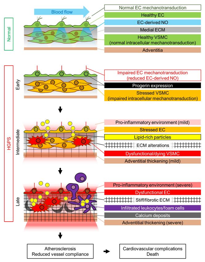
5. An Integrated Model for the Development of HGPS-Associated
Cardiovascular Dysfunction
We propose that the HGPS-associated vascular phenotype is due to progerin-induced
alteration of multiple mechanotransduction pathways in the vessel wall that affect both
VSMCs and ECs (Figure 1). The pulsatile flow of blood in the arteries subjects the arterial
wall to continuous mechanical stress. For reasons that remain elusive, progerin-expressing
VSMCs appear to be particularly sensitive to this constant cyclic insult, which induces ER
stress, dedifferentiation, calcification (and possibly acquisition of an osteogenic phenotype),
DNA damage, and eventually VSMC death, probably due to progerin-mediated altered nu-
cleocytoskeletal connections and defective intracellular force transmission. VSMC damage
results in the production of proinflammatory cytokines and ECM remodeling in the arte-
rial adventitial, medial, and probably intimal layers. This proinflammatory environment,
together with the alteration of mechanical cues induced by vessel stiffening, likely affects
endothelial structure, gene expression, and function. Additionally, progerin-expressing
ECs fail to properly mechanotransduce atheroprotective and antifibrotic laminar flow-
mediated signals, resulting in decreased eNOS expression. Subsequent reduction in the
production of endothelial NO (and probably other angiocrine factors) also contributes to
VSMC dedifferentiation and the acquisition of a pro-fibrotic phenotype, thereby establish-
ing a detrimental feedback loop of vascular damage. The reduction in vessel compliance
can induce cardiac problems, which may be worsened by the pro-fibrotic environment
generated by progerin-expressing cardiac ECs. EC alterations may also further promote
atherosclerosis development by increasing endothelial permeability. We propose that the
main difference between the mechanisms that regulate atherosclerosis onset in physiolog-
ical aging and in HGPS is the primary factor inducing endothelial dysfunction. During
normal aging, the primary causes of endothelial stress are mainly vessel-extrinsic factors
such as dyslipidemia (e.g., hypecholesterolemia, hypertriglyceridemia) and hyperglycemia,
which compromise endothelial function and trigger and sustain the vascular damage loop
that involves VSMC dysfunction. Conversely, in HGPS patients this cycle is started by
VSMC damage, which promotes endothelial dysfunction (Figure 1). Both scenarios ulti-
mately lead to atherosclerosis development and cardiovascular complications, the main
cause of death in both the elderly population and HGPS patients.Cells 2021, 10, x FOR PEER REVIEW 14 o
Cells 2021, 10, 1157 14 of 20
cardiovascular complications, the main cause of death in both the elderly population a
HGPS patients.
Figure 1.Figure
Proposed mechanism
1. Proposed of HGPS-associated
mechanism cardiovascular
of HGPS-associated dysfunction.
cardiovascular Progerin expression
dysfunction. is represented by a
Progerin expression
black nuclear rim. EC, endothelial cell; ECM, extracellular matrix; NO, nitric oxide; VSMC, vascular smooth
is represented by a black nuclear rim. EC, endothelial cell; ECM, extracellular matrix; NO, nitric muscle cell.
oxide; VSMC, vascular smooth muscle cell.
6. Conclusions and Open Questions
6. Conclusions and Open
DespiteQuestions
the extremely low number of HGPS patients worldwide, several studies o
Despite thethe past decades
extremely have defined
low number of HGPSa characteristic cardiovascular
patients worldwide, severalphenotype associated w
studies over
the past decadesthe disease.
have definedMultiple animal models,
a characteristic together with
cardiovascular sophisticated
phenotype in vitrowith
associated approaches,
starting
the disease. Multiple to unveil
animal the cellular
models, togetherand molecular
with mechanisms
sophisticated that
in vitro control theare
approaches, onset and p
gression
starting to unveil of vascular
the cellular and cardiac
and molecular alterations induced
mechanisms by progerin
that control expression.
the onset and pro-Moreove
gression of vascular and cardiac alterations induced by progerin expression. Moreover, it is
becoming increasingly evident that VSMCs and ECs both play key roles in the acquisition of
the HGPS cardiovascular phenotype. Further studies are needed to characterize the micro-
and macrovascular endothelium in more detail in different tissues from HGPS animal
models. These studies need to address multiple aspects of the endothelial phenotype in situYou can also read