Moria M2 Single Use Microkeratome Head in 100 Consecutive LASIK Procedures
←
→
Page content transcription
If your browser does not render page correctly, please read the page content below
Moria M2 Single Use Microkeratome Head
in 100 Consecutive LASIK Procedures
A. John Kanellopoulos, MD; Lawrence H. Pe, MD; Lynda Kleiman, MD

                                                            L
                                                                      aser in situ keratomileusis (LASIK) has become the
                     ABSTRACT
                                                                      preferred procedure for the correction of myopia and
PURPOSE: To evaluate the safety and efficacy of the
                                                                      moderate degrees of hyperopia.1 Its advantages must
Moria M2 single use 130 microkeratome head in con-          be weighed, however, against the possible intraoperative cor-
secutive laser in situ keratomileusis (LASIK) procedures    neal flap related complications such as irregular flaps, but-
for correction of myopia and myopic astigmatism.            tonhole flaps, and free caps,2-5 as well as postoperative flap
                                                            complications such as infection and diffuse lamellar kerati-
METHODS: One hundred eyes of 55 patients underwent          tis.6-13 These complications mostly rely on surgeon and mi-
LASIK in which the flaps were created with the Moria M2
microkeratome using the single use 130 head and ex-
                                                            crokeratome factors.
cimer laser ablation was done with the Allegretto Wave-        Several studies have evaluated the consistency and repro-
light laser. Flap parameters measured were: thickness,      ducibility of flap thickness and flap diameter cut by various
diameter, hinge length, and overall quality. Preoperative   microkeratomes as well as the induction of wavefront chang-
uncorrected visual acuity (UCVA), best spectacle-cor-       es.14-18 This study evaluated the safety and flap consistency
rected visual acuity (BSCVA), refraction, wavefront aber-
rations, and low contrast sensitivity were compared to
                                                            of the Moria M2 Single Use 130 microkeratome head (Moria,
postoperative values at 6-month follow-up.                  Antony, France) as well as the postoperative quality of vision
                                                            and wavefront changes.
RESULTS: Mean flap thickness was 145⫾17.5 µm,
mean flap diameter was 8.5⫾0.40 mm, and mean                                      PATIENTS AND METHODS
hinge cord length was 4.05⫾0.35 mm. At 6-month
follow-up, UCVA improved from 20/200 (⫾0.24) to
20/18.5 (⫾0.12) and BSCVA improved from 20/20.5
                                                            PATIENT POPULATION
(⫾0.18) to 20/17.5 (⫾0.11).                                    One hundred consecutive LASIK procedures of 55 patients
                                                            for myopia and/or myopic astigmatism were evaluated. All
CONCLUSIONS: The Moria 130 single use head for              cases were routine procedures with a wide range of myopia
the M2 microkeratome appears to be safe and effec-          and cylinder correction. Inclusion criteria were myopia of
tive in performing LASIK procedures. [J Refract Surg.       ⫺1.00 to ⫺12.00 diopters (D) and astigmatism up to ⫺5.00
2005;21:xxx-xxx.]
                                                            D. Patients with previous corneal surgery, history of herpetic

                                                            From the Department of Ophthalmology, Manhattan Eye, Ear and Throat
                                                            Hospital, New York, NY (Kanellopoulos, Pe, Kleiman); the Department
                                                            of Ophthalmology, New York University Medical School, New York, NY
                                                            (Kanellopoulos, Pe, Kleiman); and LaserVision.gr Eye Institute, Athens, Greece
                                                            (Kanellopoulos, Pe).
                                                            The authors have no proprietary interest in the materials presented herein.
                                                            Partly presented as a poster at Association for Research in Vision and
                                                            Ophthalmology Meeting; May 4-9, 2003; Ft Lauderdale, Fla.
                                                            Correspondence: A. John Kanellopoulos, MD, LaserVision.gr Eye Institute,
                                                            Pyrgos Athinon, Mesogeion 2 & Vasilissis Sofias, Ampelokipoi, 11527,
                                                            Athens, Greece. Tel: 30 210 7472 777; Fax 30 210 7472 789; E-mail:
                                                            laservision@internet.gr
                                                            Received: June 10, 2004
                                                            Accepted: December 23, 2004

Journal of Refractive Surgery Volume 21 September/October 2005                                                                            1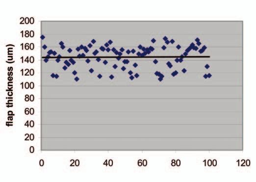
Evaluation of the Moria M2 Microkeratome/Kanellopoulos et al

                         TABLE 1
     Mean Flap Dimensions of 100 Eyes
    Undergoing LASIK With the M2 Single
       Use 130 Head Microkeratome
    Flap Thickness   Flap Diameter    Hinge Length
    (µm)             (mm)             (mm)
    155⫾17.5         8.5⫾0.4          4.05⫾0.35

eye disease, corneal dystrophy, corneal scarring, kera-
toconus, severe dry eye, and collagen vascular diseases
were excluded.
   Preoperative evaluation included uncorrected visual
acuity (UCVA), best spectacle-corrected visual acuity       Figure 1. Scatterplot of flap thickness.
(BSCVA), refraction (manifest, dilated, wavefront refrac-
tions), keratometry, slit-lamp examination with fundus         The ablations were then performed with the Alle-
evaluation, corneal topography (Orbscan II; Bausch &        gretto Wave excimer laser. After the ablation, the stro-
Lomb, Rochester, NY), and ultrasonic pachymetry (US-        mal bed was irrigated with balanced salt solution to
1800 Echoscan; Nidek, Achi, Japan).                         wash out any debris or epithelial cells. Flap position
                                                            and centration were checked using the gentian violet
OPERATIVE TECHNIQUE                                         pre-markings on the cornea and additionally with a
   A drop of proparacaine 1% (Alcaine; Alcon, Ft            drop of prednisolone acetate 1% (Pred Forte; Allergan,
Worth, Tex) was instilled into the patient’s eye just       Santa Ana, Calif) to check the flap edge alignment.
before the procedure. Eyelids were painted with povi-       Patients were examined under a slit-lamp 30 minutes
done iodine antiseptic 5% (Betadine; Purdue Pharma          postoperatively to check the corneal flaps.
L.P., Stamford, Conn), and the eyelashes were isolated         Postoperatively, patients were given prednisolone
with sterile plastic adhesive drapes (Tegaderm; 3M          acetate 1% and ofloxacin 0.3% (Exocin; Allergan,
Health Care, St Paul, Minn). The corneal epithelium         County Mayo, Ireland) drops 4 times a day for 1 week.
near the limbus was marked with gentian violet.                All surgeries were performed by a single sur-
   The flaps were created with the Moria M2 micro-           geon (A.J.K.) in a refractive surgery center in Athens,
keratome with the single use 130 head; the hinges were      Greece.
positioned superiorly in all cases. A single head-blade        Follow-up examinations were scheduled for 1 day,
assembly was used for both eyes, and the right eye was      1 week, and 1, 3, 6, and 12 months postoperatively.
done first in all bilateral cases. After the microkeratome   Wavefront measurement was repeated during 6-month
pass, the corneal flap was reflected and an ultrasound        follow-up.
pachymetry measurement of the residual stromal bed             Main outcome measures were corneal flap thick-
was done with the same corneal pachymeter that had          ness, flap diameter, and hinge cord length. The cor-
been used preoperatively. Three measurements were           relation between flap thickness and preoperative
taken and the median was used. This value was sub-          corneal thickness and between flap thickness and
tracted from the preoperative corneal thickness, and the    keratometry values were tested.
difference was taken as the corneal flap thickness (sub-        The data were analyzed using Wilcoxon signed
traction pachymetry). The flap and hinge diameters were      rank test, Pearson’s correlation, and paired Student
also measured intraoperatively with the use of a caliper.   t test.
Intraoperative flap complications were recorded.
   This microkeratome system uses four different suc-                             RESULTS
tion rings with three different flap-stop adjustments to       This study included 100 eyes of 55 patients with an
accommodate various keratometry. The specific nomo-          average age of 32.5⫾6.83 years. The mean preoperative
gram provided by the manufacturer was used in each          sphere was 5.25⫾2.56 D and mean cylinder 1.50⫾0.78
procedure to select the appropriate ring and flap-stop       D. Mean preoperative keratometry was 43.5⫾1.43 D.
setting.                                                    Mean corneal thickness was 548⫾0.34 µm (Table 1).

2                                                                                                      journalofrefractivesurgery.com
Evaluation of the Moria M2 Microkeratome/Kanellopoulos et al
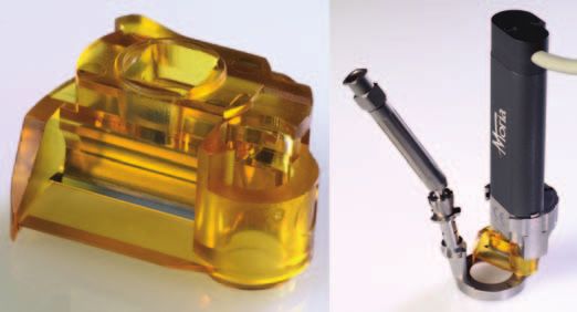
A B
Figure 3. A) Moria M2 130 single use head. B) Microkeratome head
assembled onto the motorized hand-piece and suction ring.
Figure 2. Scatterplot of flap diameter.
The mean flap thickness was 145⫾17.5 µm (Fig 1), has several significant advantages over a conventional
mean flap diameter was 8.5⫾0.40 mm (Fig 2), and mean reusable microkeratome head. First, sterilization is not
hinge length was 4.05⫾0.35 mm. necessary, as it is distributed sterile by the manufac-
A positive correlation was noted between flap turer. This avoids possible contamination by microbial
thickness and preoperative corneal thickness (P⬍.01). pathogens and bacterial endotoxins from the autoclave
A negative correlation was noted between preopera- water and sterilizing trays.6,7,10-13 Second, no wear and
tive average keratometry and resulting flap thickness tear occurs, as a new head is provided for every pa-
(P⬍.01). Both were tested using Pearson’s correlation. tient. Third, minimal technical manipulation and as-
At 6-month follow-up, UCVA improved from 20/200 sembly is required, as the microkeratome blade is pre-
(⫾0.24) to 20/18.5 (⫾0.12) and BSCVA improved from assembled with the disposable head. This reduces the
20/20.5 (⫾0.18) to 20/17.5 (⫾0.11) (P⬍.01, Wilcoxon possibility of human error in blade insertion and mi-
signed rank test). Of the 100 eyes, 92% saw ⭓20/20, crokeratome assembly. And lastly, the single use head
47% saw ⭓20/15, and 31% attained 20/10. is made of translucent plastic, which facilitates easier
No major flap complications occurred. placement and locking of the head onto the suction
ring and allows better visualization of the procedure.
DISCUSSION The surgeon can take advantage of the translucent head
We previously reported our clinical experience with to guide it directly on to the stylus (Fig 3).
the Allegretto Wave excimer laser and the M2 micro- The M2 130 in this series cuts consistent flaps with the
keratome with the standard reusable head for myopia diameter of 8.5⫾0.40 mm. The standard deviation ⫾17.5-
and myopic astigmatism (Stein and A.J. Kanellopoulos, µm in flap thickness in this study compares closely to the
unpublished data 2003). In this study, we used the same findings of ⫾23.5 µm in a previous study by Miranda et
surgical technique, but instead of the standard reusable al14 with the standard (re-usable) M2 110.
M2 microkeratome head, we used the single use 130 We found a positive correlation between the preop-
head. erative eventual flap thickness and the preoperative
Creating the corneal flap is one of the most critical corneal thickness. Patients who had thicker preopera-
steps for a successful LASIK procedure. Areas of con- tive pachymetry tend to have thicker corneal flaps. An
cern for the LASIK surgeon are microkeratome steril- inverse correlation was noted between preoperative
ization, assembly, and blade handling. Several studies keratometry and flap thickness. Corneas with higher
have reported diffuse lamellar keratitis and infectious keratometry tend to have thinner flaps. These associa-
keratitis complications related to the microkeratome tions are in contrast to previous studies dealing with
sterilization process and major fluctuations or devia- the same microkeratome by Miranda et al14 and other
tions of flap parameters due to microkeratome head microkeratomes,17 where they report no correlation be-
wear and tear.2-17 tween the preoperative corneal thickness, keratometry,
The concept of a disposable microkeratome has been or patient age.
investigated and brought to clinical practice by a num-
ber of manufacturers. None of these has been commer- REFERENCES
cially successful because of inadequate design choice 1. Sugar A, Rapuano CJ, Culbertson WW, Huang D, Varley GA,
Agapitos PJ, de Luise VP, Koch DD. Laser in situ keratomileu-
and plastic material. The disposable microkeratome
Journal of Refractive Surgery Volume 21 September/October 2005 3Evaluation of the Moria M2 Microkeratome/Kanellopoulos et al
sis for myopia and astigmatism: safety and efficacy: a report mellar keratitis after laser in situ keratomileusis. J Refract Surg.
by the American Academy of Ophthalmology. Ophthalmology. 2004;20:72-75.
2002;109:175-187. 11. Nakano EM, Nakano K, Oliveira MC, Portellinha W, Simonelli
2. Tham VM, Maloney RK. Microkeratome complications of laser R, Alvarenga LS. Cleaning solutions as a cause of diffuse lamel-
in situ keratomileusis. Ophthalmology. 2000;107:920-924. lar keratitis. J Refract Surg. 2002;18:S361-S363.
3. Tabbara KF, El-Sheikh HF, Vera-Cristo CL. Complications 12. Yuhan KR, Nguyen L, Wachler BS. Role of instrument cleaning
of laser in situ keratomileusis (LASIK). Eur J Ophthalmol. and maintenance in the development of diffuse lamellar kerati-
2003;13:139-146. tis. Ophthalmology. 2002;109:400-403.
4. Pallikaris IG, Katsanevaki VJ, Panagopoulou SI. Laser in situ 13. Johnson JD, Harissi-Dagher M, Pineda R, Yoo S, Azar DT. Diffuse
keratomileusis intraoperative complications using one type of lamellar keratitis: incidence, associations, outcomes, and a new
microkeratome. Ophthalmology. 2002;109:57-63. classification system. J Cataract Refract Surg. 2001;27:1560-1566.
5. Jacobs JM, Taravella MJ. Incidence of intraoperative flap compli- 14. Miranda D, Smith SD, Krueger RR. Comparison of flap thick-
cations in laser in situ keratomileusis. J Cataract Refract Surg. ness reproducibility using microkeratomes with a second mo-
2002;28:23-28. tor for advancement. Ophthalmology. 2003;110:1931-1934.
6. Alio JL, Perez-Santonja JJ, Tervo T, Tabbara KF, Vesaluoma M, 15. Flanagan GW, Binder PS. Precision of flap measurements
Smith RJ, Maddox B, Maloney RK. Postoperative inflammation, for laser in situ keratomileusis in 4428 eyes. J Refract Surg.
microbial complications, and wound healing following laser in 2003;19:113-123.
situ keratomileusis. J Refract Surg. 2000;16:523-538. 16. Spadea L, Cerrone L, Necozione S, Balestrazzi E. Flap mea-
7. Solomon R, Donnenfeld ED, Azar DT, Holland EJ, Palmon FR, surements with the Hansatome microkeratome. J Refract Surg.
Pflugfelder SC, Rubenstein JB. Infectious keratitis after laser 2002;18:149-154.
in situ keratomileusis: results of an ASCRS survey. J Cataract 17. Genth U, Mrochen M, Walti R, Salaheldine MM, Seiler T.
Refract Surg. 2003;29:2001-2006. Optical low coherence reflectometry for noncontact measure-
8. Verma S, Watson SL, Dart JK, Eykyn SJ. Bilateral Mycobac- ments of flap thickness during laser in situ keratomileusis.
terium chelonae keratitis following LASIK. J Refract Surg. Ophthalmology. 2002;109:973-978.
2003;19:379-380. 18. Oshika T, Klyce SD, Applegate RA, Howland HC, El Dana-
9. Pushker N, Dada T, Sony P, Ray M, Agarwal T, Vajpayee RB. soury A. Comparison of corneal wavefront aberrations after
Microbial keratitis after laser in situ keratomileusis. J Refract photorefractive keratectomy and laser in situ keratomileusis.
Surg. 2002;18:280-286. Am J Ophthalmol. 1999;127:1-7.
10. Noda-Tsuruya T, Toda I, Asano-Kato N, Hori-Komai Y, Fuku-
moto T, Tsubota K. Risk factors for development of diffuse la-
4 journalofrefractivesurgery.comEvaluation of the Moria M2 Microkeratome/Kanellopoulos et al AUTHOR QUERIES per Dr Waring The title has been changed. Okay as edited? Original: Prospective Evaluation of the M2 Single Use 130 Head in 100 Consecutive LASIK Procedures Edited: Moria M2 Single Use Microkeratome Head in 100 Consecutive LASIK Procedures Please be consistent when describing the equipment throughout. Should it be referred to as the Moria M2 Single Use 130 Microkeratome Head or the Moria 130 Single Use Head for the M2 microkeratome? Please indicate if the 130 head is intended to give a 130-µm thick flap. Please provide the dates of the surgeries. Was 100% follow-up achieved at 6 months for all 100 eyes enrolled in the trial initially? How were the average visual acuities attained? How did you measure visual acuity and what visual acuity chart was used? You state no major complications occurred. What minor complications occurred? Please identify when and where you previously reported the “unpublished data.” Please provide Stein’s first initial and the month of the unpublished data. Please provide detailed figure captions, ie, interpret what is shown. Please indicate where the cutting blade is on Figure 3. Table 2 has been omitted. Making conclusions about contrast sensitivity and corneal aberrations and attributing them to the microkera- tome seems inappropriate when the majority of those changes would be induced by the excimer laser ablation itself. Your paper is about the microkeratome, and therefore I limited your conclusions to findings with the mi- crokeratome specifically, and not conclusions that combine the outcome of the microkeratome and the excimer laser, as your paper is not about the overall outcomes in this series of eyes based on the laser itself. Journal of Refractive Surgery Volume 21 September/October 2005 5
You can also read