Neutrophil and Eosinophil DNA Extracellular Trap Formation: Lessons From Pathogenic Fungi - Frontiers
←
→
Page content transcription
If your browser does not render page correctly, please read the page content below
REVIEW
                                                                                                                                                published: 18 February 2021
                                                                                                                                           doi: 10.3389/fmicb.2021.634043

                                             Neutrophil and Eosinophil DNA
                                             Extracellular Trap Formation:
                                             Lessons From Pathogenic Fungi
                                             Juliana da Costa Silva 1 , Glaucia de Azevedo Thompson-Souza 2 ,
                                             Marina Valente Barroso 1,2 , Josiane Sabbadini Neves 2 and Rodrigo Tinoco Figueiredo 3*
                                             1
                                              Institute of Microbiology Paulo de Góes, Federal University of Rio de Janeiro, Rio de Janeiro, Brazil, 2 Institute of Biomedical
                                             Sciences, Federal University of Rio de Janeiro, Rio de Janeiro, Brazil, 3 Campus Duque de Caxias, Federal University of Rio
                                             de Janeiro, Rio de Janeiro, Brazil

                                             Fungal infections represent a worldwide health problem. Fungal pathogens are
                                             responsible for a variety of conditions, including superficial diseases, allergic pathologies
                                             and potentially lethal invasive infections. Neutrophils and eosinophils have been
                                             implicated as effector cells in several pathologies. Neutrophils are major effector
                                             cells involved in the control of fungal infections and exhibit a plethora of antifungal
                                             mechanisms, such as phagocytosis, reactive oxygen species production, degranulation,
                          Edited by:
              Constantin Felix Urban,        extracellular vesicle formation, and DNA extracellular trap (ET) release. Eosinophils are
             Umeå University, Sweden         polymorphonuclear cells classically implicated as effector cells in the pathogenesis of
                          Reviewed by:       allergic diseases and helminthic infections, although their roles as immunomodulatory
                           Jeniel E. Nett,
                                             players in both innate and adaptive immunity are currently recognized. Eosinophils are
     University of Wisconsin-Madison,
                           United States     also endowed with antifungal activities and are abundantly found in allergic conditions
                         Olaf Kniemeyer,     associated with fungal colonization and sensitization. Neutrophils and eosinophils have
   Leibniz Institute for Natural Product
       Research and Infection Biology,       been demonstrated to release their nuclear and mitochondrial DNA in response to many
                                Germany      pathogens and pro-inflammatory stimuli. ETs have been implicated in the killing and
                   *Correspondence:          control of many pathogens, as well as in promoting inflammation and tissue damage.
            Rodrigo Tinoco Figueiredo
                                             The formation of ETs by neutrophils and eosinophils has been described in response
            figueiredo.rt@xerem.ufrj.br
                                             to pathogenic fungi. Here, we provide an overview of the mechanisms involved in the
                     Specialty section:      release of neutrophil and eosinophil ETs in response to fungal pathogens. General
          This article was submitted to
                    Infectious Diseases,
                                             implications for understanding the formation of ETs and the roles of ETs in fungal
                a section of the journal     infections are discussed.
              Frontiers in Microbiology
                                             Keywords: neutrophil extracellular traps, eosinophil extracellular traps, pathogenic fungi, neutrophils, eosinophils
       Received: 26 November 2020
        Accepted: 01 February 2021
       Published: 18 February 2021
                                             INTRODUCTION
                             Citation:
     Silva JC, Thompson-Souza GA,            Fungi represent major human pathogens. Estimates indicate that approximately 300 different
           Barroso MV, Neves JS and
                                             fungal species are pathogenic to humans (Taylor et al., 2001; Wardeh et al., 2015), with fungal
     Figueiredo RT (2021) Neutrophil
and Eosinophil DNA Extracellular Trap
                                             infections contributing to nearly 1.5–1.6 million deaths annually (Brown et al., 2012). The number
Formation: Lessons From Pathogenic           of individuals affected by fungal diseases is increasing worldwide. Risk factors for the development
  Fungi. Front. Microbiol. 12:634043.        of severe invasive mycoses include immunosuppression, cancer, bone marrow or solid organ
    doi: 10.3389/fmicb.2021.634043           transplantation, and the aging (Suleyman and Alangaden, 2016). Although fungal infections

Frontiers in Microbiology | www.frontiersin.org                                      1                                          February 2021 | Volume 12 | Article 634043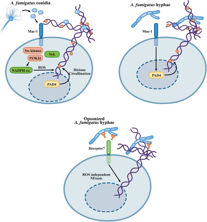
Silva et al.                                                                                                               NET/EET Formation to Fungi

contribute greatly to human morbidity and mortality, the true                crystalloid granules, primary granules, and secretory vesicles.
magnitude of these diseases in humans is unknown due to                      Among these, crystalloid granules are the largest, and unique
underreporting or misdiagnosis (Vallabhaneni et al., 2016). In               to eosinophils, they contain a variety of preformed granule-
cases of invasive fungal infections, the mortality rate of patients          stored proteins, including cationic proteins, such as major
can exceed 50% (Brown et al., 2012; Vallabhaneni et al., 2016). In           basic protein (MBP), which is the most abundant granular
addition to acting as causative agents of infections, fungi are also         protein in eosinophils; eosinophilic peroxidase (EPO); eosinophil
implicated in allergic pathologies. Exposure to environmental                cationic protein (ECP); and eosinophil-derived neurotoxin
fungi and their antigens promotes allergic pathologies such                  (EDN) (Muniz et al., 2012; Acharya and Ackerman, 2014). The
as asthma and rhinitis (Knutsen et al., 2012), and fungal                    abundance of cationic proteins makes eosinophils stainable by
colonization is a common complication of asthma resulting                    acidic dyes, such as eosin, which can be used to distinguish
in allergic bronchopulmonary mycoses (ABPMs) (Agarwal and                    them from other polymorphonuclear leukocytes. Classically,
Chakrabarti, 2013; Ishiguro et al., 2014).                                   eosinophils are considered effector cells in allergic pathologies
    Neutrophils and eosinophils have been implicated as effector             and helminthic infections and have been implicated in tissue
cells in infections caused by fungal pathogens and in diseases               damage in allergic inflammatory pathologies. However, evolving
associated with allergic sensitization to fungi (Yoon et al., 2008;          knowledge in the field has revealed that eosinophils exhibit
Lilly et al., 2014; Gazendam et al., 2016a; Figueiredo and Neves,            inflammatory and immunomodulatory functions and play roles
2018). Conditions resulting in neutropenia or deficiencies in                in tissue remodeling (Lee et al., 2004; Jacobsen et al., 2008;
neutrophil responses are major risk factors for the development              Fulkerson and Rothenberg, 2013).
of severe systemic mycoses (Lilic, 2012; Gazendam et al.,                        The increase in eosinophil blood counts, as well as
2016a). Neutrophils and eosinophils are promptly recruited to                tissue eosinophilia in allergic diseases associated with fungal
inflammatory settings where their activation contributes to tissue           sensitization/colonization, has been widely recognized (Knutsen
damage and immunopathology through the release of toxic                      et al., 2012; Figueiredo and Neves, 2018). Exposure and
components of their granules, the generation of reactive oxygen              sensitization to fungal allergens is an important factor in
species (ROS), the production of inflammatory mediators and the              patients with respiratory allergies, and in this context, fungi play
formation of DNA extracellular traps (ETs) (Amulic et al., 2012;             important roles in the development, severity and persistence
Fulkerson and Rothenberg, 2013).                                             of allergic lung diseases, especially asthma (Knutsen et al.,
    Neutrophils are peripheral blood cells of the myeloid lineage            2012). ABPMs are characterized by robust inflammation due to
and the most abundant leukocytes in human blood, constituting                fungal colonization of the airways, particularly in patients with
approximately 70% of the leukocytes in human peripheral blood                asthma or cystic fibrosis (Knutsen et al., 2012). One of the main
(and approximately 30% of the leukocytes in mouse blood).                    characteristics indicating a diagnosis of ABPM is eosinophilia,
Neutrophils can be distinguished from other granulocytes by                  in addition to increased serum IgE levels and colonization of
the absence of granule staining upon exposure to acidic or                   the airways by fungi (Asano et al., 2020). Although ABPMs
basic dyes (neutral property for which these cells were named),              have a profile of a Th2 inflammatory response with eosinophilic
and in addition, the presence of multilobed nuclei allows                    infiltrates, the nature of the interactions between eosinophils and
their identification as polymorphonuclear leukocytes (PMNs)                  fungi is unclear, and the participation of eosinophils in fungal
(Amulic et al., 2012). Neutrophils are essential for the clearance           infections continues to be extensively discussed, as eosinophils
of fungal pathogens. Neutrophils kill fungi by a variety of                  played no role in certain aspects of the pulmonary pathology
mechanisms, including degranulation, phagocytosis, oxidative                 in the experimental ABPM induced by Aspergillus fumigatus
burst, the release of extracellular vesicles, and the formation of           exposure (Dietschmann et al., 2020). However, whether these
neutrophil DNA extracellular traps (NETs) (Urban et al., 2006;               findings are relevant clinically or are limited to the experimental
Gazendam et al., 2016a; Shopova et al., 2020). Neutrophils express           model utilized remains to be elucidated. In the last decade, new
pattern recognition receptors (PRRs) involved in the recognition             data have shown an important antifungal role of eosinophils
of fungi, such as Toll-like receptor (TLR) 2 and TLR4, C-type                against several species of fungi, such as Alternaria alternata,
lectin receptors (CLRs), such as Dectin-1, Dectin-2, and Mincle,             Cryptococcus neoformans, and A. fumigatus, the latter being the
the β2 -integrin Mac-1 (macrophage-1 antigen, also known as                  main cause of ABPMs (Yoon et al., 2008; Garro et al., 2011;
complement receptor 3/CR3, αM β2 integrin or CD11b/CD18)                     Lilly et al., 2014).
(Futosi et al., 2013; Patin et al., 2019). In summary, these receptors           Extracellular traps are produced by neutrophils and
trigger signaling pathways that coordinate neutrophil responses              eosinophils in response to fungal pathogens and are involved
involved in the killing of fungal pathogens (Gazendam et al.,                in the killing and/or entrapment of fungi (Muniz et al., 2018;
2016a; Lehman and Segal, 2020).                                              Urban and Nett, 2019). Considerable effort has been dedicated to
    Eosinophils constitute a minor leukocyte population in the               the elucidation of the mechanisms of neutrophil and eosinophil
bloodstream, comprising from 1 to 5% of circulating cells,                   ET formation. While this work has focused on the formation
and a sudden increase in eosinophil blood counts in certain                  of DNA extracellular traps in response to fungi, more detailed
pathological conditions indicates that eosinophils are linked to             information on the basic aspects of NET and eosinophil
the onset and maintenance of inflammatory processes (Hogan                   extracellular trap (EET) formation can be found in recent
et al., 2008; Fulkerson and Rothenberg, 2013). Eosinophils are               publications (Mukherjee et al., 2018; Papayannopoulos, 2018).
characterized by their numerous cytoplasmic granules, including              Here, we review the mechanisms by which ETs are released from

Frontiers in Microbiology | www.frontiersin.org                          2                                   February 2021 | Volume 12 | Article 634043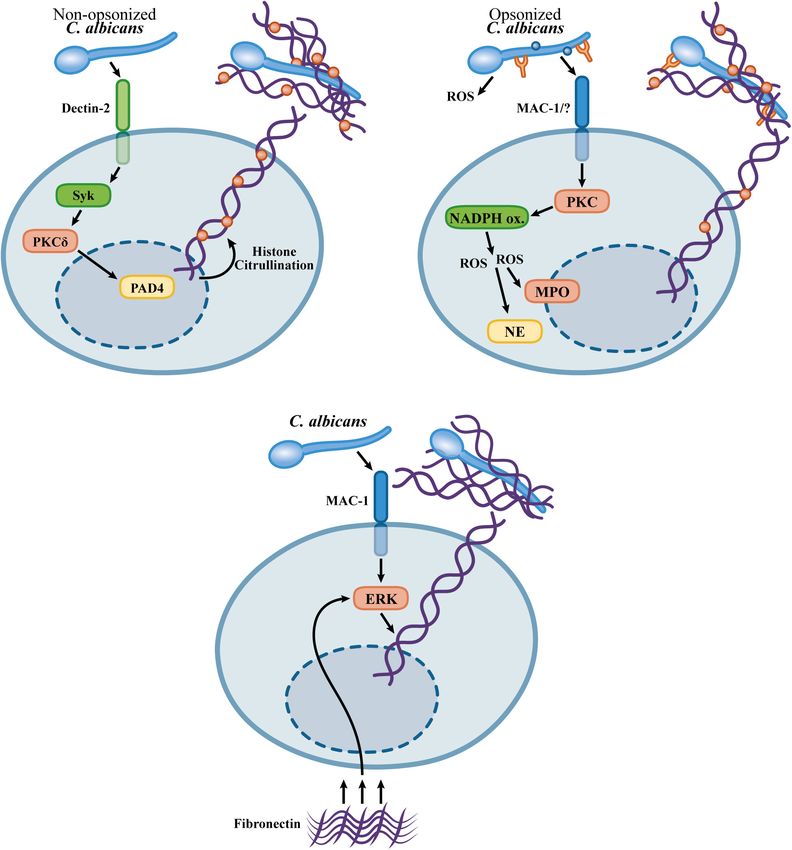
Silva et al.                                                                                                          NET/EET Formation to Fungi

neutrophils and eosinophils in response to fungal pathogens and          A/C and serine 10 in histone 3, modifications characteristic
the role of these ETs in pathologies caused by fungal infections         of cell cycle entry. NET inducers, however, do not promote
or hypersensitivity responses to fungi and their antigens.               neutrophil DNA replication or cell division. Neutrophils express
                                                                         CDK4 and 6, and CDK6 is required for NET formation
                                                                         (Amulic et al., 2017). Thus, pathways in NETosis induction
NEUTROPHIL EXTRACELLULAR TRAPS                                           and mitosis are shared, with the activation of pre-mitotic
                                                                         machinery inducing a neutrophil-specific cell death program
Takei et al. (1996) characterized a previously undiscovered              that leads to nuclear disruption and chromatin release into the
process of cell death in human neutrophils stimulated with               extracellular environment.
phorbol-12-myristate-13-acetate (PMA). Neutrophil death                     Autophagy has been described as a process required for
induced by PMA is characterized by morphological changes in              NETosis based on the observation of autophagic vesicles and
the shape of the nucleus, chromatin decondensation, and further          the inhibition of NET formation by class III phosphatidylinositol
leakage of the nuclear envelope without markers of apoptosis             3-kinase (PI3K) inhibitors, such as wortmannin and 3-
or necrosis (Takei et al., 1996). Later, it was demonstrated that        methyladenine (3-MA) (Mitroulis et al., 2011; Remijsen et al.,
the PMA-induced neutrophil death represents a novel effector             2011). The role of autophagy in the NET release, however, has
mechanism that culminates with the release of DNA fibers                 not been confirmed in ATG5 mice conditionally deficient in
involved in the killing gram-negative and gram-positive bacteria         neutrophils and eosinophils or by the pharmacological inhibition
(Brinkmann et al., 2004). These DNA web-like structures released         of autophagosome acidification (Germic et al., 2017). Thus, it
by neutrophils are called NETs and contain histones and granular         seems that the effects of inhibitors of class III PI3Ks, such
proteins such as neutrophil elastase (NE) and myeloperoxidase            as wortmannin, reflect other pharmacological targets, such as
(MPO) (Brinkmann et al., 2004). The process involved in the              class I PI3Ks, the activity of which is required for NETosis
NET release represents a novel type of cell death, called NETosis,       induced by Leishmania amazonensis and A. fumigatus (DeSouza-
characterized by the loss of the lobulated nuclear structure,            Vieira et al., 2016; Silva et al., 2020). Alternatively, since
fragmentation of the nuclear envelope, interaction of granular           Germic et al. investigated the formation of ETs by a rapid
proteins with decondensed chromatin and the subsequent                   mechanism demonstrated for stimuli that induce the formation
extrusion of nuclear content into the extracellular compartment          of mitochondrial ETs (Germic et al., 2017), they could not
(Fuchs et al., 2007).                                                    exclude the participation of autophagic mechanisms in the ETosis
    However, further investigations demonstrated that NET                in response to inducers that promote nuclear fragmentation
release can also occur without membrane rupture and can occur            and release of chromatin, by a process that takes hours to
independent of neutrophil death (Yousefi et al., 2009; Yipp              occur, resulting in cell death (Fuchs et al., 2007). Thus, it
et al., 2012). The origin of the DNA released has also been              remains to be established whether autophagy is a consequence of
investigated. Neutrophils primed with granulocyte/macrophage             neutrophil activation and occurs in parallel but is not involved
colony-stimulating factor (GM-CSF) and then further stimulated           in the mechanisms triggering ETosis, or whether autophagy is
with lipopolysaccharide (LPS) or complement factor 5a (C5a)              necessary under some conditions leading to ETosis, particularly
release mitochondria-derived DNA by a process that maintains             ET formation originating from nuclear chromatin (Fuchs et al.,
the integrity of the nuclear membrane and neutrophil viability.          2007; Thiam et al., 2020).
Mitochondrial NETs do not present nuclear components, such                  Several inflammatory stimuli have been described as inducers
as lamin B or nuclear DNA; however, these structures contain             of NET formation, including calcium ionophores, PMA, a
mitochondrial DNA in association with granular proteins, such            protein kinase C activator, bacteria, fungi, protozoans, viruses,
as NE and MPO. The release of mitochondrial NETs does not                and immune complexes (Brinkmann et al., 2004; Urban et al.,
involve cell death, membrane rupture or nuclear disruption and is        2006; Bruns et al., 2010; McCormick et al., 2010; Saitoh et al.,
faster than the NET formation involving the nuclear DNA release,         2012; Neeli and Radic, 2013; Muraro et al., 2018; Veras et al.,
with mitochondrial NETs released within 15 min of stimulation            2020). NETosis induced by PMA, a protein kinase C (PKC)
in a mechanism that requires the generation of ROS by NADPH              activator, bacteria, fungi, or immune complexes, requires the
oxidase (Yousefi et al., 2009). The general contribution of              activity of NADPH oxidase in a mechanism that relies on the
mitochondrial NETs, however, is still unclear since the NETs             generation of ROS (Fuchs et al., 2007; Ermert et al., 2009; Behnen
formed in response to several relevant stimuli and pathogens are         et al., 2014; Kenny et al., 2017). A role for oxidants generated
composed basically of nuclear DNA (Pilsczek et al., 2010; Kenny          by the NADPH oxidase complex has been demonstrated by
et al., 2017). Furthermore, in many experimental models and              the impairment in NET formation by neutrophils obtained
clinical samples, histones are associated with NETs, indicating          from individuals suffering from chronic granulomatous disease
that nuclear chromatin is the source of these NETs (Brinkmann            (CGD), which show impaired NADPH oxidase activity (Fuchs
et al., 2004; Guimarães-Costa et al., 2009; Röhm et al., 2014).          et al., 2007). The role of ROS generation downstream of
    Neutrophils are terminally differentiated cells unable to            NADPH oxidase activity has been supported by considerable
initiate mitosis; however, signaling pathways involved in the            evidence: (1) exogenous H2 O2 or the extracellular generation
mitotic program are involved in NETosis. Induction of NETosis            of H2 O2 by the incubation of neutrophils with glucose oxidase
leads to the expression of the mitotic marker Ki-67 and                  induces NETosis in the absence of NADPH oxidase activity,
the phosphorylation of the retinoblastoma protein pRb, lamin             and (2) antioxidants are effective inhibitors of NET formation

Frontiers in Microbiology | www.frontiersin.org                      3                                  February 2021 | Volume 12 | Article 634043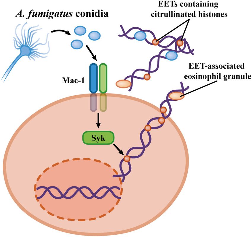
Silva et al.                                                                                                             NET/EET Formation to Fungi

in response to many neutrophil activators (Fuchs et al., 2007;             and chromatin decondensation that precede DNA release, and
Kenny et al., 2017). In addition, NADPH oxidase-independent                nuclear rupture requires PAD4 nuclear localization and activity
mechanisms for NET formation have been described in response               (Thiam et al., 2020). The relevance of PAD4-mediated histone
to a toxin-producing Staphylococcus aureus strain (Pilsczek et al.,        citrullination for NET formation has been demonstrated in
2010). NET formation by ROS-independent mechanisms has                     PAD4-KO mice (Li et al., 2010). PAD4-deficient neutrophils do
also been observed for a calcium ionophore and nigericin                   not show histone citrullination or NET release when stimulated
(Kenny et al., 2017).                                                      with calcium ionophores, LPS, PMA, H2 O2 , or S. flexneri,
    In addition to the roles of ROS generated by NADPH oxidase,            and PAD4-KO mice show an increased susceptibility to a
NETosis requires the activity of NE and MPO. Genetic deficiency            DNAse-deficient Streptococcus pyogenes strain (Li et al., 2010).
of MPO results in impairment of NET release in response to PMA             A role for PAD4 in the formation of NETs in experimental
and other NET inducers (Metzler et al., 2011). During NETosis,             models of inflammatory pathologies and infections has also been
NE and MPO are released from azurophilic granules into the                 demonstrated (Hemmers et al., 2011; Martinod et al., 2013, 2017).
cytosol (Papayannopoulos et al., 2010; Metzler et al., 2014). NE               However, the role of PAD4-mediated histone citrullination as
is then translocated to the nucleus, where it degrades histones.           a universal promoter of NETosis has been questioned. PMA does
This process is amplified by MPO, which also shows nuclear                 not promote histone citrullination in human neutrophils under
localization upon the induction of NETosis (Papayannopoulos                conditions in which NETs are formed (Neeli and Radic, 2013).
et al., 2010). The inhibition of NE results in impaired NET                Furthermore, PMA inhibits the histone citrullination induced
formation, and NE-deficient mice do not exhibit NETs in                    by calcium ionophores without interfering with the NET release
the lungs in an experimental model of infection by Klebsiella              promoted by these stimuli (Neeli and Radic, 2013). NETosis
pneumoniae (Papayannopoulos et al., 2010). The cellular                    in response to many pathogens, such as group B streptococci,
compartmentalization of NE during neutrophil activation is                 K. pneumoniae and fungi, does not require PAD4-mediated
a determinant of the induction of NETosis. Recognition of                  histone citrullination (Kenny et al., 2017; Claushuis et al., 2018;
pathogens under conditions favorable to phagocytosis does not              Guiducci et al., 2018; Silva et al., 2020; Thompson-Souza et al.,
result in NETosis, while fungal morphotypes, such as hyphae,               2020). PAD4-dependent histone citrullination has been suggested
that are not phagocytosed by neutrophils induce NET formation              to be an essential process for the induction of NETosis in response
(Branzk et al., 2014). The trigger for NET release has been                to calcium ionophores (Li et al., 2010; Lewis et al., 2015), but
attributed to the release of NE from azurophilic granules to the           this supposition has been questioned (Kenny et al., 2017). The
cytosol in response to the recognition of pathogens in the absence         reasons for the discrepancies in the roles of PAD4 in NETosis
of internalization by neutrophils. In contrast, phagocytosis results       are not clear. Although the data seem to indicate that PAD4-
in the targeting of elastase to phagosomes, which turns off the            mediated histone citrullination is the mechanism responsible for
NETosis program due to the reduction in the amount of cytosolic            the induction of NETosis, PAD4 can mediate other signaling
NE required for nuclear translocation and histone degradation              mechanisms, possibly through the citrullination of non-histone
(Branzk et al., 2014).                                                     proteins (Loos et al., 2008, 2009; Proost et al., 2008; Jang et al.,
    Histone citrullination by peptidyl arginine deiminase 4                2015; Sun et al., 2017). Thus, other roles for PAD4 that do
(PAD4) has also been implicated in the induction of NETosis                not involve chromatin decompaction by histone citrullination
(Wang et al., 2009; Li et al., 2010). Peptidyl arginine deiminases         may be involved indirectly in the induction of NETosis in vitro
constitute a group of enzymes that modify arginine residues                and in vivo. Alternatively, the involvement of PAD4-mediated
in polypeptide chains by deimination or demethylimination,                 histone citrullination in the NETosis must not reflect a common
resulting in the formation of citrulline (Wang and Wang,                   process for NET formation but must be dispensable depending
2013). PAD4 is highly expressed in neutrophils, and stimuli,               on the NET inducer.
such as calcium ionophores, TNF and LPS, promote histone                       While NADPH oxidase-induced ROS generation and PAD4
citrullination (Neeli et al., 2008; Wang et al., 2009). The role of        histone citrullination have been identified as downstream
PAD4 in NET formation was first revealed by the observation                mediators of NET formation, the receptors and upstream
that HL-60 leukemia cells when differentiated to a neutrophil-             signaling pathways critical for triggering NETosis are less
like phenotype undergo histone citrullination in response to a             understood. PMA has been used as a prototypical NET inducer
calcium ionophore that is associated with ET formation (Wang               requiring ROS generation by the NADPH oxidase complex
et al., 2009). The release of ETs by the HL-60 cell line requires          (Fuchs et al., 2007). PMA-induced NETosis involves the activity
the activity of PAD4 since its inhibition results in the absence           of conventional PKCβ isoforms (Neeli and Radic, 2013). In
of ET formation and histone citrullination. The role of PAD4 in            contrast, calcium ionophores require the atypical PKCζ isoform
the HL-60 model was attributed to chromatin decompaction by                for the induction of histone citrullination and NETosis (Neeli
histone citrullination and was also observed when HL-60 cells              and Radic, 2013). Interestingly, PMA not only fails to promote
were primed with CXCL8 followed by stimulation with Shigella               histone citrullination but also inhibits the histone citrullination
flexneri (Wang et al., 2009). Following these observations, the            induced by a calcium ionophore by means of PKCα/β activity
involvement of PAD4 in the process of NET formation has been               (Neeli and Radic, 2013). Thus, divergent programs of NETosis
described in many experimental settings (Li et al., 2010; Lewis            can be initiated through the activity of distinct PKC isoforms.
et al., 2015). A recent study demonstrated that NETosis proceeds           A large screening of small-molecule compounds revealed that a
through actin cytoskeleton dismantling, nuclear envelope rupture           selective inhibitor of c-Raf impaired PMA-induced NETosis at

Frontiers in Microbiology | www.frontiersin.org                        4                                   February 2021 | Volume 12 | Article 634043
Silva et al. NET/EET Formation to Fungi
an early stage of nuclear disruption and chromatin expansion. response to PAF, IgG/IgA immune complexes or PMA also relied
Furthermore, PMA-induced NETosis involves PKC activation on ROS generation by NADPH oxidase as did EET formation
of the MEK/ERK pathway, with the c-Raf/MEK/ERK pathway induced by TSLP (Yousefi et al., 2008; Morshed et al., 2012; Ueki
upstream of NADPH oxidase activation (Hakkim et al., 2011). et al., 2013). However, lysophosphatidylserine (LysoPS) induces
EET formation by a ROS-independent mechanism (Kim et al.,
2020). As observed for neutrophils (Asaga et al., 2001; Nakashima
EOSINOPHIL EXTRACELLULAR TRAPS et al., 2002), human eosinophils also express the enzyme PAD4
(Asaga et al., 2001), and PAD4-mediated histone citrullination is
The release of DNA extracellular traps by eosinophils (named necessary for LysoPS-induced EET formation (Kim et al., 2020).
EETs) was first described by Yousefi et al. (2008), 4 years after the However, the role of PAD4-mediated histone citrullination in
characterization of ET release from neutrophils. In the first study, EET formation is unknown for several ETT inducers, such as
the authors observed multiple extracellular DNA fibers associated PMA, immune complexes, PAF and monosodium urate crystals
with MBP and ECP in colon biopsy samples taken from patients (Schorn et al., 2012; Ueki et al., 2013).
with Crohn’s disease, schistosomiasis, or intestinal spirochetosis EETs and/or EETosis have been implicated in various
(Yousefi et al., 2008). In vitro, it was observed that human eosinophil-associated allergic diseases, including rhinosinusitis
eosinophils primed with IFN-γ or IL-5 and activated with LPS, with nasal polyps, eosinophilic esophagitis, allergic asthma,
C5a, or eotaxin/CCL11 released EETs through a ROS-dependent eosinophilic bronchopulmonary aspergillosis, eosinophilic otitis,
mechanism. Interestingly, the origin of the DNA in the EETs was and chronic obstructive pulmonary disease (Simon et al.,
characterized as mitochondrial, and the phenomenon did not 2015; Ueki et al., 2016; Uribe Echevarría et al., 2017;
involve cell death (Yousefi et al., 2008). Yet in this same study, Muniz et al., 2018; Hwang et al., 2019; Persson et al.,
the authors showed that human eosinophils released EETs in 2019). EETs have also been characterized as pro-inflammatory
response to opsonized Escherichia coli, and these EETs presented entities in non-allergic processes, such as atherosclerotic plaque
bactericidal activity. A few years later, a subsequent study showed formation and thrombosis (Marx et al., 2019), sepsis and colitis
that the stimulation of human eosinophils with thymic stromal (Yousefi et al., 2008).
lymphopoietin (TSLP), a cytokine secreted by epithelial cells
and known to contribute to the promotion of Th2 responses,
also induced the release of EETs of mitochondrial origin in a NETs AND PATHOGENIC FUNGI
mechanism independent of cell death but dependent on the
activation of NADPH oxidase and a β2 -integrin (Morshed et al., In the context of pathogenic fungi, the NET release phenomenon
2012). Non-cytolytic mitochondrial EET formation occurred has been described for Candida albicans and other Candida
independently of autophagy in eosinophils primed with GM-CSF spp. A. fumigatus, Paracoccidioides brasiliensis, Scedosporium
and stimulated with C5a or LPS or with low concentrations of apiospermum, and Histoplasma capsulatum (Urban et al., 2006;
PMA without cell priming (Germic et al., 2017). Bruns et al., 2010; McCormick et al., 2010; Svobodová et al., 2012;
Afterward, the process of EET release involving cell death Mejía et al., 2015; Rocha et al., 2015; Campos-Garcia et al., 2019;
was described, introducing the concept of EETosis (similar to Luna-Rodríguez et al., 2020; Negoro et al., 2020; Thompson-
the mechanism observed for neutrophils), where eosinophils Souza et al., 2020). Fungi present great diversity in terms of
undergo a cytolytic process with nuclear disruption, DNA mixing interactions with host cells and tissue colonization, forms of
with intact granules and release of chromatin and associated development and immune evasion strategies. Neutrophils and
granules into the extracellular medium in an NADPH oxidase- other immune cells must deal with small fungal structures,
dependent mechanism (Ueki et al., 2013). In this study, the such as conidia and yeast cells, which can be phagocytosed,
EETs released by human eosinophils were observed in vitro after as well as large multicellular forms, such as hyphae, that
stimulation with immobilized immunoglobulins (IgG and IgA), require non-phagocytic effector mechanisms. Most of the current
PAF, calcium ionophore or PMA. The DNA that constituted knowledge on the induction of NETosis and EETosis is based
the EETs was characterized as having nuclear origin, and on investigations using non-physiological inducers, such as PMA
histones were found to be associated with these EET structures. or calcium ionophores, which are useful for the elucidation of
Thus, EETs can be either released from live eosinophils, after the basic mechanisms of NET formation, but do not account for
mitochondrial DNA mobilization, associated with ECP and EPO the true complexity of pathogens. In this sense, fungal pathogens
(Yousefi et al., 2008), or as part of a slower process that results have provided valuable information about the mechanisms of ET
in the death of eosinophils by cytolysis and extrusion of histone- formation and the roles of ETs in immunity and pathology.
enriched nuclear DNA associated with clusters of intact granules
(Ueki et al., 2013).
Eosinophil extracellular traps are formed in either an Candida albicans-INDUCED NET
oxidative NADPH oxidase-dependent or oxidative-independent FORMATION
manner. NADPH oxidase-dependent mechanisms are required
for mitochondrial EET formation in response to IFN-γ/IL-5 cell The dimorphic fungus C. albicans is a component of the human
priming followed by stimulation with LPS, C5a or eotaxin/CCL11 microbiota colonizing tissues such as skin, the genitourinary
(Yousefi et al., 2008). The release of nuclear-derived EETs in tract, gastrointestinal tract and oral mucosa. C. albicans is
Frontiers in Microbiology | www.frontiersin.org 5 February 2021 | Volume 12 | Article 634043Silva et al. NET/EET Formation to Fungi
the major causative agent of mucosal fungal infections in candidates for C. albicans opsonization for NET formation
healthy individuals and a relevant pathogen causing life- include iC3b or fibrinogen, well-established ligands for Mac-
threatening disseminated infections in immunocompromised 1 (Diamond et al., 1993). Furthermore, in the context of
patients worldwide, particularly neutropenic patients (Poulain, extracellular matrix adhesion upon tissue migration, signaling
2015). C. albicans hyphae and yeast cells induce NET release by fibronectin recognition must be associated to the direct
in human neutrophils, and these extracellular traps ensnare and recognition of fungal β-glucans by Mac-1 to trigger NET release
kill both morphotypes (Urban et al., 2006). While C. albicans in the absence of an oxidative burst.
hyphae are efficient inducers of NETosis, in the absence of the The contribution of the distinct mechanisms for NETosis
morphological transition to hyphae, yeast cells are unable to in the context of C. albicans infection remains a point of
induce NET release (Branzk et al., 2014; Guiducci et al., 2018; discussion. In an experimental model of C. albicans peritonitis,
Wu et al., 2019). The inability of C. albicans yeast cells to induce pharmacological inhibition of PAD4 abolished NET formation
NETosis is a consequence of yeast phagocytosis, which reroutes in vivo, leading to C. albicans dissemination to the kidneys (Wu
NE from azurophilic granules to phagosomes, while hyphae that et al., 2019). In contrast, in an intravenous model of C. albicans
are not internalized trigger NET formation, with the release of NE infection, PAD4-KO mice showed a slight increase in C. albicans
to the cytosol and its subsequent nuclear translocation, leading load in the kidneys without increased kidney pathology or
to the NE-mediated degradation of histones (Branzk et al., increased fungal burden at later timepoints, indicating that
2014). NETs contain associated calprotectin, a cytosolic protein PAD4 is dispensable for immunity in a model of systemic
released during NETosis but not during neutrophil degranulation candidiasis (Guiducci et al., 2018). Moreover, PAD4-deficient
(Urban et al., 2009). NET-associated calprotectin chelates zinc, neutrophils show similar fungicidal activity and NETosis in
which inhibits C. albicans growth, and immunodepletion of response to C. albicans (Guiducci et al., 2018). The mechanistic
calprotectin abolishes the fungicidal effect of NETs on C. albicans. differences in C. albicans-induced NETosis in vivo are not clear,
Corroborating the role of calprotectin in vitro, calprotectin- but it is possible that, in the context of a local infection, in
deficient mice show increased susceptibility in experimental the absence of C. albicans opsonization, the Dectin-2/PAD4-
models of C. albicans infection (Urban et al., 2009). dependent pathway is critical for NETosis (Wu et al., 2019),
Different mechanisms have been proposed for C. albicans- while a PAD4-independent mechanism leads to NET formation
induced NET formation. NETosis induced by C. albicans in in systemic infection in which C. albicans must be exposed to
mouse neutrophils requires an oxidative burst by NAPDH serum opsonins (Guiducci et al., 2018). Figure 1 summarizes the
oxidase, as demonstrated by the impaired NET release from mechanisms described for C. albicans-induced NET release.
gp91−/− neutrophils in response to C. albicans hyphae
(Ermert et al., 2009). C. albicans-induced NETosis by human
neutrophils requires the generation of ROS; however, neutrophils Aspergillus fumigatus-INDUCED
obtained from CGD patients show similar NET formation. The NETosis
requirement for a ROS-dependent mechanism for C. albicans-
induced NETosis was attributed to ROS generation by opsonized Aspergillus spp. are saprophytic mold fungi with a worldwide
C. albicans, as the preincubation of C. albicans hyphae with distribution. Aspergillus species produce small spores (2–3 µm),
a ROS scavenger abolished NET release; thus, ROS generation conidia, which are critical for the fungal dispersion. Under
by C. albicans cells can supply the oxidant environment adequate conditions, conidia germinate, giving rise to the
necessary for the induction of NETosis (Kenny et al., 2017). filamentous multicellular morphotype, the hyphae. Aspergillus
NADPH oxidase-independent NETosis has been described for spp. conidia are easily dispersed in the air which results
C. albicans in the absence of serum opsonization by a mechanism in continuous human exposure to the inhalation of these
involving Dectin-2 recognition, Syk, PKCδ, calcium mobilization conidia (Latgé and Chamilos, 2019). A. fumigatus is an
and PAD4-mediated histone citrullination (Wu et al., 2019). environmental fungus and is also a common opportunistic agent
Adding more complexity to the role of NADPH oxidase- causing invasive human respiratory diseases, such as allergic
dependent ROS generation in C. albicans-induced NETosis, bronchopulmonary aspergillosis (ABPA) and a severe pulmonary
a faster process (30–50 min) for NET formation has been infection, invasive aspergillosis (IA) (Kosmidis and Denning,
described for neutrophils adhered to fibronectin in response 2015). The major predisposing factors for the development of
to β-glucans or C. albicans (Byrd et al., 2013). Crosstalk invasive aspergillosis are immunosuppression associated with
involving fibronectin-derived signals and β-glucan recognition neutropenia or deficiencies in neutrophil effector functions
by Mac-1/CR3 results in NET formation via a ROS-independent (Brown et al., 2012; Gazendam et al., 2016a).
pathway in response to β-glucans and C. albicans (Byrd et al., Aspergillus fumigatus resting conidia, swollen conidia, germ
2013). Thus, neutrophils exhibit flexible mechanisms for NET tubes and hyphae induce the formation of NETs (Bruns et al.,
formation. In the absence of serum opsonization, Dectin-2 must 2010; McCormick et al., 2010). Web-like DNA structures are
recognize α-mannans on the surface of C. albicans hyphae (Saijo also formed in the lungs of A. fumigatus-infected mice and
et al., 2010); in contrast, in the presence of serum opsonins, are absent in neutrophil-depleted mice, indicating that they
C. albicans recognition by Mac-1 leads to NADPH oxidase- are a consequence of NETosis in vivo and not a result of
dependent NETosis (Wu et al., 2019). The opsonins involved in cell damage during infection (Bruns et al., 2010). In contrast
the NETosis in response to C. albicans are unknown. Possible to the NET formed against C. albicans, the NETs are not
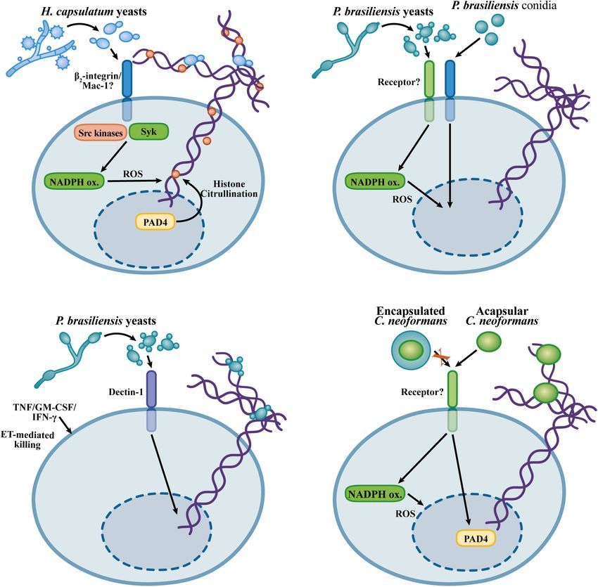
Frontiers in Microbiology | www.frontiersin.org 6 February 2021 | Volume 12 | Article 634043Silva et al. NET/EET Formation to Fungi FIGURE 1 | Mechanisms proposed for NET formation in response to Candida albicans. (A) Dectin-2 mediates NETosis in response to non-opsonized C. albicans via a pathway involving Syk, PKCδ, and the PAD4-mediated histone citrullination (Wu et al., 2019). (B) Opsonized C. albicans triggers NETosis by a ROS-dependent pathway involving NADPH oxidase activity (mouse neutrophils) (Ermert et al., 2009) or ROS generation by C. albicans (human neutrophils) (Kenny et al., 2017). PAD4-mediated citrullination is not required for NETosis in response to opsonized C. albicans, however, histone citrullination occurs during NETosis (Kenny et al., 2017; Guiducci et al., 2018). (C) Fast NETosis induced by the crosstalk upon the adhesion to fibronectin and recognition of C. albicans β-glucans by Mac-1 (Byrd et al., 2013). fungicidal for A. fumigatus or A. nidulans conidia; instead, seems that although they do not play roles in the killing of these NETs exert fungistatic effects that have been attributed A. fumigatus, NETs must inhibit fungal growth and germination. to zinc chelation by calprotectin (McCormick et al., 2010; Furthermore, NETs capture A. fumigatus conidia and hyphae, Bianchi et al., 2011). Neutrophils cause a slight decrease in the which prevents tissue dissemination. Interestingly, two-photon metabolic rate of A. fumigatus hyphae, and DNAse I addition confocal analyses reveal neutrophils carrying swollen conidia restores hyphal metabolic activity (Bruns et al., 2010). Thus, it and small hyphae in proximity to other neutrophils, which Frontiers in Microbiology | www.frontiersin.org 7 February 2021 | Volume 12 | Article 634043
Silva et al.                                                                                                           NET/EET Formation to Fungi

results in the entrapment of fungal structures, a process that           the ITAM motifs in DAP12 and the FcRγ chain, which recruits
must maximize neutrophil effector activity, such as NETosis and          Syk, resulting in Syk phosphorylation (Mócsai et al., 2006). This
oxidative killing (Bruns et al., 2010).                                  signaling module is required for ROS generation and adhesion
   NETosis in response to A. fumigatus requires superoxide               resulting from the β2 -integrin adhesion (Lowell et al., 1996;
generation by the NADPH oxidase complex. In an experimental              Mócsai et al., 1999, 2006). Interestingly, neither Src kinases,
model of A. fumigatus pulmonary infection, NADPH oxidase-                Syk, DAP12 nor the FcRγ chain are required for chemotaxis
deficient (Ncf1 KO, p47-deficient) mice did not form NETs                and neutrophil migration in vitro and in vivo (Mócsai et al.,
in lung tissue, and p47-deficient neutrophils were unable to             2002, 2006; Kovács et al., 2014), indicating that Src kinases
release NETs in response to A. fumigatus hyphae (Röhm et al.,            and Syk are involved in specific neutrophil responses, such as
2014). In vitro results confirmed the role of NADPH oxidase              ROS generation, adhesion and NETosis. Interestingly, Syk is
in NETosis in response to A. fumigatus conidia (Bruns et al.,            required for NETosis in response to immune complexes and
2010; Silva et al., 2020). The role of NADPH oxidase in                  S. aureus, indicating that Syk is a convergent signaling molecule
NETosis in response to A. fumigatus hyphae seems to differ               for NETosis induction by several stimuli (Van Ziffle and Lowell,
according to the experimental conditions. Opsonization of                2009; Behnen et al., 2014).
A. fumigatus hyphae with human serum induces NET formation                  Class I PI3Ks are involved in neutrophil responses, including
in a NADPH oxidase-independent way (Gazendam et al., 2016b),             oxidative burst, adhesion and chemotaxis (Hirsch et al., 2000;
while in other experimental settings without human serum,                Sasaki et al., 2000; Sadhu et al., 2003; Boyle et al., 2011).
NETosis in response to hyphae requires NADPH oxidase activity            Class IB PI3Kγ is activated downstream of G coupled-protein
(Bruns et al., 2010).                                                    receptors (GPCRs), while class IA PI3 kinases α, β, and δ
   TLR2, TLR4, Dectin-1, and the β2 -integrin Mac-1 are involved         are activated by tyrosine kinase pathways (Luo and Mondal,
in the immune recognition of A. fumigatus (Mambula et al.,               2015). Selective inhibition of class IA PI3Kδ abolishes ROS
2002; Meier et al., 2003; Hohl et al., 2005; Steele et al., 2005;        generation and NETosis in response to A. fumigatus conidia.
Gersuk et al., 2006; Gazendam et al., 2016b). While, TLR2,               In contrast, class I PI3Kγ inhibition exerts only partial effects
TLR4, and Dectin-1 are involved in cytokine production by                on ROS generation and does not affect A. fumigatus-induced
macrophages in response to A. fumigatus, these receptors are             NET release at concentrations in which the selective effects of
dispensable for NETosis induced by A. fumigatus (Clark et al.,           class I PI3Kγ are observed (Silva et al., 2020). NADPH oxidase
2018; Silva et al., 2020). Mac-1 recognizes fungal β-glucans             activation in response to A. fumigatus hyphae requires the
through a lectin domain distinct from the so-called I-domain             associated activity of class I PI3Kδ and β; however, the role of
involved in the recognition of iC3b, fibrinogen and ICAM-1               class I PI3Kβ in A. fumigatus-induced NETosis has not been
(Thornton et al., 1996; Xia and Ross, 1999). NETosis in response         established; thus, it remains to be investigated whether class IA
to the A. fumigatus hyphal extracts, and curdlan, a particulate          PI3Kδ cooperates with another class IA PI3K to provide signaling
preparation of β-glucans, requires the recognition mediated by           for NETosis induction by A. fumigatus (Boyle et al., 2011). How
Mac-1 through its lectin domain, as evaluated by the blockade            class I PI3Ks trigger NETosis and ROS generation in response
with a monoclonal antibody that targets this domain (Clark               to A. fumigatus remain unclear. Class I PI3Ks phosphorylate
et al., 2018). This mechanism differs from the mechanisms of             membrane phosphoinositides (PIs) at the 3rd position of the
NET release and ROS generation induced by A. fumigatus live              inositol moiety, generating PtdIns(3,4,5)P3 (Luo and Mondal,
conidia, which are mediated by Mac-1 through the I-domain                2015). Akt/PKB serine-threonine kinases are activated upon the
without the participation of the lectin domain (Silva et al.,            translocation to the cell membrane and their PH domain interacts
2020). The I-domain is the region in the αM chain critical               with PtdIns(3,4,5)P3 produced by the activity of class I PI3Ks
for binding to fibrinogen, ICAM-1 and iC3b (Diamond et al.,              (Luo and Mondal, 2015). In neutrophils, Akt2 phosphorylates
1993). Recognition of β-glucans by the lectin domain in Mac-1            and activates the NADPH oxidase subunit p47 in response to
promotes an active conformational change in the Mac-1 complex            C5a and fMLP, which is required for the assembly of NADPH
that shows increased binding through the I-domain. Priming               oxidase and its subsequent activity (Chen et al., 2010). In contrast,
of Mac-1 by the lectin domain also induces the exposure of an            opsonized zymosan induces ROS generation in the absence of
activation epitope in the I-domain that is the binding site of the       Akt activity, indicating that Akt activation must not be a general
monoclonal antibody CBRM1/5 (O’Brien et al., 2012). Thus, it             mechanism for the NADPH oxidase activation (Chen et al., 2010).
seems possible that distinct mechanisms function during fungal           Thus, it remains to be established whether Akt isoforms are
recognition mediated by Mac-1: (1) a priming by the β-glucan             downstream effectors of class I PI3Ks for NETosis and ROS
recognition through the lectin domain that must promote the              generation in response to A. fumigatus and other fungi.
I-domain recognition of A. fumigatus and (2) direct recognition             PAD4-mediated histone citrullination occurs during
of A. fumigatus molecules by the I-domain in living conidia.             neutrophil activation with curdlan, a preparation of particulate
   NETosis and neutrophil ROS generation induced by                      β-glucans, A. fumigatus hyphae and conidia (Clark et al., 2018;
A. fumigatus conidia require the activity of Src kinases, Syk,           Silva et al., 2020). NET formation in response to curdlan is
and class I PI3Kδ (Silva et al., 2020). Mac-1 signaling involves         partially dependent on histone citrullination mediated by
the immunoreceptor tyrosine-based activation motif (ITAM)-               PAD4, and in an experimental model of A. fumigatus corneal
containing membrane proteins DAP-12 and the FcRγ chain                   infection, histone citrullination was abolished in PAD4-KO mice
(Mócsai et al., 2006). Src kinases promote the phosphorylation of        (Clark et al., 2018). NET formation in response to A. fumigatus

Frontiers in Microbiology | www.frontiersin.org                      8                                   February 2021 | Volume 12 | Article 634043
Silva et al.                                                                                                                NET/EET Formation to Fungi

conidia, however, does not require PAD4 activity (Silva et al.,               effect did not require uptake of yeast cells (Newman et al., 1993).
2020). The reason for the discrepant role of PAD4 in β-glucan-                Thus, phagocytosis does not explain the fact that neutrophils have
and A. fumigatus-induced NETosis is unknown. Since PAD4                       antifungal capabilities against H. capsulatum yeast cells.
histone citrullination makes only a partial contribution to                       Thompson-Souza et al. described that H. capsulatum yeast
β-glucan-induced NETosis, another pathway contributes to                      cells induce NET release by human neutrophils, and the
NET release (Clark et al., 2018). Furthermore, while curdlan is               extracellular killing of H. capsulatum requires NET formation
a particulate β-glucan preparation, A. fumigatus conidia express              (Thompson-Souza et al., 2020). This process occurred through a
a variety of different molecules on their surface, which must                 signaling pathway mediated by NADPH oxidase-dependent ROS
trigger NETosis through a mechanism that overcomes a possible                 generation, β2 -integrin-mediated recognition, Src kinases and
requirement for PAD4 activity. Figures 2A–C summarizes the                    Syk, and culminated in the loss of neutrophil viability. Neutrophil
mechanisms described for A. fumigatus-induced NET formation.                  ROS production in response to H. capsulatum required Src
                                                                              kinase and Syk activity, demonstrating a role for NADPH
                                                                              oxidase-dependent oxidative burst downstream of the signaling
                                                                              cascade promoting NETosis (Thompson-Souza et al., 2020).
NET RELEASE IN RESPONSE TO THE                                                In addition, NETs formed in response to H. capsulatum show
DIMORPHIC ENDEMIC FUNGI                                                       bona fide NET markers, such as associated NE and citrullinated
H. capsulatum var. capsulatum AND                                             histones. However, extrusion of H. capsulatum-induced NETs
P. brasiliensis                                                               occurs independently of PAD4-mediated histone citrullination
                                                                              (Thompson-Souza et al., 2020).
Histoplasmosis is an endemic disease whose etiologic agent is                     The fact that NETs exhibit fungicidal activity against
the fungus H. capsulatum, a thermally dimorphic fungus in the                 H. capsulatum may explain the apparent contradiction found
Americas and Africa that can affect both immunocompromised                    by Newman et al. (1993) who showed that the uptake of yeast
and immunocompetent individuals (Wheat et al., 2016; Oladele                  cells was not required for the antifungal activities of neutrophils.
et al., 2018). H. capsulatum lives in the soil as a filamentous               While previous works have evaluated total killing through
fungus. H. capsulatum mycelia produce sporulated structures,                  neutrophil lysis and H. capsulatum CFU counts after relatively
macroconidia and microconidia (8–14 and 2–5 µm diameter,                      short periods of incubation, Thompson-Souza et al. (2020)
respectively). Inhalation of H. capsulatum conidia and mycelial               investigated extracellular H. capsulatum killing by propidium
fragments results in pulmonary infections that can, in some cases,            iodide entry into yeast cells after 6 h. Thus, it seems that
become disseminated, particularly in individuals presenting                   H. capsulatum yeast cells are able to survive the intracellular
deficiencies in T-cell-mediated immune responses. Once in the                 microbicidal activity of neutrophils, while the extracellular yeasts
                   ◦
host tissues at 37 C, H. capsulatum undergoes a morphological                 trapped by NETs are vulnerable to the toxic components present
transition, giving rise to yeast cells that reside in an intracellular        in the NETs. The presence of NETs, as well as the role of
niche when phagocytosed by macrophages (Woods, 2016).                         these structures, in infections caused by H. capsulatum have
    The interaction of neutrophils with H. capsulatum var                     not been investigated; therefore, it remains to be established
capsulatum yeast cells results in phagocytosis and NET extrusion              whether NETs contribute to the control of H. capsulatum in the
(Schnur and Newman, 1990; Newman et al., 1993, 2000;                          context of infection or even whether NETs contribute to the
Thompson-Souza et al., 2020). Although neutrophils are capable                immunopathogenesis during histoplasmosis. Figure 3A shows
of phagocytosing H. capsulatum cells, phagocytosis does not seem              the mechanisms involved in H. capsulatum-induced NETosis.
to underlie their antifungal activities (Brummer et al., 1991;                    Paracoccidioides brasiliensis is a pathogenic dimorphic fungus
Kurita et al., 1991; Newman et al., 1993). It was previously                  that causes paracoccidioidomycosis, a systemic disease prevalent
shown that H. capsulatum yeast cell phagocytosis by neutrophils               in Latin America (Colombo et al., 2011). Infections caused by
triggers an oxidative burst process that is interestingly unable to           P. brasiliensis seem to be established upon the inhalation of
contribute to fungal killing (Schnur and Newman, 1990; Kurita                 conidia that subsequent differentiate in the host environment to
et al., 1991). The capacity of H. capsulatum to neutralize the                establish intracellular yeast parasitism in macrophages (Cezar-
fungicidal effect of the oxidative burst is attributed to its efficient       Dos-Santos et al., 2020). Human neutrophils form NETs in
system of antioxidative enzymes (Youseff et al., 2012; Holbrook               response to P. brasiliensis conidia and yeast cells, and NETs
et al., 2013). Another observation that corroborates this finding             are found in the lesions obtained from patients suffering
is that neutrophils obtained from patients with CGD show                      from paracoccidioidomycosis (Della Coletta et al., 2015; Mejía
fungistatic activity similar to that of neutrophils that generate             et al., 2015). NETosis induced by P. brasiliensis yeast cells
an oxidative burst (Newman et al., 1993). Neutrophils can bind                requires ROS generation by the NADPH oxidase complex,
opsonized H. capsulatum through Mac-1 (CD11b/CD18), CR1,                      while conidia trigger NET release by an NADPH oxidase-
and FcγRIII in a cooperative manner (Newman et al., 1993). As                 independent mechanism. In any case, neutrophils are unable
mentioned, ROS do not seem to be a relevant mechanism for                     to kill P. brasiliensis yeast cells. Consistent with the lack
H. capsulatum killing by neutrophils; however, components of                  of fungicidal activity of neutrophils, NET degradation by
primary granules such as cathepsin G, bactericidal/permeability-              DNAse I, inhibition of NADPH oxidase or phagocytosis do
increasing protein (BPI) and defensins seem to play fungistatic               not affect the viability of this fungus during the interaction
roles (Newman et al., 1993, 2000). Interestingly, this fungistatic            with neutrophils (Mejía et al., 2015). NETosis in response

Frontiers in Microbiology | www.frontiersin.org                           9                                   February 2021 | Volume 12 | Article 634043
Silva et al. NET/EET Formation to Fungi FIGURE 2 | Mechanisms proposed for the NETosis in response to Aspergillus fumigatus. (A) Recognition of A. fumigatus triggers NETosis by a Mac-1/Src kinase/Syk and class I PI3Kδ mechanism, inducing a NET-releasing pathway that requires ROS generation by the NADPH oxidase but occurs independently of the PAD4-mediated histone citrullination (Silva et al., 2020). (B) A. fumigatus extracts, as well as β-glucans, induce NETosis through a Mac-1 and PAD4-dependent mechanism (Clark et al., 2018). (C) A. fumigatus opsonized hyphae promote NETosis through a NADPH oxidase-independent pathway (Gazendam et al., 2016b). to P. brasiliensis is partially dependent on Dectin-1, while neutrophil activation induced by pro-inflammatory cytokines, TLR2 and TLR4 are dispensable (Bachiega et al., 2016). leading to NET-associated fungicidal activity. It would be Although human neutrophils exhibit reduced fungicidal activity interesting to evaluate whether pro-inflammatory cytokines lead against P. brasiliensis, neutrophil primming with TNF, GM- to a different NET composition or whether cooperation of CSF, or IFN-γ results in P. brasiliensis killing by a mechanism NETs with other microbicidal mechanisms can be induced by requiring NET formation (Bachiega et al., 2016). Thus, although neutrophil-activating cytokines, thus enabling fungal killing. P. brasiliensis evades neutrophil killing, their ability to escape Figures 3B,C illustrates the findings concerning the NET from neutrophil fungicidal mechanisms can be overcome by formation in response to P. brasiliensis. Frontiers in Microbiology | www.frontiersin.org 10 February 2021 | Volume 12 | Article 634043
Silva et al.                                                                                                                                 NET/EET Formation to Fungi

  FIGURE 3 | Overview of the mechanisms described for the NET induction in response to Histoplasma capsulatum, Cryptococcus neoformans, and
  Paracoccidioides brasiliensis. (A) H. capsulatum NETosis relies on β2 -integrin recognition, Src and Syk kinase, via a NADPH oxidase-dependent process, but in a
  histone citrullination-independent manner (Thompson-Souza et al., 2020). (B) NET formation in response to P. brasiliensis yeast cells and conidia occurs through
  NADPH oxidase-dependent and NADPH oxidase -independent mechanisms, respectively (Mejía et al., 2015; Bachiega et al., 2016). (C) Dectin-1 mediated
  recognition of P. brasiliensis yeast cells promotes NET formation. Although P. brasiliensis yeast cells are resistant to the neutrophil killing, neutrophil-activating
  cytokines promote fungicidal activity against P. brasiliensis through NET-mediated fungal damage (Bachiega et al., 2016). (D) Mechanisms of NETosis in response to
  C. neoformans. Encapsulated C. neoformans evades NETosis, while acapsular yeast cells induce NET formation by NADPH oxidase- and PAD4-dependent
  mechanisms (Rocha et al., 2015).

Cryptococcus gattii AND Cryptococcus                                                    nervous system leading to meningitis. While C. gattii has
neoformans                                                                              been identified as a primary pathogen causing infections
                                                                                        in immunocompetent individuals, C. neoformans infections
Cryptococcus gattii and C. neoformans are environmental fungi                           are mostly associated with deficiencies in T cell-mediated
able to grow as encapsulated yeast cells. Infections caused by                          immunity, especially in HIV-infected patients (May et al.,
C. neoformans and C. gattii are initiated by the inhalation                             2016). Human neutrophils exhibit fungicidal activity against
of environmental yeast cells or spores, which establishes a                             C. neoformans by either oxidative or non-oxidative mechanisms
pulmonary infection and subsequent dissemination to the central                         (Mambula et al., 2000). Through the fractionation of human

Frontiers in Microbiology | www.frontiersin.org                                    11                                         February 2021 | Volume 12 | Article 634043
Silva et al. NET/EET Formation to Fungi
neutrophils, in cytosolic and granular preparations, the (Lilly et al., 2014). Thus, despite different studies exploring the
antifungal components calprotectin and defensins (human mechanisms of fungal recognition by eosinophils, there is still
neutrophil proteins 1 and 3, HNP-1 and HNP-3), have been a shortage of evidence describing how eosinophils contribute to
identified as anticryptococcal molecules present in the cytosol fungal infections by releasing EETs.
and primary granules, respectively (Mambula et al., 2000). Bronchial secretions of patients with ABPA show EETs in
Killing of C. gattii by neutrophils requires serum opsonization, association with large numbers of eosinophils with clear nuclear
phagocytosis and serine protease activity, but the NADPH characteristics of EETosis. EETs in the mucus obtained from
oxidative burst is dispensable (Ueno et al., 2019). ABPA individuals show citrullinated histone 3 (Muniz et al.,
Cryptococcus gattii yeast cells growing on plant-derived 2018). Human eosinophils form EETs in response to A. fumigatus
material produce extracellular fibrils. The formation of conidia through a cytolytic process. EETs show labeling for
extracellular fibrils results in increased resistance of C. gattii MBP, a granular protein; however, in contrast to NETs that
yeast cells to killing by neutrophils. Paradoxically, the production exhibit an association with free granule proteins, such as NE
of extracellular fibrils by C. gattii results in increased NET and MPO, EETs exhibit large punctuated immunostaining for
formation, indicating that although extracellular fibrils induce cationic proteins, suggesting that these proteins are not freely
neutrophil activation, these structures confer resistance to attached to these structures (Muniz et al., 2018). The presence
neutrophil effector mechanisms (Springer et al., 2010). NETs of clusters of intact free eosinophil granules associated with
show fungicidal activity against C. neoformans yeast cells EETs has been described for other stimuli, such as a calcium
due to the microbicidal effects of MPO, elastase, histones ionophore and LysoPS (Ueki et al., 2013; Kim et al., 2020).
and collagenase (Urban et al., 2009; Rocha et al., 2015). Recent evidence indicates that intact granules are associated
While encapsulated C. neoformans yeast cells do not induce with A. fumigatus-induced EETs (Muniz et al., 2018), which
NET formation, a capsule-deficient strain leads to NETosis must reflect a general mechanism by which many stimuli can
via an NADPH oxidase- and PAD4-dependent mechanism lead to the cytolytic process in EET formation. In contrast,
(Rocha et al., 2015). Glucuronoxylomannan (GXM) is the the presence of clusters of eosinophil granules attached to
major component of the C. neoformans capsule (O’Meara EETs was not observed in studies reporting mitochondrial-
and Alspaugh, 2012). Purified GXM inhibits NET formation derived EET release (Yousefi et al., 2008; Morshed et al.,
and neutrophil ROS generation, and incubation of a non- 2012). In addition, there is no evidence showing that faster
encapsulated C. neoformans strain with GXM results in inhibition non-cytolytic mitochondrial EETs are released in response to
of NETosis, thus indicating that GXM is the component in the fungal exposure. However, this is an interesting possibility that
C. neoformans capsule critical for the inhibition of NETosis. cannot be discarded.
Interestingly, glucuronoxylomannogalactan (GXMGal), a minor In neutrophils, NE and MPO are directed to the nucleus
component of the C. neoformans capsule, is an inducer of NET and have roles in the chromatin decondensation that precedes
formation through a ROS-independent mechanism (Rocha et al., nuclear membrane rupture, mixture of nuclear content and
2015). Thus, the secretion of extracellular polysaccharides by NET extrusion, as previously mentioned (Fuchs et al., 2007;
Cryptococcus spp. represents a major evasion mechanism for the Papayannopoulos et al., 2010). However, for eosinophils, it is
fungicidal activity of NETs. Figure 3D illustrates the knowledge uncertain whether granular proteins have roles in the chromatin
about NET formation in response to C. neoformans. decompaction and nuclear rupture that precede EETosis, or
which molecules are involved in this process. Thus, the
mechanisms that underlie EET extrusion considering the roles
EETs AND FUNGI of granular proteins and possible mediators of EETosis remain
to be elucidated.
Different studies have characterized eosinophils as capable of Reactive oxygen species generation has traditionally been
recognizing fungi, as well as fungal molecular patterns, which suggested as an important mechanism of host defense against
promote eosinophil activation and antifungal responses (Inoue pathogens and a downstream signal for the induction of
et al., 2005; Yoon et al., 2008; Garro et al., 2011; Lilly et al., NETosis (Fuchs et al., 2007; Winterbourn et al., 2016). However,
2014). Human eosinophils respond to A. alternata hyphae by pharmacological inhibition of NADPH oxidase or mitochondrial
releasing their granular eosinophilic content, which reduces ROS generation does not interfere with A. fumigatus-induced
fungal viability (Yoon et al., 2008). Eosinophil degranulation EET formation, indicating that EETosis occurs independently
was induced by β-glucans, but not chitin, through recognition of the major systems critical for ROS production in leukocytes
mediated by the β2 -integrin Mac-1 (Yoon et al., 2008). In addition (Muniz et al., 2018). Furthermore, eosinophils do not exhibit
to the recognition of β-glucans, eosinophils also recognize ROS production in response to A. fumigatus, while PMA is
proteases secreted by A. alternata in a mechanism that involves able to induce an oxidative burst in eosinophils in an NADPH
protease-activated receptor-2 (PAR-2) (Matsuwaki et al., 2009, oxidase-dependent manner. This difference is interesting because
2011). Eosinophils are required for the clearance of A. fumigatus the release of EETs differs from NETs formed by neutrophils
in an experimental model of pulmonary infection (Lilly et al., responding to A. fumigatus, and also from other neutrophil
2014). In addition, in vitro murine bone marrow-differentiated responses in which ROS involvement is clearly important (Bruns
eosinophils present fungicidal properties against A. fumigatus et al., 2010; Röhm et al., 2014; Winterbourn et al., 2016; Muniz
conidia in a mechanism that does not depend on contact et al., 2018; Silva et al., 2020).
Frontiers in Microbiology | www.frontiersin.org 12 February 2021 | Volume 12 | Article 634043You can also read