Patient-derived xenograft: a developing tool for screening biomarkers and potential therapeutic targets for human esophageal cancers
←
→
Page content transcription
If your browser does not render page correctly, please read the page content below

www.aging-us.com                                                               AGING 2021, Vol. 13, No. 8
                                                                                                                Review
Patient-derived xenograft: a developing tool for screening
biomarkers and potential therapeutic targets for human esophageal
cancers
Tianfeng Lan1, Xia Xue1,2, Louisa Chard Dunmall3, Jinxin Miao1,4, Yaohe Wang1,3
1
 Sino-British Research Center for Molecular Oncology, National Center for the International Research in Cell and
Gene Therapy, School of Basic Sciences, Academy of Medical Sciences, Zhengzhou University, Zhengzhou, Henan,
P.R. China
2
 The Academy of Medical Science, Precision Medicine Center of the Second Affiliated Hospital of Zhengzhou
University, Zhengzhou University, Henan, P.R. China
3
 Centre for Cancer Biomarkers and Biotherapeuitcs, Barts Cancer Institute, Queen Mary University of London,
London, UK
4
 Academy of Chinese Medicine Science, Henan University of Chinese Medicine, Zhengzhou, Henan, P.R. China

Correspondence to: Jinxin Miao, Yaohe Wang; email: jinxin.miao@yahoo.com, https://orcid.org/0000-0003-1688-3066;
yaohe.wang@qmul.ac.uk
Keywords: patient-derived xenograft, esophageal cancer, biomarkers, therapeutic targets, immunodeficient mice
Received: September 4, 2020       Accepted: March 23, 2021           Published: April 26, 2021

Copyright: © 2021 Lan et al. This is an open access article distributed under the terms of the Creative Commons Attribution
License (CC BY 3.0), which permits unrestricted use, distribution, and reproduction in any medium, provided the original
author and source are credited.

ABSTRACT
Esophageal cancer (EC) represents a human malignancy, diagnosed often at the advanced stage of cancer and
resulting in high morbidity and mortality. The development of precision medicine allows for the identification
of more personalized therapeutic strategies to improve cancer treatment. By implanting primary cancer tissues
into immunodeficient mice for expansion, patient-derived xenograft (PDX) models largely maintain similar
histological and genetic representations naturally found in patients’ tumor cells. PDX models of EC (EC-PDX)
provide fine platforms to investigate the tumor microenvironment, tumor genomic heterogeneity, and tumor
response to chemoradiotherapy, which are necessary for new drug discovery to combat EC in addition to
optimization of current therapeutic strategies for EC. In this review, we summarize the methods used for
establishing EC-PDX models and investigate the utilities of EC-PDX in screening predictive biomarkers and
potential therapeutic targets. The challenge of this promising research tool is also discussed.

INTRODUCTION                                                      EC can be divided into esophageal squamous cell
                                                                  carcinoma (ESCC) and esophageal adenocarcinoma
Esophageal cancer (EC) is an aggressive and invasive              (EAC) based on the different cell origins. ESCC
disease and early diagnosis is clinically challenging. It         originates from squamous cells, while EAC originates
is associated with one of the highest mortality rates             predominantly from Barrett mucosa [3]. It is known that
(500,000 per year) and incidence rates (570,000 new               the incidences of ESCC and EAC vary geographically.
cases per year) [1] and the global incidence and                  ESCC is predominant in East Asia and parts of Africa
mortality of EC are predicted to increase in the coming           and accounts for 90% of the new cases of EC every year
decades [1, 2]. The growing risk from this malignancy             [4]. The major causes of ESCC include smoking and
presents a heavy burden on health care providers in               excessive drinking. Other risk factors are dietary
almost every population, particularly in Eastern Asia,            deficiencies, hot beverage intake, achalasia, history of
the world leader in tobacco use, which is one of the              head and neck squamous cell cancer, and radiation
most important risk factors for EC [2].                           therapy [5, 6]. EAC is found more frequently in Europe

www.aging-us.com                                        12273                                                     AGING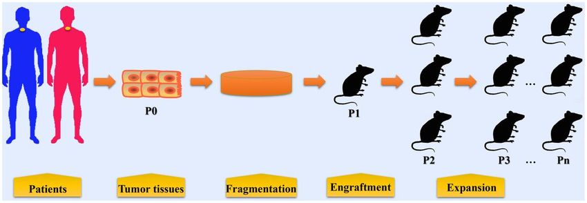
and North America and is related to chronic                       examination for pathological confirmation. Tumor
inflammation,      intestinal     metaplasia     (Barrett’s       tissues or biopsy specimens are then fragmented and
esophagus) in the distal esophageal epithelium and                these tissue fragments will be directly implanted or
obesity [7–9]. Notably, compared to ESCC, the                     blended with Matrigel before implanting into
incidence of EAC has increased persistently in some               immunocompromised mice for tumor growth and
developed countries in recent years [10]. Although age            expansion. Esophageal tumor cell populations isolated
has not been listed as a risk factor of EC, age may affect        from ESCC tissues have also been implanted to
patient survival and treatment methods [11, 12]. One              establish PDX model [18].
study demonstrated that overall survival of patients ≥70
years old was shorter, while length of stay was longer            Tumor tissues or biopsy specimens from EC patients
than those
cells that fail in SCID mice. IL-2 receptor subunit             [15]. A study showed that intramuscular engraftment
gamma (IL-2Rγ) is indispensable for high-affinity               might improve the success rate of esophageal PDX
signaling for the IL-2, IL-4, IL-7, IL-9, IL-15, and            establishment (intramuscular vs subcutaneous, 72% vs
IL-21 receptors. A lack of IL-2Rγ cripples both the             16%) [33]. They attributed the improvement to a more
adaptive and innate immune system. NSG mice                     abundant blood supply in the muscles than cutaneous
combine the characters of NOD-SICD mice and                     tissue. This novel method in tumor tissue engraftment
IL-2Rγnull mice, and are highly receptive to engraftment        may optimize the process of testing therapeutic drugs
of human primary tumors. Nevertheless, no significant           for EC. However, lymphomatous transformation
improvement in primary EC engraftment has been                  occurred in some xenografts when using the
found using NSG mice compared with NOD-SCID                     intramuscular method [33]. Intramuscular engraftment
mice [27]. Similarly to NSG mice, NOG mice also                 has also been used in establishing xenograft models
lack T and B lymphocytes and NK cells and are                   for canine osteosarcoma and human ovarian tissues
compatible with human cells and tissues [28]. The               [34, 35]. The feasibility of this engraftment approach
engraftment rate of human hematopoietic cells in NOG            should be further validated by more studies. The
mice are significantly elevated when compared with              procedures in establishing PDX models of EC are
NOD-SCID mice [29]. However, there is no evidence               summarized in Figure 1.
indicating NOG mice are superior recipients for EC-
PDX.                                                            Characteristics of EC-PDX
                                                                Distinguishing features of EC-PDX models support
Most studies that establish a EC-PDX model used                 them become useful tools in translational cancer
animals aged 6-8 weeks for engraftment of patient-
                                                                research. Firstly, the morphology and histology in EC-
derived xenografts, aging mice might not be suitable for
                                                                PDX remained consistent when compared that of the
xenografts implantation. The reasons may include: (1)
                                                                corresponding primary tumor tissues [22]. Through 3-
The activity of T cell in athymic nude mice tends to
                                                                4 passages, the degree of differentiation in tumor
increase with the age. Therefore, engraftment rate of
                                                                xenografts varied slightly [22]. Importantly, drug
tumor cells or tissues could be enhanced in younger
                                                                sensitivity, including paclitaxel and cisplatin, in PDX
mice (5-10 weeks) (reviewed by Szadvari et al [23]); (2)
In some aging mice, such as SCID mice, spontaneous              models correlates well with the clinical response in
thymic and non-thymic tumors may develop and                    corresponding patients [22]. Thus, EC-PDX model
seriously affect their survival, even they are maintained       provided a realistic model for drug sensitivity selection
in an SPF, barrier-protected environment [30]; (3) the          for EC patients [22, 36, 37]. Secondly, EC-PDXs are
life spans of immunodeficient mice vary across                  able to mimic the current clinical genetic setting of
different species. The median life span of NOD-SCID             EC, including mutations in PIK3CA, EGFR, K-Ras, B-
mice has been reported as 37 weeks, while that of NSG           Raf and HER2 amplification [19]. These models may
mice was 89 weeks (range, 59–95 weeks) [26, 31]. (4)            support further investigation of the effect of driver
Inflammatory conditions are also present in aging NSG           gene mutation on treatment response. For instance, the
female mice and contribute to morbidity and mortality           efficacy of Trastuzumab has been developed for the
in these mice [32].                                             treatment of HER2 positive breast cancer [38].
                                                                Likewise, Trastuzumab caused tumor regression in
The engraftment methods                                         HER2 positive EC-PDX models [39]. However, when
Currently,      subcutaneous,     orthotopic,     and           PIK3CA mutation was present in the models,
intramuscular implanting are three methods employed             Trastuzumab lost the ability to suppress tumor growth,
by researchers in the establishment of PDX models               which suggest PIK3CA mutation may be a mechanism
for EC (Table 1). Subcutaneous engraftment is a well-           of Trastuzumab resistance [39]. Clinical response to
established technique employed by most researchers              chemotherapy using 5-FU and cisplatin was also
in establishing PDX models. Both resected tumor                 compromised in EC-PDX models with PIK3CA
tissues or biopsy derived from human ESCC or EAC                mutation [19]. Additionally, cancer associated
could be engrafted subcutaneously into immuno-                  fibroblasts (CAFs) constitute the majority of the tumor
deficient mice. Orthotopic implantation of human                microenvironment (TME) [8]. CAFs may promote
primary EC tissues is scarcely reported. Veeranki et            tumor growth through their mechanical contributions
al. [15] transabdominally implanted a biopsy sample             to the stroma and cytokines secretion [40]. Unlike in
of EAC at the distal esophagus/gastroesophageal                 cell line xenograft, patient-derived CAFs are preserved
junction to mimic tumor growth patterns in patients.            well in PDX models and contribute to the therapy
The orthotopic mouse model closely mimics tumor                 resistance of EAC [41]. Therefore, PDX models are
growth patterns seen in patients and recapitulated the          superior in studying the interaction between EC and
response to radiation treatment in patients with EAC            TME.

www.aging-us.com                                        12275                                                   AGING
Table 1. A summary of PDX models for esophageal cancer.
Implantation Xenograft success rate
Histology Tissue type Mouse strain Refs.
method (%)
ESCC Resected SC SCID mice 37/96 (38.5) [19]
ESCC 4/12 (25) EAC
ESCC/EAC Resected SC NSG mice [20]
13/49 (33)
ESCC Resected SC SCID mice 14/26 (53.6) [21]
ESCC Biopsy SC NOD-SCID mice 25/188 (13.3) [22]
ESCC 5/16(31) EAC
ESCC / EAC Biopsy SC NOD-SCID/NSG mice [27]
8/54(33)
GEJ adenocarcinoma Biopsy Ort SCID mice 1/1 (100) [15]
SCID/NOD-SCID/NSG IM 13/18 (72) SC 1/6
GEJ/ESCC/EAC Resected/biopsy IM/SC [33]
mice (16)
ESCC Resected SC Athymic nude mice 61/110 (55.5) [36]
ESCC Resected SC NOD-SCID mice 23/54 (42.6) [42]
GEJ/ ESCC/EAC Resected SC NOD-SCID mice 21/55 (38) [43]
ESCC Resected SC SICD mice 25/54(46.3) [39]
ESCC: esophageal squamous cell carcinoma; GEJ: gastroesophageal junction; EAC: esophageal adenocarcinoma; SC:
Subcutaneous; Ort: orthotopic; IM: Intramuscular; SCID: C.B17-Prkdcscid; NOD-SCID: NOD.C.B17-Prkdcscid; NSG: NOD.Cg-
PrkdcscidIl2rgtm1Wjl.
Table 2. The success rate of PDX for other tumor types.
Tumor types Methods Recipient Success rates% Refs.
neuroblastoma Ort NSG/ athymic nude mice 24 [44]
osteosarcoma Ort NSG/ athymic nude mice 48 [44]
rhabdomyosarcoma Ort NSG/ athymic nude mice 65 [44]
retinoblastoma Ort SCID/athymic nude mice 70 [44]
Wilms tumour Ort NSG/ athymic nude mice 78 [44]
desmoplastic small round-cell tumour Ort NSG/ athymic nude mice 22 [44]
Ewing sarcoma Ort NSG/ athymic nude mice 29 [44]
high-grade sarcoma Ort NSG/ athymic nude mice 83 [44]
Colorectal cancer SC athymic nude mice 52 [45]
Prostate cancer SC SCID/NSG/ C57BL/6 pfp/rag2 mice 100-66 [46]
SC: Subcutaneous; Ort: orthotopic; SCID: C.B17-Prkdcscid; NOD-SCID: NOD.C.B17-Prkdcscid; NSG: NOD.Cg-PrkdcscidIl2rgtm1Wjl.
Application of PDX models in screening For instance, CAFs derived from EAC PDXs were
predictive biomarkers for chemoradiotherapy shown to play important roles in inducing resistance to
chemoradiotherapy [41]. Interleukin-6 (IL-6) produced
Although multidisciplinary approaches have been from/by CAFs drives EMT and enhances cell migration
developed for the treatment of locally advanced EC, and survival in EAC [41]. Therefore, IL-6 expression
only a small percentage (less than 40%) of patients from CAFs may provide value in prediction of patient
respond well to these treatments [47]. Many resistance to chemotherapy and radiotherapy [41].
nonresponsive patients may suffer severe adverse TP53-induced glycolysis and apoptosis regulator
effects and even lose the option of surgical resection (TIGAR) is a downstream regulator of p53 and highly
[48]. Therefore, predictive biomarkers are critical in expressed in many hematologic and solid tumors,
determining whether chemoradiotherapy solutions are including leukemia, breast cancer, and EC [49, 50].
suitable and effective in preventing EC progression in TIGAR remodels energy metabolism in ESCC cells and
patients. Identification of predictive biomarkers would promotes cell proliferation and colony formation [51].
facilitate accurate risk stratification of patients for Compared to ESCC-PDXs with low TIGAR expression,
therapy and avoid potential morbidity due to ineffective those with TIGAR overexpression were more resistant
treatment. The employment of PDXs in screening to 5-fluorouracil/Cisplatin, whereas they were sensitized
biomarkers has been carried out by many researchers. by a glutaminase inhibitor, CB-839 [51]. Therefore,
www.aging-us.com 12276 AGINGTIGAR expression in EC tissues might be a predictive (HCPT) is another topoisomerase I inhibitor isolated
biomarker in guiding chemotherapeutic strategies [51]. from Camptotheca cuminata. HCPT suppresses the
Furthermore, NAD(P)H quinone dehydrogenase 1 enzymatic activity of topoisomerase I, impedes cell
(NQO1), an enzyme involved in cellular reactive proliferation, and induces cell cycle arrest and apoptosis
oxygen species clearance [52], showed enhanced in ESCC cells [59]. The tumor growth of PDX models
expression in ESCC cells during the treatment of a was also suppressed by HCPT, supporting its anti-tumor
preparation of curcumin (THC) and was associated with activity [59]. Both studies validated the antitumor
THC resistance [53]. However, the combination of THC efficacy of topoisomerase I inhibitors in ESCC cells and
and NQO1 inhibitor exerted a superior effect on tumor PDX models, which may pave the way for the clinical
growth than THC monotherapy in ESCC-PDX, use of these drugs in the treatment of EC.
suggesting that NQO1 expression might be a critical
biomarker of THC response in ESCC patients [53]. EGFR and HER2
The HER family of receptor tyrosine kinases contains
Application of PDX models in evaluating epidermal growth factor receptor (EGFR/ErbB1/HER1),
therapeutic targets for chemotherapy erb-b2 receptor tyrosine kinase 2 (ERBB2/HER2/Neu),
erb-b2 receptor tyrosine kinase 3 (ERBB3/HER3), and
Topoisomerase I erb-b2 receptor tyrosine kinase 4 (ERBB4/HER4) [62,
Topoisomerase I binds to the supercoiled DNA and 63]. The aberrant activation of these receptor tyrosine
cleaves the phosphate backbone of the DNA to release kinases facilitates the tumorigenesis and progression of
supercoiled DNA [54]. It functions as a critical nuclear multiple malignant tumors, such as EC, lung cancer,
enzyme that facilitates DNA replication, transcription, gastric cancer, and colon cancer [64]. EGFR and HER2
recombination and repair [55–57]. High expression of are overexpressed in human primary EC tissues and
topoisomerase I can be found in human ESCC tissues significantly associated with overall survival in EC [65,
and is related to poor prognosis, while topoisomerase I 66]. The effect and mechanism of inhibitors targeting
expression is relatively low in the normal squamous EGFR and HER2 have been evaluated using EC-PDX
epithelium [58, 59]. Gimatecan is a modified lipophilic models. Theliatinib is a potent and highly selective
analog of camptothecin [60], which exerts anti-tumor EGFR inhibitor currently in Phase I clinical study in
activity through specifically inhibiting topoisomerase I China (NCT02601248). Theliatinib was effective in
activity. Gimatecan can induce DNA damage, S-phage restraining the tumor growth of ESCC-PDX models with
arrest and apoptosis in ESCC cells in cell-line-derived EGFR gene amplification [42]. However, PIK3CA
xenograft (CDX) models as well as in PDX models mutation or FGFR1 over-expression in PDX attenuated
through suppressing the expression and function of the effect of theliatinib, suggesting care to apply
topoisomerase I [61]. (S)-10-Hydroxycamptothecin theliatinib to only responsive subsets of patients is
Figure 1. The procedures in establishing patient-derived xenograft models of esophageal cancer. Tumor tissues or biopsy are
obtained from patients with EC during surgery or endoscopic examination. These tumor tissues and biopsy are termed P0 and are then
fragmented before implantation. In some condition, cell populations are isolated from tumor tissues for PDX model establishment.
Fragmented samples or primary tumor cells are then implanted into immunocompromised mice (termed P1), either subcutaneously or
orthotopically. When P1 tumors reached 500~1500 mm3, fresh tumor fragments are harvested from mice and then subsequently re-
implanted into other mice for expansion (P2, P3, and so on).
www.aging-us.com 12277 AGINGrequired [42]. Cetuximab is a mouse-human chimeric clinical treatment of breast cancer has been approved in
antibody that functions through binding with EGFR, the United States [84, 85]. These inhibitors may interrupt
leading to an inhibition of EGFR phosphorylation and the hyperactive cyclin D associated kinases in Rb
activation [67]. Zhu et al [36] tested the response of positive tumor cells, resulting in cell cycle arrest [84].
ESCC to cetuximab via PDX models. It is notable that The effectiveness of CDK4/6 inhibitors for EC therapy
EGFR amplification, EGFR mRNA levels and EGFR has been also investigated in preclinical and clinical trials
protein expression could be significantly correlated with [86–88]. The results of high-throughput sequencing in
the ESCC-PDXs response to cetuximab treatment [36]. EACs showed that more than half of EACs contained
biomarkers of response to CDK4/6 inhibitors [89].
The anti-HER2 monoclonal antibody, trastuzumab has SHR6390 is an orally bioavailable inhibitor of CDK4/6.
been shown its efficacy in prolonging overall survival of Suppression of proliferation of EC cells and tumor
patients with HER2-positive advanced gastric or gastro- growth of PDX model were observed following
oesophageal junction cancer [68, 69]. Trastuzumab SHR6390 treatment [90]. The combination of SHR6390
resistance was observed in PDX models of ESCC with a with paclitaxel or cisplatin synergistically inhibited tumor
dose-dependent decrease in HER2 expression and a growth in a PDX model [90]. The effects of another two
significant increase of HER3 and HER4 expression [70]. CDK4/6 inhibitors, palbociclib (PD-0332991) and
The HER3 might be a potential therapeutic targets for ribociclib (LEE011) on human ESCC were validated in
trastuzumab resistant cancer, as inhibition of HER3 could vitro and using CDX and PDX models [37]. ESCC cells
reverse trastuzumab resistance in ESCC and EAC cells and PDX models with cyclin dependent kinase inhibitor
[70]. Afatinib is a pan-HER inhibitor for clinical treatment 2A (CDKN2A) or CDKN2B loss were more sensitive to
of lung cancer and breast cancer [71, 72]. EGFR gene palbociclib and ribociclib treatment than cells with wild-
amplification or overexpression was a predictor for type genes [37]. Intriguingly, through using a mouse
afatinib sensitivity of ESCC [73]. Afatinib inhibited the avatar model of ESCC, the authors demonstrated that
phosphorylation of EGFR, S6, and ERK and induced G1 CDKN2A and CDKN2B loss were critical biomarkers
phase arrest and apoptosis in ESCC cells and PDX models for CDK4/6 inhibitor therapy [37]. Lastly, researchers
[73]. In the PDX model of esophagogastric cancer, examined the expression of CDK9 in human EAC tissues
afatinib resistance could be caused by MET amplification, and Barrett's esophagus and found that CDK9 was
which might be overcome by MET inhibitor [74]. overexpressed in EAC [91]. Pharmaceutical inhibition of
Additionally, lapatinib, a dual tyrosine kinase inhibitor of CDK9 by Flavopiridol and CAN508 diminished cell
EGFR2 and HER2, was able to contain tumor growth of proliferation and promoted apoptosis in EAC cells [91].
PDX in combination with 5-fluorouracil [75]. Furthermore, the treatment using a CDK9 inhibitor might
enhance the cell-killing effect of radiation on EAC.
Aurora-A and -B Synergetic effect of the CDK9 inhibitor, BAY1143572
Aurora-A and -B are two members of Aurora family and radiation were assessed in EAC cell lines and
kinases that are implicated in the control of mitosis [76, PDX models [92]. By inhibiting CDK9 activation,
77]. Aurora-A is required for centrosome maturation and BAY1143572 could sensitize EAC cells and PDX of
separation and bipolar spindle assembly, while Aurora-B EAC to radiation [92]. The precise mechanism by which
regulates cytokinesis and acts as a member of the CDK4/6/9 inhibitors suppress EC progression remains
chromosome passenger complex [77]. Both Aurora-A unclear and needs further investigation.
and -B can be found overexpressed in human EC tissues
[78, 79] and act to enhance cell invasion and malignant JAK/STAT3 signaling pathway
phenotypes in ESCC [80, 81]. Treatment of APIO-EE-9, Janus kinase (JAK)/ signal transducer and activator of
an Aurora kinase inhibitor, resulted in inhibition of cell transcription 3 (STAT3) signaling activation frequently
growth and proliferation, induction of apoptosis, and present in primary ESCC and is associated with poor
reduction of Aurora-A and -B activities in ESCC cell prognosis in patients [93]. JAK/STAT3 can be recruited
lines [82]. In PDX models of ESCC, APIO-EE-9 by EGFR and contributes to esophageal keratinocyte
effectively inhibited tumor growth with minimal toxicity migration [94]. Aberrant activated STAT3 in cancer cells
[82]. The inhibition of Aurora-A and -B might be induces epithelial mesenchymal transition and facilitated
effective in reducing uncontrolled proliferation in ESCC, metastasis [95]. Suppressor of cytokine signaling 1
thus contributing to tumor suppression. (SOCS1) is a multifunction protein that functions as a
signal inhibitor and a regulator in the process of
Cyclin-dependent kinase 4/6/9 ubiquitination [96]. SOCS1 negatively regulates
Cyclin-dependent kinases (CDKs) can integrate many JAK/STAT3 signaling transduction via interaction with
extracellular signaling pathways to drive cell cycle JAK proteins [97]. Overexpression of SOCS1 using
transition [83]. The employment of CDK4/6 inhibitors, recombinant adenoviral vectors reduced cell proliferation
such as palbociclib, ribociclib, and abemaciclib for and inactivated JAK/STAT3 signaling in ESCC cells as
www.aging-us.com 12278 AGINGwell as in ESCC PDX models [98]. By restraining the overexpression is associated with cisplatin resistance
JAK/STAT3/c-MYC pathway, metformin could inhibit and promotes malignant transition of ESCC via the
the transition of normal endothelial cells toward tumor PTEN/AKT/β-catenin signaling pathway [117, 118].
endothelial cells induced by tumor conditioned medium Glypican-1 expression is relatively weaker in human
[99]. In the human ESCC PDX model, metformin normal heart, kidney, small intestine, colon and
prevented tumor growth and tumor angiogenesis [99]. esophageal tissues compared to ESCC tissues [119].
The phosphorylation of STAT3 in ESCC cells could be Knockdown of glypican-1 inhibits cell growth and the
blocked by a small molecular STAT3 inhibitor, Stattic activation of EGFR, AKT and p44/42-MAPK signaling
[100]. Stattic alone or in combination with 5-fluorouracil pathways [119]. Targeting glypican-1 using anti-
markedly suppressed tumor growth of ESCC-PDX, with glypican-1 monoclonal antibody restrained tumor growth
less cell proliferation and increased apoptosis in and promoted apoptosis in ESCC PDX models [119].
xenografts [100].
Hedgehog signaling
The MAPK cascades Hedgehog signaling pathway is critical for tissue
Mitogen-activated protein kinase kinase (MEK)/ development, injury repair and tumorigenesis [120, 121].
extracellular signal-regulated kinase (ERK) signaling is The Hedgehog signaling cascade contains 3 ligands,
an essential component of the mitogen-activated protein Sonic (SHH), Indian, and Desert Hedgehog, which
kinase (MAPK) cascades [101]. Mutations of MEK/ERK activate downstream signal transducer protein
signaling are frequently seen in many human tumors, smoothened (SMO) and subsequently the GLI protein
including EC [102], lung cancer [103], and breast cancer family (GLI1, GLI2, and GLI3) by binding with the
[104]. Researchers have taken efforts to develop transmembrane receptor Patched-1 [122]. Aberrant
inhibitors of MEK and ERK as cancer therapeutic agents activation of Hedgehog signaling is linked to cancer
[105, 106]. Purpurogallin, a phenol distilled from oak progression and chemoresistance. GLI1 activity was
nutgalls, inhibits the function of MEK1 and MEK2 by elevated in EAC and correlated with EAC differentiation
binding within their ATP-binding pocket [107]. as well as the response to neoadjuvant chemotherapy
Purpurogallin inhibited the malignant phenotypes of [123]. In the established EAC PDX models, upregulation
ESCC cells and tumor growth of ESCC PDX model by of hedgehog ligands (e.g. SHH) was found in tumor
targeting MEK1 and MEK2 [107]. Ethyl gallate (EG) is a epithelium and upregulation was further enhanced by
natural phenolic compound obtained from herbs like radiation treatment [20, 124]. Unlike EAC and Barrett’s
Galla Rhois, Longan and Acacia nilotica Wild [108– Esophagus, SHH expression in ESCC is relatively rare
110]. EG directly interacts with ERK1/2 and negatively [125]. Hence, researchers concentrated on developing
regulated ERK1/2 activities in ESCC cells, leading to the inhibitors targeting SHH signaling for invasive EAC [126,
inhibition of cell proliferation, interruption of cell cycle, 127]. Evidence has shown that the Hedgehog signaling
and increase of cell apoptosis [111]. In ESCC PDX pathway is a target for improving chemoradiation therapy
models, EG administration suppressed tumor growth via in EC [128]. SHH inhibition using a monoclonal antibody
the inactivation of ERK1/2 [111]. MSK2 acts as a 5E1 augmented the growth delay of PDX tumors
downstream of the ERK1/2 or p38 MAPK pathways and following radiation [124]. Likewise, SMO inhibition with
has a regulatory effect on CREB and histone H3 [112, an SMO inhibitor, LDE225, also increased growth delay
113]. MSK2 activation as well as downstream CREB- induced by radiation [124].
Bcl-2 pathway could be dampened by sulforaphene,
leading to the induction of apoptosis and cell cycle arrest PI3K/AKT signaling pathway
and inhibition of cell migration and invasion in EC cells The phosphatidylinositol-4,5-bisphosphate 3-kinase
[114] and using EC-PDX models, the anti-tumor effect of (PI3K)/ serine/threonine kinase 1 (AKT) signaling
sulforaphene was validated [114]. Finally, MKK3/6 acts pathway plays a critical role in modulating cellular
as an upstream activator of p38 MAPK. A hexa- processes such as cell proliferation, survival, protein
hydroxylated flavonoid named gossypetin reduces cell synthesis and glucose homeostasis [129, 130]. The
viability and anchorage-independent growth and induces PI3K/AKT signaling pathway can be activated by
apoptosis in ESCC through binding with MKK3 and different receptor tyrosine kinases (RTKs) including the
MKK6 [115]. Using an ESCC PDX model, the anti- EGFR family, insulin-like growth factor 1 (IGF-1)
tumor activity of gossypetin was further demonstrated in receptor, and fibroblastic growth factor [131]. The
vivo [115]. strategies that target the PI3K/AKT pathway help to
inhibit cadherin switching, diminish cell proliferation and
Glypican-1 migration, alleviate inflammation, restore chemo-
Glypican-1 is a cell surface proteoglycan that presents sensitivity, and increase radiosensitivity in EC cells. For
in a variety of solid tumors and modulates tumor instance, the combination of a clinical PI3Kα-selective
growth, invasion and progression [116]. Glypican-1 inhibitor CYH33 and radiation promoted DNA damage,
www.aging-us.com 12279 AGINGcell cycle arrest and apoptosis in ESCC cells [132]. In the instance, the inhibitor of HSP90 Ganetespib (STA-
PDX model, CYH33 and radiation inhibited tumor 9090) could inhibit cell proliferation and induce
growth, lowered Akt phosphorylation and M2-like apoptosis in ESCC cells and PDX models [148].
macrophage infiltration [132]. Oridonin, Xanthohumol, Interestingly, the effect of HSP90 inhibition seemed to
and Scutellarin are natural compounds isolated from be dependent on MYC expression. ESCC cells and
herbs. Their activities in targeting AKT activation in xenografted primary tumors overexpressing MYC were
ESCC cells and ESCC-PDX have been reported [133– more sensitive to STA-9090 [148].
135]. These AKT inhibitors were effective in suppressing
cell growth and inducing cell cycle arrest in ESCC cells as Notch signaling pathway
well as decreasing PDX tumor growth in vivo. Dysregulation of notch signaling due to NOTCH1,
Importantly, the effects of these inhibitors were dependent NOTCH2 or NOTCH3 gene mutation has been shown in
on AKT protein level in ESCC cells [134, 135]. ESCC [149, 150]. Nuclear accumulation of notch
intracellular domain (NICD) is closely linked to tumor
VEGFR2 grade and stage in human ESCC [151]. Higher expression
The vascular endothelial growth factor (VEGF)/vascular of NICD is detected in human EAC tissues compared
endothelial growth factor receptor 2 (VEGFR2) system with the normal esophageal mucosa and the normal
plays an important role in tumor angiogenesis. Patients gastric cardia [152] and NICD expression correlates with
with solid tumors have demonstrated benefit from drugs the stage of EAC. Notch signaling regulates EAC cell
targeting VEGF and/or VEGFR2 [136, 137]. proliferation and transformation of normal esophageal
Ramucirumab is an anti-VEGFR2 monoclonal antibody epithelial cells [152, 153]. DAPT treatment suppressed
that may prevent VEGFR2 dimerization and thus tumor growth and promoted apoptosis in EAC CDX
suppress downstream signaling transduction [138]. models and PDX models [152]. Inhibition of Notch
VEGFR2 expression was found to be significantly signaling also decreased the expression of cancer stem-
elevated in EC tissues and correlated with poor efficacy cell markers in EAC cells [152].
of cytotoxic treatment [139]. Ramucirumab has been
approved by FDA for treating gastric and GEJ Other targets and utilities
adenocarcinomas either as a single agent or in Microtubules are composed of alpha- and beta-tubulin
combination with paclitaxel [140, 141]. Apatinib, a broad heterodimers, the basic structures that are essential for
inhibitor of VEGFR2, RET, c-Kit and c-Src, induced cell cell shape and behavior. Microtubules are highly
apoptosis and cell cycle arrest, inhibited malignant dynamic structures that change during the cell cycle.
transformation and sensitized EC to cisplatin [139]. The Clinically, tubulin binding agents (TBA) can suppress
efficacy of apatinib monotherapy as second- or further- microtubule dynamics and induce cell cycle arrest, thus
line treatment for advanced EC has been validated in a contributing to tumor growth inhibition [154, 155].
Phase II study [142]. Apatinib also exhibited its potential PPMP (2-[4-(3,4-dimethoxyphenyl)-3-methyl-1H-
efficacy in patients with metastatic ESCC when pyrazol-5-yl]-5-[(2-methylprop- 2-en-1-yl)oxy]phenol),
combined with docetaxel [143]. Moreover, inhibition of a novel TBA, reduced cell viability, caused cell cycle
VEGFR2 using DC101, a murine VEGFR2 inhibitor, arrest and apoptosis in ESCC cell lines [156]. PPMP
delayed tumor growth and prolonged survival of animals might occupy the colchicine binding site of tubulin and
with EAC xenografts [144]. However, vascular inhibit tubulin polymerization in ESCC cells [156]. In
regression induced by DC101 impaired the uptake of vivo, PPMP effectively suppressed tumor growth in
intraperitoneally administered nab-paclitaxel [144]. This animals bearing ESCC PDX [156].
study suggested the limits of the combination of anti-
angiogenesis and cytotoxic agents in EAC therapy [144]. With the extensive application of next-generation
sequencing to cancer transcriptomes, the role of long
HSP90 non-coding RNAs (LncRNAs) in tumor progression has
A significant correlation between heat shock protein 90 increasingly drawn people’s attention [157]. LncRNAs
(HSP90) expression and Her2 status has been found in act as tumor suppressors or oncogenes by modulating
EAC [145]. Serum HSP90a level was a significant tumor-suppressive or oncogenic pathways [158].
predictor for definitive chemoradiotherapy in patients LncRNA AGPG is highly enhanced in human ESCC
with ESCC [146]. The reduction ratio of HSSP90a tissues and cell lines. AGPG interacts with PFKFB3,
could be an independent prognostic factor for ESCC contributing to metabolism remodeling in ESCC cells.
patients [146]. The detailed role of HSP90 as a Administration of an AGPG inhibitor to ESCC PDX
therapeutic target in EC has been reviewed in a previous models markedly reduced tumor growth [159].
study [147]. Drugs targeting HSP90 alone or combined
with other chemotherapeutic drugs (i.e. cisplatin) and Additionally, PDXs are also of great value in
radiation play inhibitory roles in EC cell survival. For establishing chemoresistant cell lines and identity new
www.aging-us.com 12280 AGINGTable 3. Agents and their targets tested in PDX models of esophageal cancer.
Agent Target Histology Administration method Mouse strain Reference
Gimatecan Topoisomerase I ESCC Oral gavage NOD-SCID mice [61]
HCPT Topoisomerase I ESCC Paraneoplastic injection SCID mice [59]
Cetuximab EGFR ESCC Intraperitoneal injection Athymic nude mice [36]
Theliatinib EGFR ESCC Oral gavage NOD-SCID mice [42]
Trastuzumab HER2 EAC Intraperitoneal injection NSG mice [70]
Trastuzumab/
HER2/HER3 EAC Intraperitoneal injection NSG mice [160]
pertuzumab
Afatinib EGFR /Src family
ESCC Oral gavage NOD-SCID mice [73]
dasatinib kinase
Afatinib/AMG 337 HER2/MET EG - - [74]
Lapatinib EGFR/HER2 ESCC Oral gavage Athymic nude mice [75]
APIO-EE-9 Aurora A and B ESCC - SCID mice [82]
SHR6390 CDK4/6 ESCC Oral gavage NOD-SCID mice [90]
Palbociclib CDK4/6 ESCC Oral gavage BALB/c nude mice [37]
BAY1143572 CDK9 EAC Intraperitoneal injection Athymic nude mice [92]
AdSOCS1 SOCS1 ESCC Intratumoral injection NOD-SCID [98]
Metformin JAK/STAT3 ESCC - SCID mice [99]
Stattic STAT3 ESCC Intraperitoneal injection SCID mice [100]
Purpurogallin MEK1/2 ESCC Oral gavage SCID mice [107]
Ethyl gallate ERK1/2 ESCC Oral gavage SCID mcie [111]
Sulforaphene MSK2 ESCC Intraperitoneal injection SCID mice [114]
Gossypetin MKK3/6 ESCC Oral gavage SCID mice [115]
Anti-Glypican-1 mAb Glypican-1 ESCC Intraperitoneal injection NOG/SCID mice [119]
5E1 SHH Intraperitoneal injection NOD-SCID/NSG
EAC [124]
LDE225 SMO Oral gavage mice
CYH33 PI3Kα ESCC Oral gavage BALB/c nude mice [132]
Oridonin Akt ESCC Oral gavage SCID mice [133]
Xanthohumol Akt ESCC Oral gavage SCID mice [134]
Scutellarin Akt1/2 ESCC Oral gavage SCID mice [135]
DC101 VEGFR2 EAC Intraperitoneal injection Athymic nude mice [144]
Ganetespib HSP90 ESCC Intraperitoneal injection NSG mice [148]
DAPT Notch signaling EAC Intraperitoneal injection NSG [152]
PPMP Tubulin ESCC Intraperitoneal injection SCID mice [156]
Antisense
LncRNA AGPG ESCC Intratumoral injection Athymic nude mice [159]
oligonucleotides
HCPT: (S)-10-Hydroxycamptothecin; EGFR: epidermal growth factor receptor; HER2: erb-b2 receptor tyrosine kinase 2;
HER3: erb-b2 receptor tyrosine kinase 3; MET: MET proto-oncogene, receptor tyrosine kinase; CDK4/6/9:cyclin dependent
kinase 4/6/9; SOCS1:suppressor of cytokine signaling 1; JAK: Janus kinase; STAT3:signal transducer and activator of
transcription 3; MEK1/2:mitogen-activated protein kinase kinase 1/2; ERK1/2: extracellular signal-regulated kinase 1/2;
MSK2:ribosomal protein S6 kinase A4; MKK3/6:mitogen-activated protein kinase kinase 3/6; SHH: sonic hedgehog; SMO:
smoothened, frizzled class receptor; PI3Kα:phosphatidylinositol-4,5-bisphosphate 3-kinase catalytic subunit alpha; Akt:
serine/threonine kinase 1; VEGFR2: vascular endothelial growth factor receptor 2; HSP90: heat shock protein 90; EAC:
esophageal adenocarcinoma; ESCC: esophageal squamous cell carcinoma; EG: esophagogastric cancer; SCID: C.B17-Prkdcscid;
NOD-SCID: NOD.C.B17-Prkdcscid; NOG/SCID: NODShi.Cg-Prkdcscid Il2rgtm1Sug; NSG: NOD.Cg-PrkdcscidIl2rgtm1Wjl.
therapeutic targets. Liu et al [18] established cisplatin- CONCLUSIONS
resistant ESCC cell lines through repeatedly treating
ESCC-PDX models with cisplatin. With these cisplatin- PDX models of EC are increasingly utilized for
resistant ESCC cells, they were able to pick out studying tumor biology, investigating genetic
microRNA-455-3p as a potential therapeutic target to heterogeneity, and screening predictive biomarkers and
overcome drug resistance in EC patients [18]. therapeutic targets (Table 3). Indeed, investigators have
www.aging-us.com 12281 AGINGtested various drugs or radiation therapy on mice have become the roadblocks for the popularization
bearing PDX and screened predictive biomarkers/ of this tool;
therapeutic targets that may guide for EC therapy in
patients (Figure 2). 3) A replacement of human stromal cells by mouse
stroma occurs in the initial stage of PDX
However, there are still several problems that need to be establishment [20], which blocks the study of the
solved in the establishment and usage of EC-PDX: interaction between EC cells and stromal cells due
to the loss of human stromal cells in PDX;
1) The engraftment rates of EC-PDX remain relatively
low with the current methods, and only a minority 4) A lack of a functional immune system also prevents
of tumor tissues derived from patients can be the analysis of immunotherapeutic approaches to
successfully engrafted. As a result, there is a high EC therapy.
cost in establishing successful PDX models. To
solve this issue, novel immunodeficient animals are Although the drawbacks exist in the current EC-PDX
needed, such as gene-modified rats and hamsters models, the development of novel immunodeficient
[161–163]; animals may help accelerate their usage in a preclinical
study. For instance, tumor cells in immunodeficient
2) Although subcutaneous engraftment is commonly Syrian hamster can communicate with host fibroblasts,
employed by most researchers [164], subcutaneous which may provide growth factors to keep human
models less accurately reflect tumor progression cancer and stromal cells survive longer [163].
compared with orthotopic methods and hinder the Moreover, humanized animal models with reconstituted
investigation of tumor metastasis, angiogenesis and human immune cells will be more meaningful, which
tumor microenvironment in EC. The difficulties in allow the investigation of the interaction between
establishing and examining orthotopic PDX models cancer cells and various human immune cells.
Figure 2. The application of patient-derived xenograft (PDX) in screening predictive biomarkers and therapeutic targets for
esophageal cancer therapy. Esophageal cancer tissues are obtained from patients and implanted into immunodeficient mice for PDX
models establishment. With the PDX models, the treatment response of chemotherapeutic drugs, radiotherapeutic methods or targeted
drugs are tested on these tumor xenografts. Subsequently, genome-wide sequencing techniques and expressional analysis are carried out to
screen genes with differential expression, which are related to various therapeutic methods. Through bioinformatic analysis, potential
biomarkers are selected from differentially expressed genes. Finally, clinical trials are designed and performed to validate the feasibility of
these biomarkers.
www.aging-us.com 12282 AGINGSearch strategy fuels esophageal cancer development by inducing a
distinct inflammatory signature. Oncogene. 2012;
Searching databases include PubMed, Medline, and 31:4550–58.
Web of Science by using “patient derived xenograft” https://doi.org/10.1038/onc.2011.592
and “esophagus*”, or “mouse avatar”, “xenograft”, PMID:22179833
“primary esophageal cancer”.
6. Chen Y, Tong Y, Yang C, Gan Y, Sun H, Bi H, Cao S, Yin X,
Lu Z. Consumption of hot beverages and foods and the
AUTHOR CONTRIBUTIONS risk of esophageal cancer: a meta-analysis of
observational studies. BMC Cancer. 2015; 15:449.
TL, JM, and YW: manuscript concept and design. TL:
https://doi.org/10.1186/s12885-015-1185-1
manuscript writing. XX, JM, LCD, and YW: manuscript
PMID:26031666
revising.
7. Wang RH. From reflux esophagitis to Barrett’s
CONFLICTS OF INTEREST esophagus and esophageal adenocarcinoma. World J
Gastroenterol. 2015; 21:5210–19.
The authors declare that they have no conflicts of https://doi.org/10.3748/wjg.v21.i17.5210
interest. PMID:25954094
8. Lin EW, Karakasheva TA, Hicks PD, Bass AJ, Rustgi AK.
FUNDING The tumor microenvironment in esophageal cancer.
Oncogene. 2016; 35:5337–49.
This project is supported by the National Key R&D
https://doi.org/10.1038/onc.2016.34
Program of China (NO. 2016YFE0200800), Natural
PMID:26923327
Science Foundation of Henan Province (No.
202300410259), Postdoctoral Research Startup Project 9. Arnold M, Soerjomataram I, Ferlay J, Forman D. Global
in Henan Province (No.202001043), the Nature incidence of oesophageal cancer by histological
Sciences Foundation of China (NO. 81771776 and subtype in 2012. Gut. 2015; 64:381–87.
U1704282) and The MRC (NO. MR/M015696/1). https://doi.org/10.1136/gutjnl-2014-308124
PMID:25320104
REFERENCES
10. Gupta B, Kumar N. Worldwide incidence, mortality and
time trends for cancer of the oesophagus. Eur J Cancer
1. Bray F, Ferlay J, Soerjomataram I, Siegel RL, Torre LA,
Prev. 2017; 26:107–18.
Jemal A. Global cancer statistics 2018: GLOBOCAN
https://doi.org/10.1097/CEJ.0000000000000249
estimates of incidence and mortality worldwide for 36
PMID:27014938
cancers in 185 countries. CA Cancer J Clin. 2018;
68:394–424. 11. Domper Arnal MJ, Ferrández Arenas Á, Lanas Arbeloa
https://doi.org/10.3322/caac.21492 PMID:30207593 Á. Esophageal cancer: Risk factors, screening and
2. Malhotra GK, Yanala U, Ravipati A, Follet M, endoscopic treatment in Western and Eastern
Vijayakumar M, Are C. Global trends in esophageal countries. World J Gastroenterol. 2015; 21:7933–43.
cancer. J Surg Oncol. 2017; 115:564–79. https://doi.org/10.3748/wjg.v21.i26.7933
https://doi.org/10.1002/jso.24592 PMID:26185366
PMID:28320055 12. Huang FL, Yu SJ. Esophageal cancer: Risk factors,
3. Liu K, Zhao T, Wang J, Chen Y, Zhang R, Lan X, Que J. genetic association, and treatment. Asian J Surg. 2018;
Etiology, cancer stem cells and potential diagnostic 41:210–15.
biomarkers for esophageal cancer. Cancer Lett. 2019; https://doi.org/10.1016/j.asjsur.2016.10.005
458:21–28. PMID:27986415
https://doi.org/10.1016/j.canlet.2019.05.018 13. Madhavan A, Kamarajah SK, Navidi M, Wahed S,
PMID:31125642 Immanuel A, Hayes N, Griffin SM, Phillips AW. The
4. Abnet CC, Arnold M, Wei WQ. Epidemiology impact of age on patients undergoing transthoracic
of Esophageal Squamous Cell Carcinoma. esophagectomy for cancer. Dis Esophagus. 2021;
Gastroenterology. 2018; 154:360–73. 34:doaa056.
https://doi.org/10.1053/j.gastro.2017.08.023 https://doi.org/10.1093/dote/doaa056
PMID:28823862 PMID:32556151
5. Taccioli C, Chen H, Jiang Y, Liu XP, Huang K, Smalley KJ, 14. Zeng Y, Liang W, Liu J, He J, Ng CS, Liu CC, Petersen RH,
Farber JL, Croce CM, Fong LY. Dietary zinc deficiency Rocco G, D’Amico T, Brunelli A, Chen H, Zhi X, Dong X,
www.aging-us.com 12283 AGINGet al, and written on behalf of the AME Thoracic to explore new therapeutic strategies for
Surgery Collaborative Group. Esophageal cancer in esophageal squamous cell carcinoma. Oncol Rep.
elderly patients: a population-based study. J Thorac 2016; 35:785–92.
Dis. 2018; 10:448–57. https://doi.org/10.3892/or.2015.4459 PMID:26718633
https://doi.org/10.21037/jtd.2018.01.89 22. Zou J, Liu Y, Wang J, Liu Z, Lu Z, Chen Z, Li Z, Dong B,
PMID:29600077
Huang W, Li Y, Gao J, Shen L. Establishment and
15. Veeranki OL, Tong Z, Mejia A, Verma A, Katkhuda R, genomic characterizations of patient-derived
Bassett R, Kim TB, Wang J, Lang W, Mino B, Solis L, esophageal squamous cell carcinoma xenograft models
Kingsley C, Norton W, et al. A novel patient-derived using biopsies for treatment optimization. J Transl
orthotopic xenograft model of esophageal Med. 2018; 16:15.
adenocarcinoma provides a platform for translational https://doi.org/10.1186/s12967-018-1379-9
discoveries. Dis Model Mech. 2019; 12:dmm041004. PMID:29370817
https://doi.org/10.1242/dmm.041004 PMID:31732509
23. Szadvari I, Krizanova O, Babula P. Athymic nude mice
16. Hori T, Yamashita Y, Ohira M, Matsumura Y, as an experimental model for cancer treatment.
Muguruma K, Hirakawa K. A novel orthotopic Physiol Res. 2016 (Suppl 4); 65:S441–53.
implantation model of human esophageal carcinoma in https://doi.org/10.33549/physiolres.933526
nude rats: CD44H mediates cancer cell invasion in vitro PMID:28006926
and in vivo. Int J Cancer. 2001; 92:489–96.
24. Budzynski W, Radzikowski C. Cytotoxic cells in
https://doi.org/10.1002/ijc.1234 PMID:11304682
immunodeficient athymic mice. Immunopharmacol
17. Ohara T, Takaoka M, Sakurama K, Nagaishi K, Takeda Immunotoxicol. 1994; 16:319–46.
H, Shirakawa Y, Yamatsuji T, Nagasaka T, Matsuoka J, https://doi.org/10.3109/08923979409007097
Tanaka N, Naomoto Y. The establishment of a new PMID:7528237
mouse model with orthotopic esophageal cancer
25. Taghian A, Budach W, Zietman A, Freeman J, Gioioso D,
showing the esophageal stricture. Cancer Lett. 2010;
Ruka W, Suit HD. Quantitative comparison between
293:207–12.
the transplantability of human and murine tumors into
https://doi.org/10.1016/j.canlet.2010.01.017
the subcutaneous tissue of NCr/Sed-nu/nu nude and
PMID:20153104
severe combined immunodeficient mice. Cancer Res.
18. Liu A, Zhu J, Wu G, Cao L, Tan Z, Zhang S, Jiang L, Wu J, 1993; 53:5012–17.
Li M, Song L, Li J. Antagonizing miR-455-3p inhibits PMID:8402692
chemoresistance and aggressiveness in esophageal
26. Shultz LD, Schweitzer PA, Christianson SW, Gott B,
squamous cell carcinoma. Mol Cancer. 2017; 16:106.
Schweitzer IB, Tennent B, McKenna S, Mobraaten L,
https://doi.org/10.1186/s12943-017-0669-9
Rajan TV, Greiner DL. Multiple defects in innate and
PMID:28633632
adaptive immunologic function in NOD/LtSz-scid mice.
19. Zhang J, Jiang D, Li X, Lv J, Xie L, Zheng L, Gavine PR, Hu J Immunol. 1995; 154:180–91.
Q, Shi Y, Tan L, Ge D, Xu S, Li L, et al. Establishment and PMID:7995938
characterization of esophageal squamous cell
carcinoma patient-derived xenograft mouse models 27. Dodbiba L, Teichman J, Fleet A, Thai H, Sun B, Panchal
for preclinical drug discovery. Lab Invest. 2014; D, Patel D, Tse A, Chen Z, Faluyi OO, Renouf DJ, Girgis
94:917–26. H, Bandarchi B, et al. Primary esophageal and gastro-
https://doi.org/10.1038/labinvest.2014.77 esophageal junction cancer xenograft models:
PMID:24999713 clinicopathological features and engraftment. Lab
Invest. 2013; 93:397–407.
20. Damhofer H, Ebbing EA, Steins A, Welling L, Tol JA, https://doi.org/10.1038/labinvest.2013.8
Krishnadath KK, van Leusden T, van de Vijver MJ, PMID:23399854
Besselink MG, Busch OR, van Berge Henegouwen MI,
van Delden O, Meijer SL, et al. Establishment of 28. Ito M, Kobayashi K, Nakahata T. NOD/Shi-scid
patient-derived xenograft models and cell lines for IL2rgamma(null) (NOG) mice more appropriate for
malignancies of the upper gastrointestinal tract. J humanized mouse models. Curr Top Microbiol
Transl Med. 2015; 13:115. Immunol. 2008; 324:53–76.
https://doi.org/10.1186/s12967-015-0469-1 https://doi.org/10.1007/978-3-540-75647-7_3
PMID:25884700 PMID:18481452
21. Jiang Y, Wu Q, Yang X, Zhao J, Jin Y, Li K, Ma Y, Chen 29. Ito M, Hiramatsu H, Kobayashi K, Suzue K, Kawahata M,
X, Tian F, Zhao S, Xu J, Lu J, Yin X, et al. A method Hioki K, Ueyama Y, Koyanagi Y, Sugamura K, Tsuji K,
for establishing a patient-derived xenograft model Heike T, Nakahata T. NOD/SCID/gamma(c)(null) mouse:
www.aging-us.com 12284 AGINGan excellent recipient mouse model for engraftment of of predictors of drug sensitivity using patient-derived
human cells. Blood. 2002; 100:3175–82. models of esophageal squamous cell carcinoma. Nat
https://doi.org/10.1182/blood-2001-12-0207 Commun. 2019; 10:5076.
PMID:12384415 https://doi.org/10.1038/s41467-019-12846-7
PMID:31700061
30. Huang P, Westmoreland SV, Jain RK, Fukumura D.
Spontaneous nonthymic tumors in SCID mice. Comp 38. Cameron D, Piccart-Gebhart MJ, Gelber RD, Procter M,
Med. 2011; 61:227–34. Goldhirsch A, de Azambuja E, Castro G Jr, Untch M,
PMID:21819692 Smith I, Gianni L, Baselga J, Al-Sakaff N, Lauer S, et al,
and Herceptin Adjuvant (HERA) Trial Study Team. 11
31. Shultz LD, Lyons BL, Burzenski LM, Gott B, Chen X,
years’ follow-up of trastuzumab after adjuvant
Chaleff S, Kotb M, Gillies SD, King M, Mangada J,
chemotherapy in HER2-positive early breast cancer:
Greiner DL, Handgretinger R. Human lymphoid and
final analysis of the HERceptin Adjuvant (HERA) trial.
myeloid cell development in NOD/LtSz-scid IL2R
Lancet. 2017; 389:1195–205.
gamma null mice engrafted with mobilized human
https://doi.org/10.1016/S0140-6736(16)32616-2
hemopoietic stem cells. J Immunol. 2005;
PMID:28215665
174:6477–89.
https://doi.org/10.4049/jimmunol.174.10.6477 39. Wu X, Zhang J, Zhen R, Lv J, Zheng L, Su X, Zhu G,
PMID:15879151 Gavine PR, Xu S, Lu S, Hou J, Liu Y, Xu C, et al.
Trastuzumab anti-tumor efficacy in patient-derived
32. Santagostino SF, Arbona RJ, Nashat MA, White JR, esophageal squamous cell carcinoma xenograft
Monette S. Pathology of Aging in NOD scid gamma (PDECX) mouse models. J Transl Med. 2012; 10:180.
Female Mice. Vet Pathol. 2017; 54:855–69. https://doi.org/10.1186/1479-5876-10-180
https://doi.org/10.1177/0300985817698210 PMID:22935382
PMID:28355107
40. Karakasheva TA, Lin EW, Tang Q, Qiao E, Waldron TJ,
33. Read M, Liu D, Duong CP, Cullinane C, Murray WK, Soni M, Klein-Szanto AJ, Sahu V, Basu D, Ohashi S, Baba
Fennell CM, Shortt J, Westerman D, Burton P, Clemons K, Giaccone ZT, Walker SR, et al. IL-6 Mediates Cross-
NJ, Phillips WA. Intramuscular Transplantation Talk between Tumor Cells and Activated Fibroblasts in
Improves Engraftment Rates for Esophageal Patient- the Tumor Microenvironment. Cancer Res. 2018;
Derived Tumor Xenografts. Ann Surg Oncol. 2016; 78:4957–70.
23:305–11. https://doi.org/10.1158/0008-5472.CAN-17-2268
https://doi.org/10.1245/s10434-015-4425-3 PMID:29976575
PMID:25691278
41. Ebbing EA, van der Zalm AP, Steins A, Creemers A,
34. Coomer AR, Farese JP, Milner R, Taylor D, Salute ME, Hermsen S, Rentenaar R, Klein M, Waasdorp C, Hooijer
Rajon DA, Bova FJ, Siemann DW. Development of an GK, Meijer SL, Krishnadath KK, Punt CJ, van Berge
intramuscular xenograft model of canine Henegouwen MI, et al. Stromal-derived interleukin 6
osteosarcoma in mice for evaluation of the effects of drives epithelial-to-mesenchymal transition and
radiation therapy. Am J Vet Res. 2009; 70:127–33. therapy resistance in esophageal adenocarcinoma.
https://doi.org/10.2460/ajvr.70.1.127 PMID:19119958 Proc Natl Acad Sci USA. 2019; 116:2237–42.
35. Oktay K. Ovarian tissue cryopreservation and https://doi.org/10.1073/pnas.1820459116
transplantation: preliminary findings and PMID:30670657
implications for cancer patients. Hum Reprod 42. Ren Y, Zheng J, Fan S, Wang L, Cheng M, Shi D,
Update. 2001; 7:526–34. Zhang W, Tang R, Yu Y, Jiao L, Ni J, Yang H, Cai H, et
https://doi.org/10.1093/humupd/7.6.526 al. Anti-tumor efficacy of theliatinib in esophageal
PMID:11727860 cancer patient-derived xenografts models with
36. Zhu H, Wang C, Wang J, Chen D, Deng J, Deng J, Fan J, epidermal growth factor receptor (EGFR)
Badakhshi H, Huang X, Zhang L, Cai J, Guo S, Qian W, overexpression and gene amplification. Oncotarget.
et al. A subset of esophageal squamous cell carcinoma 2017; 8:50832–44.
patient-derived xenografts respond to cetuximab, https://doi.org/10.18632/oncotarget.17243
which is predicted by high EGFR expression and PMID:28881608
amplification. J Thorac Dis. 2018; 10:5328–38. 43. Dodbiba L, Teichman J, Fleet A, Thai H, Starmans MH,
https://doi.org/10.21037/jtd.2018.09.18 Navab R, Chen Z, Girgis H, Eng L, Espin-Garcia O, Shen
PMID:30416780 X, Bandarchi B, Schwock J, et al. Appropriateness of
37. Su D, Zhang D, Jin J, Ying L, Han M, Chen K, Li B, Wu J, using patient-derived xenograft models for
Xie Z, Zhang F, Lin Y, Cheng G, Li JY, et al. Identification pharmacologic evaluation of novel therapies for
www.aging-us.com 12285 AGINGesophageal/gastro-esophageal junction cancers. PLoS remodeling by TIGAR overexpression is a therapeutic
One. 2015; 10:e0121872. target in esophageal squamous-cell carcinoma.
https://doi.org/10.1371/journal.pone.0121872 Theranostics. 2020; 10:3488–502.
PMID:25826681 https://doi.org/10.7150/thno.41427 PMID:32206103
44. Stewart E, Federico SM, Chen X, Shelat AA, Bradley C, 52. Ross D, Siegel D. Functions of NQO1 in Cellular
Gordon B, Karlstrom A, Twarog NR, Clay MR, Bahrami Protection and CoQ10 Metabolism and its Potential
A, Freeman BB 3rd, Xu B, Zhou X, et al. Orthotopic Role as a Redox Sensitive Molecular Switch. Front
patient-derived xenografts of paediatric solid tumours. Physiol. 2017; 8:595.
Nature. 2017; 549:96–100. https://doi.org/10.3389/fphys.2017.00595
https://doi.org/10.1038/nature23647 PMID:28854174 PMID:28883796
45. Prasetyanti PR, van Hooff SR, van Herwaarden T, de 53. Mizumoto A, Ohashi S, Kamada M, Saito T, Nakai Y,
Vries N, Kalloe K, Rodermond H, van Leersum R, de Baba K, Hirohashi K, Mitani Y, Kikuchi O, Matsubara J,
Jong JH, Franitza M, Nürnberg P, Todaro M, Stassi G, Yamada A, Takahashi T, Lee H, et al. Combination
Medema JP. Capturing colorectal cancer inter-tumor treatment with highly bioavailable curcumin and NQO1
heterogeneity in patient-derived xenograft (PDX) inhibitor exhibits potent antitumor effects on
models. Int J Cancer. 2019; 144:366–71. esophageal squamous cell carcinoma. J Gastroenterol.
https://doi.org/10.1002/ijc.31767 PMID:30151914 2019; 54:687–98.
https://doi.org/10.1007/s00535-019-01549-x
46. Lange T, Oh-Hohenhorst SJ, Joosse SA, Pantel K, Hahn
PMID:30737573
O, Gosau T, Dyshlovoy SA, Wellbrock J, Feldhaus S,
Maar H, Gehrcke R, Kluth M, Simon R, et al. 54. Gilmour DS, Pflugfelder G, Wang JC, Lis JT.
Development and Characterization of a Spontaneously Topoisomerase I interacts with transcribed regions in
Metastatic Patient-Derived Xenograft Model of Human Drosophila cells. Cell. 1986; 44:401–07.
Prostate Cancer. Sci Rep. 2018; 8:17535. https://doi.org/10.1016/0092-8674(86)90461-7
https://doi.org/10.1038/s41598-018-35695-8 PMID:3002635
PMID:30510249
55. Li M, Liu Y. Topoisomerase I in Human Disease
47. Gebski V, Burmeister B, Smithers BM, Foo K, Zalcberg J, Pathogenesis and Treatments. Genomics Proteomics
Simes J, and Australasian Gastro-Intestinal Trials Bioinformatics. 2016; 14:166–71.
Group. Survival benefits from neoadjuvant https://doi.org/10.1016/j.gpb.2016.02.004
chemoradiotherapy or chemotherapy in oesophageal PMID:27181710
carcinoma: a meta-analysis. Lancet Oncol. 2007;
56. Pourquier P, Pommier Y. Topoisomerase I-mediated
8:226–34.
DNA damage. Adv Cancer Res. 2001; 80:189–216.
https://doi.org/10.1016/S1470-2045(07)70039-6
https://doi.org/10.1016/S0065-230X(01)80016-6
PMID:17329193
PMID:11034544
48. Blencowe NS, McNair AG, Davis CR, Brookes ST,
57. Leppard JB, Champoux JJ. Human DNA topoisomerase
Blazeby JM. Standards of outcome reporting in surgical
I: relaxation, roles, and damage control. Chromosoma.
oncology: a case study in esophageal cancer. Ann Surg
2005; 114:75–85.
Oncol. 2012; 19:4012–18.
https://doi.org/10.1007/s00412-005-0345-5
https://doi.org/10.1245/s10434-012-2497-x
PMID:15830206
PMID:22820935
58. Hanagiri T, Ono K, Kuwata T, Takenaka M, Oka S,
49. Geng J, Yuan X, Wei M, Wu J, Qin ZH. The diverse role
Chikaishi Y, Shigematsu Y, Nagata Y, Shimokawa H,
of TIGAR in cellular homeostasis and cancer. Free Radic Nakagawa M, Uramoto H, So T, Tanaka F. Evaluation of
Res. 2018; 52:1240–49.
topoisomerase I/topoisomerase IIalpha status in
https://doi.org/10.1080/10715762.2018.1489133
esophageal cancer. J UOEH. 2011; 33:205–16.
PMID:30284488
https://doi.org/10.7888/juoeh.33.205
50. GongSun X, Zhao Y, Jiang B, Xin Z, Shi M, Song L, Qin Q, PMID:21913377
Wang Q, Liu X. Inhibition of MUC1-C regulates 59. Song M, Yin S, Zhao R, Liu K, Kundu JK, Shim JH, Lee
metabolism by AKT pathway in esophageal squamous MH, Dong Z. (S)-10-Hydroxycamptothecin Inhibits
cell carcinoma. J Cell Physiol. 2019; 234:12019–28. Esophageal Squamous Cell Carcinoma Growth In Vitro
https://doi.org/10.1002/jcp.27863 and In Vivo Via Decreasing Topoisomerase I Enzyme
PMID:30523643 Activity. Cancers (Basel). 2019; 11:1964.
51. Chu J, Niu X, Chang J, Shao M, Peng L, Xi Y, Lin A, Wang https://doi.org/10.3390/cancers11121964
C, Cui Q, Luo Y, Fan W, Chen Y, Sun Y, et al. Metabolic PMID:31817790
www.aging-us.com 12286 AGINGYou can also read