Current Understanding of Exosomal MicroRNAs in Glioma Immune Regulation and Therapeutic Responses
←
→
Page content transcription
If your browser does not render page correctly, please read the page content below
REVIEW
                                                                                                                                              published: 14 January 2022
                                                                                                                                        doi: 10.3389/fimmu.2021.813747

                                            Current Understanding of
                                            Exosomal MicroRNAs in Glioma
                                            Immune Regulation and
                                            Therapeutic Responses
                                            Jinwu Peng 1,2†, Qiuju Liang 3†, Zhijie Xu 1,2,4, Yuan Cai 1, Bi Peng 1, Jianbo Li 2,
                                            Wenqin Zhang 2, Fanhua Kang 2, Qianhui Hong 2, Yuanliang Yan 3,4* and Mingyu Zhang 5*
                                            1 Department of Pathology, Xiangya Hospital, Central South University, Changsha, China, 2 Department of Pathology,

                                            Xiangya Changde Hospital, Changde, China, 3 Department of Pharmacy, Xiangya Hospital, Central South University,
                                            Changsha, China, 4 National Clinical Research Center for Geriatric Disorders, Xiangya Hospital, Central South University,
                           Edited by:       Changsha, China, 5 Department of Neurosurgery, Xiangya Hospital, Central South University, Changsha, China
                         Wen Cheng,
  The First Affiliated Hospital of China
             Medical University, China
                                            Exosomes, the small extracellular vesicles, are released by multiple cell types, including
                        Reviewed by:
                                            tumor cells, and represent a novel avenue for intercellular communication via transferring
                        Hou-Qun Ying,       diverse biomolecules. Recently, microRNAs (miRNAs) were demonstrated to be enclosed
           Second Affiliated Hospital of     in exosomes and therefore was protected from degradation. Such exosomal miRNAs can
           Nanchang University, China
                             Qiang Ju,      be transmitted to recipient cells where they could regulate multiple cancer-associated
             Qingdao University, China      biological processes. Accumulative evidence suggests that exosomal miRNAs serve
                   *Correspondence:         essential roles in modifying the glioma immune microenvironment and potentially
                         Yuanliang Yan
              yanyuanliang@csu.edu.cn
                                            affecting the malignant behaviors and therapeutic responses. As exosomal miRNAs are
                         Mingyu Zhang       detectable in almost all kinds of biofluids and correlated with clinicopathological
                   hncszmy@163.com          characteristics of glioma, they might be served as promising biomarkers for gliomas.
     †
      These authors have contributed        We reviewed the novel findings regarding the biological functions of exosomal miRNAs
                 equally to this work
                                            during glioma pathogenesis and immune regulation. Furthermore, we elaborated on their
                     Specialty section:     potential clinical applications as biomarkers in glioma diagnosis, prognosis and treatment
           This article was submitted to    response prediction. Finally, we summarized the accessible databases that can be
                        Cancer Immunity
                    and Immunotherapy,
                                            employed for exosome-associated miRNAs identification and functional exploration of
                 a section of the journal   cancers, including glioma.
                Frontiers in Immunology
                                            Keywords: exosomes, microRNAs, glioma, biomarker, therapeutic response
         Received: 12 November 2021
         Accepted: 27 December 2021
          Published: 14 January 2022

                            Citation:       INTRODUCTION
        Peng J, Liang Q, Xu Z, Cai Y,
    Peng B, Li J, Zhang W, Kang F,          Malignant gliomas, representing 80% of the whole primary brain tumors, are the most prevalent and
        Hong Q, Yan Y and Zhang M           fatal primary neoplasm of the central nervous system in adults (1). Clinicopathologically, glioma is
       (2022) Current Understanding
                                            categorized into grades I–IV based on the histologic criteria proposed by the World Health
           of Exosomal MicroRNAs in
           Glioma Immune Regulation
                                            Organization (WHO), with grade IV, glioblastoma (GBM), as the most malignant (2–4). Despite the
         and Therapeutic Responses.         enhanced understanding of the molecular mechanism of gliomas and considerable progress in
          Front. Immunol. 12:813747.        therapeutic approaches encompassing surgery, radiotherapy, chemotherapy and targeted therapy,
   doi: 10.3389/fimmu.2021.813747           recurrence is still observed in nearly all malignant gliomas, which generally causes death (5, 6).

Frontiers in Immunology | www.frontiersin.org                                       1                                        January 2022 | Volume 12 | Article 813747
Peng et al. Exosomal microRNAs in Glioma
Patients suffering from gliomas show an unsatisfactory and therapies for gliomas. In this review, we outlined the current
prognosis, with a median survival time of approximately 12-15 findings regarding exosomal miRNAs involvement in tumor
months after diagnosis (7). Apart from the rapid proliferation, initiation and progression, emphasizing glioma cancer.
high aggressiveness, genetic heterogeneity and therapeutic Moreover, we summarized the available tools and platforms
recalcitrance of glioma, the poor survival of glioma patients that help investigate the underlying regulatory mechanisms of
also results from the insufficient understanding of the specific exosomal miRNAs in gliomas. Finally, we highlighted the
molecular mechanisms controlling disease progression and potential value of exosomal miRNAs in the future clinical
shortage of reliable tools for timely diagnosis and sensitive application of gliomas.
therapeutic monitoring (8). Hence, the molecular mechanisms
correlated with glioma development and progression remain
unclear, so do the non-invasive biomarkers with high
sensitivity and specificity.
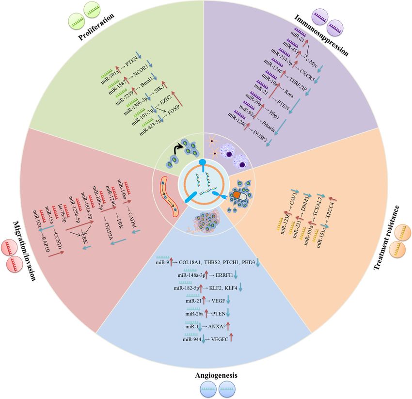
PROPOSED ROLES OF EXOSOMAL
Extracellular vesicles (EVs), membrane-encased vesicles miRNAs IN GLIOMA CANCER
secreted by cells, deliver cytoplasmic or membrane contents to
Accumulative evidence reveals that exosomes mediate the initiation
nearby cells and are detectable in biological fluids (9, 10).
and progression of gliomas by transferring biomolecules between
Although EVs were initially relegated as entities for cellular
distinct cell populations (23). Among these molecules, exosomal
waste disposal, nowadays, they work as messengers in
miRNAs are the most intriguing due to their important role in
intercellular communication (11). EVs are either buds from the
multi-respect glioma biology, encompassing proliferation,
endosomal network (exosomes) or derived from the plasma
migration and invasion, angiogenesis, immune suppression, and
membrane (microvesicles) (10). Exosomes are lipid bilayer-
treatment resistance within the glioma microenvironment (Table 1,
encapsulated vesicles with a diameter ranging from 30 to 120
Figure 1). Notably, the effects of exosomal miRNAs on glioma are
nm and are shed by diverse cell populations containing
highly similar to non-exosomal-derived miRNAs.
neoplastic cells. With multiple biologically active cargoes,
including proteins, lipids, mRNAs, and microRNAs (miRNAs),
exosomes have emerged as important players in cell-to-cell Exosomal miRNAs and Proliferation
communication (12). Exosomes are generated in endosomal of Glioma
compartments termed multivesicular bodies (MVBs) that are Proliferation is an important part of cancer progression,
late endosomes encompassing various intraluminal vesicles characterized by the alteration of expression and/or activity of
(ILVs) formed by endosomal membrane invaginations. MVBs cell cycle-associated proteins. Cell growth is also stimulated by
can subsequently integrate with plasma membranes, leading to constitutively activated signal transduction pathways (50).
the release of exosomes (13). Subsequently, exosomes are Exosomes shuttle genetic messages between cells via exosomal
internalized into neighboring or distant cells and transport miRNAs within the tumor environment, thus contributing to
their components, thereby influencing the phenotype of target glioma cell proliferation. Exosomal miRNAs regulate the
cells (14). Mounting evidence reveals that exosome-mediated proliferation of glioma cells in the following ways. Glioma
intercellular communication also serves important roles in many cells-derived exosomes are transmitted to glioma cancer cells,
respects of cancer progression, covering metastasis and drug and exosomal miRNAs modulate the proliferation of recipient
resistance as well as interfering with the immune systems within cells. For instance, Lan et al. observed the significantly enhanced
the tumor microenvironment (15, 16). It was reported that levels of exosomal miR-301a in serum of glioma patients relative
tumor cells produce high levels of exosomes, where the to healthy individuals. Moreover, miR-301a delivered by
components vary in distinct pathological and physiological exosomes derived from GBM cells promoted proliferation and
conditions (17). Recent data confirmed that circulating invasion of low-grade H4 glioma cells through directly targeting
exosomes appear as a promising means for biomarker phosphatase and tensin homolog (PTEN) to enhance the AKT
discovery since they can be noninvasively collected, and their and FAK signaling pathways (24). Non-glioma-derived
in vivo half-life is short, and their cargoes are protected from exosomes are transmitted to glioma cells and further modulate
degradation (18). their proliferation. Figueroa et al. found that miR-1587 could be
MiRNAs is a kind of small non-coding RNAs, with 19-25 transferred by glioma related-mesenchymal stem cells (GA-
nucleotides that form base pairs with the 3’-untranslated regions hMSCs) to glioma stem-like cells (GSCs) via exosomes and
(3’-UTR) of target mRNAs, which further cause either mRNA increased GSC proliferation and clonogenicity to maintain a
destabilization or translational repression (19, 20). MiRNAs GSC-supportive niche via directly targeting the expression of
function as irreplaceable intercellular communication tools, as nuclear receptor co-repressor-1 (NCOR1) (25). Li et al. also
they are transmitted between cells through exosomes and impact revealed that miR-7239-3p, released by M2 microglial exosomes,
recipient cells’ phenotype (21). Exosomal miRNAs have been could enter glioma cells via endocytosis, resulting in the
connected with glioma progression via activation and/or repression of brain and muscle ARNT-like protein-1 (Bmal1)
suppression of certain signaling pathways (22). A better expression and facilitating glioma cells proliferation and
knowledge of the biological roles of exosomal miRNAs may migration (26). In addition, the molecule mechanisms of
facilitate the exploration and development of novel diagnoses exosomal miRNA in medulloblastoma (MB), another subtype
Frontiers in Immunology | www.frontiersin.org 2 January 2022 | Volume 12 | Article 813747Peng et al. Exosomal microRNAs in Glioma
TABLE 1 | The biological roles of exosomal miRNAs in glioma biology.
Exosomal miRNAs Donor cells/recipient cells Targets Biological functions References
i
miR-301a GBM cells/low grade H4 glioma cells PTEN Promoting proliferation and invasion (24)
miR-1587 GA-hMSCsii/GSCsiii NCOR1 Promoting proliferation (25)
miR-7239-3p M2 microglial/glioma cells Bmal1 Promoting proliferation and invasion (26)
miR-130b-3p mono-macrophages/MBiv cells SIK1 Suppressing proliferation, migration and invasion (27)
miR-101-3p mono-macrophages/MB cells FOXP4 (common target) Suppressing proliferation, migration and invasion (28)
miR-423-5p EZH2 (target gene of miR- Promoting apoptosis
101-3p)
miR-148a GBM cells /GBM cells CADM1 Promoting proliferation and metastasis (29)
miR-1246 hypoxic glioma cells/normoxic glioma FRK Promoting migration and invasion (30)
miR-10b-5p cells TFAP2A
miR-181a-5p, miR-125b-5p, Group 3 MB cells/ SHH MB cells ERK Promoting invasion and migration (31)
let-7b-5p
miR-15a M2 macrophage/glioma CCND1 Suppressing migration and invasion (32)
miR-92a RAP1B
miR-1 GBM cells/HBMVECv ANXA2 Suppressing angiogenesis and invasion (33)
miR-9 glioma cells / HUVECsvi COL18A1, THBS2, Promoting proliferation, migration, invasion and (34)
PTCH1, PHD3 angiogenesis
miR-148a-3p glioma cells/HUVECs ERRFI1 Promoting angiogenesis and proliferation (35)
miR-182-5p GBM cells/HUVECs KLF2 and KLF4 Promoting angiogenesis (36)
miR-21 GSC/HBMVEC VEGF Promoting angiogenesis (37)
miR-26a GSCs/HBMECs PTEN Promoting proliferation, migration and (38)
angiogenesis
miR-944 GSCs/HUVECs VEGFC Suppressed proliferation, migration and (39)
angiogenesis
miR-1238 TMZvii-resistant cells/TMZ-sensitive cells CAV1 Promoting resistance to TMZ (40)
Enhancing anti-apoptosis
miR-151a TMZ-resistant GBM cells/TMZ-sensitive XRCC4 Increasing chemosensitivity to TMZ (41)
GBM cells
miR-221 glioma cells/glioma cells DNM3 Promoting proliferation, migration and TMZ (42)
resistance
miR-301a hypoxia glioma cells/normaxia-cultured TCEAL7 Promoting radiation resistance (43)
glioma cells
miR-451 glioma cells/microglia or macrophages c-Myc Promoting proliferation and immunosuppression (44)
miR-214-5p GBM Cells/ microglia cells CXCR5 Creating a tumor-supportive milieu (45)
miR-1246 hypoxia glioma cells /macrophages TERF2IP Promoting immunosuppression (46)
Promoting proliferation and metastasis
miR-10a hypoxic glioma cells/MDSCviii Rora/IkBa/NF-kB, Promoting immunosuppression (47)
miR-21 Pten/PI3K/AKT pathways
miR-29a hypoxia glioma cells /MDSC Hbp1 Promoting immunosuppression and proliferation (48)
mir-92a Prkar1a
miR-1246 (hypoxia) glioma cells/PBMCsix DUSP3 Promoting immunosuppression (49)
i
Glioblastoma.
ii
glioma related-mesenchymal stem cells.
iii
glioma stem-like cells.
iv
medulloblastoma.
human brain microvascular endothelial cells.
vi
human umbilical vein endothelial cells.
vii
Temozolomide.
viii
myeloid-derived suppressor cells.
ix
peripheral blood mononuclear cells.
of glioma, have been recently investigated. Based on the finding enhancer of zeste homolog 2 (EZH2) to reinforce its inhibitory
of Huang’s group, exosomal miR-130b-3p activated the p53 effects on tumors (28). In short, these studies indicated that
signaling pathway via silencing serine/threonine-protein kinase selectively transferring miRNAs via exosomes between cells
1 (SIK1), thereby suppressing the proliferation, migration and represents an essential means for intercellular communication,
invasion of MB cells (27). In line with this report, Xue et al. and for modulating the proliferation of gliomas.
demonstrated that exosomes derived from MB patients’ plasma
could shuttle miR-101-3p and miR-423-5p to MB cells and Exosomal miRNAs and Invasion/Migration
suppress the proliferation, invasion and migration of MB cells. of Glioma
Mechanically, miR-101-3p and miR-423-5p exert their It is generally believed that the invasion of cancerous cells into
suppressive effect via a common target, forkhead box P4 surrounding vasculatures and tissues is the initial step for cancer
(FOXP4), while miR-101-3p also binds to the 3’-UTR of metastasis, a leading cause of cancer-associated death (51).
Frontiers in Immunology | www.frontiersin.org 3 January 2022 | Volume 12 | Article 813747Peng et al. Exosomal microRNAs in Glioma Malignant gliomas have a unique invasion capacity: impeding then activate STAT3 pathway to facilitate the proliferation and the surgical removal of all glioma cells and making relapse metastasis of GBM cells (29). Numerous studies have illustrated inevitable (52, 53). Accumulative evidence suggests that that hypoxia leads to the epithelial-to-mesenchymal transition exosomal cargos containing nucleic acids such as miRNAs play (EMT) (54), tumor invasion (55) and metastasis (56). Qian et al. decisive roles in glioma migration and invasion. Cai et al. found found that the hypoxia microenvironment could stimulate miR- that exosomal miR-148a expression was prominently elevated in 1246 in glioma. Besides, miR-10b-5p-enriched exosomes were the serum of GBM patients and was inversely related to the then internalized by normoxia glioma cells to enhance the expression of cell adhesion molecule 1 (CADM1). MiR-148a migration and invasion of glioma through suppressing fyn- delivered by glioma-derived exosomes could target CADM1 and related kinase (FPK) and Transcription factor AP-2 alpha FIGURE 1 | The underlying molecular mechanisms of exosomal miRNAs in modulating the progression of gliomas. In the glioma environment, exosomal miRNAs are absorbed by recipient cells and subsequently exert their functions in varieties of biological processes including proliferation, migration/invasion, angiogenesis, treatment resistance, immunosuppression, etc. Frontiers in Immunology | www.frontiersin.org 4 January 2022 | Volume 12 | Article 813747
Peng et al.                                                                                                         Exosomal microRNAs in Glioma

(TFAP2A), respectively (30). Similarly, exosomal miRNAs could            1a/VEGF signaling transduction (34). Besides, glioma-derived
also drive the migration and invasion of MB cells. Transfer of           exosomal miR-148a-3p reinforced angiogenesis through
Group 3 MB-derived exosomal miRNAs (miR-181a-5p/miR-                     activating the EGFR/MAPK signaling pathway with the ERBB
125b-5p/let-7b-5p) could promote the invasion and migration              receptor feedback inhibitor 1 (ERRFI1) (35).
of less invasive SHH MB cells by enhancing extracellular                    Hypoxia is a universal manifestation of solid tumors. It is
regulated kinases (ERK) activities in Ras/MAPK pathway (31).             caused by the rapid tumor growth that runs out the supply of
Finally, the exosomes released by cancer cells and the exosomes          oxygen, and impairment of blood flow results from the aberrant
secreted from other cell types participated in glioma invasion and       blood vessels in the tumor (66). It can promote angiogenesis and
migration. MiR-15a and miR-92a were poorly expressed in M2               tumor progression via altering the tumor microenvironment
macrophages exosomes. It has been verified that M2                        (TME) (67, 68). In a recent study by Li et al., hypoxic GBM-
macrophages could secret miR-15a and miR-92a via exosomes,               secreted miR-182-5p could be taken up by human umbilical vein
and subsequently, miR-15a and miR-92a separately repressed               ECs (HUVECs) via exosomes, which could promote
cyclin D1 (CCND1) and Ras-related protein Rap1b (Rap1b).                 angiogenesis by directly suppressing Kruppel-like Factor 2 and
Thus, the PI3K/AKT/mTOR signaling pathway was interfered,                4 (KLF2 and KLF4) and subsequently elevating VEGF receptors
and glioma migration and invasion was suppressed (32).                   (VEGFR) expression (36).
                                                                            Glioma stem cells (GSCs) are tumor cells that exist in primary
Exosomal miRNAs and Angiogenesis                                         GBM with stem-cell-like properties such as self-renew capacity
in Glioma                                                                and producing heterogeneous offspring (69). Moreover, GSCs
As a pivotal hallmark of tumors, angiogenesis is a requisite for         have been elucidated to participate in tumor growth and
cancers to satisfy their needs for nutrients and oxygen (57).            angiogenesis (70, 71). GSCs trigger tumor angiogenesis by
Aberrant angiogenesis could lead to malignant phenotypes and             releasing factors that facilitate ECs proliferation and tube
promote cancer metastasis (58). It is also established that              formation (72). Existing evidence demonstrated that GSCs
angiogenesis relates to the progression of glioma (59).                  secrete extracellular vesicles (containing exosomes) that include
Nevertheless, the molecular mechanisms that regulate glioma              pro-angiogenic proteins, mRNA and miRNA, which are
angiogenesis are still unclear and will be the focus of current          absorbed by human brain microvascular ECs (HBMVECs)
research. Since Folkman first discovered a theory about the               (37–39, 73, 74). Sun et al. identified that overexpressed miR-21
correlation between angiogenesis and tumor growth, anti-                 in GSCs-derived exosomes could activate the angiogenic capacity
angiogenic gene therapy has attracted the attention of many              of ECs by augmenting levels of VEGF, which further interplayed
scientists with its apparent advantages (60). The development of         with VEGFR2 to trigger downstream PI3-kinase/Akt pathway
angiogenesis inhibitors targeting pro-angiogenic signaling               (37). Furthermore, Wang et al. showed that GSCs-derived
pathways regulated by vascular endothelial growth factor                 exosomes carrying miR-26a promoted the tube formation of
(VEGF) has exhibited clinical benefits in the treatment of                HBMECs in vitro by targeting and suppressing PTEN, which
varieties of cancers. While the resistance of patients to anti-          activated the PI3K/Akt signaling pathway (38). Finally, a recently
VEGF therapy impeded cancer treatment (61). Therefore, a                 published report confirmed that exosome-mediated diffusion of
better understanding of the molecular mechanisms of tumor                miR-944 from GSCs to HUVECs decreased proliferation and
angiogenesis is necessary for a more effective anti-                     angiogenesis by decreasing VEGFC expression and remarkably
Angiogenic therapy.                                                      restraining Akt/ERK pathways (39). Thus, GSCs play a dual role
    The critical roles of miRNA in regulating tumor angiogenesis         in regulating angiogenesis in ECs, which depends on the
(62) were highlighted in studies. Exosomes that selectively              exosomal miRNA.
packed miRNA and shed from tumor cells can be internalized
by endothelial cells (ECs), thus facilitating the growth of new          Exosomal miRNAs and Treatment
blood vessels (63, 64). Increasing evidence has revealed that            Resistance of Glioma
exosomes regulate angiogenesis partially through delivering              The mainstay treatments against glioma remain surgical resection,
miRNAs in the glioma microenvironment. MiR-1 is a well-                  which is followed by chemotherapy and radiotherapy. Although
recognized tumor suppressor in several cancers (65). Loading             these methods can remove the majority of the tumor masses, tumor
miR-1 into GBM-derived EV could alleviate angiogenesis,                  recurrence remains inevitable, which raises great challenges to
invasion, and neurosphere formation of GBM cells through                 clinical management owing to treatment resistance (75). Evidence
directly targeting and inhibiting annexin A2 (ANXA2).                    revealed that GSCs with intrinsic resistance to therapy cause the
ANXA2 was evidenced to be an important oncogene whose                    recurrence (76). Exosomes secreted by both tumor and stromal cells
expression was inversely correlated with miR-1 in GBM cells              play an integral role in treating resistance because of their nature as
(33). In addition, miR-9 derived from glioma cells was                   mediators of intercellular communication (77). Moreover, it has
internalized by vascular endothelial cells, which enhanced               been suggested that the key exosomal miRNA could target diverse
angiogenesis. Mechanically, miR-9 could target collagen type             signaling pathways or affect regulatory proteins and their
XVIII alpha 1 chain (COL18A1), thrombospondin 2 (THBS2),                 corresponding genes, thereupon modulating GBM drug
patched 1 (PTCH1), and egl-9 family hypoxia-inducible factor 3           resistance (78). Exploring the machinery of treatment resistance
(PHD3) to degrade these mRNA, contributing to initiating HIF-            is relevant for eliminating these aggressive tumors.

Frontiers in Immunology | www.frontiersin.org                        5                                   January 2022 | Volume 12 | Article 813747
Peng et al. Exosomal microRNAs in Glioma
Temozolomide (TMZ), as a DNA alkylating agent, is the first- relevant molecules appears as a novel and promising therapy
line treatment for GBM (79) and has been confirmed to exert its for gliomas.
anti-tumor role through inducing DNA damage (80). TMZ GAMs occupy the largest proportion in tumor-infiltrating cells
treatment significantly prolongs the survival of GBM patients, for glioma and take up 30% of the whole glioma mass (89). The
while the existence of resistance limited the clinic efficacy (81). appearance and intensity of GAMs are closely related to gliomas
Relevant studies indicated that exosomes derived from TMZ- progression, and conclusive evidence reveals the indispensable role
resistant glioma could confer TMZ chemoresistance to the of interplay between GAMs and gliomas in creating an
recipient TMZ-sensitive cells via exosomes (40, 41). Thus, drug immunosuppressive milieu, consequently favoring glioma growth
resistance increased with the secretion of exosomes (82). Yin and invasion (90). Nevertheless, the specific functioning
et al. reported that bioactive miR-1238 could be incorporated mechanisms of tumor-infiltrated GAMs are yet to be addressed.
into TMZ-resistant GBM cells-derived exosomes and absorbed Based on the report by Van der Vos, GBM-derived EVs were
by TMZ-sensitive cells, which disseminated TMZ resistance by absorbed into microglia and monocyte/macrophage via in vivo
directly targeting and suppressing caveolin-1 (CAV1) and combined with in vivo methods. That resulted in the transfer of
subsequently activating EGFR-PI3K-Akt-mTOR pathway. The miR-21 and miR-451, two abundant miRNAs within GBM-EVs
epidermal growth factor receptor (EGFR) played a pivotal role in with known oncogenic properties, into the latter cell types and
TMZ resistance (40). Additionally, Zeng et al. demonstrated that decreased the level of c-Myc mRNA, a shared target of both
miR-151a showed downregulated expression in TMZ-resistant miRNAs. The absorption of GBM-EVs was accompanied by
GBM cells and recurrent specimens and was correlated with alterations in microglia phenotype, covering enhanced
TMZ resistance. Further study revealed that TMZ-resistant GBM proliferation and a shift in their cytokines profile towards
cells spread TMZ chemoresistance to TMZ-responsive GBM immunosuppression (44). Furthermore, Yang and his colleagues
cells under an exosomal miR-151a loss-dependent condition. demonstrated that the expression of miR-214-5p was aberrantly
Mechanically, X-ray cross-complementing gene 4 (XRCC4) was enhanced in GBM cells. It could be transferred into recipient
directly targeted by miR-151a, and the repression of miR-151a microglia through exosomes, leading to the repression of C-X-C
elevated XRCC4 levels, activating DNA repair and increasing the motif chemokine receptor 5 (CXCR5) and ameliorating the
resistance of glioma to TMZ (41). Finally, the level of serum expression of pro-inflammatory cytokines such as interleukin 6
exosomal miR-221 increased with the glioma grades, and (IL-6), interleukin 8 (IL-8) and tumor necrosis factor-a (TNF-a) to
gliomas with higher grades displayed a higher level of miR- regulate the inflammatory response of microglial cells, which helps
221. Exosomal miR-221 was connected with decreased TMZ create a tumor-supportive milieu (45). Besides, tumor-associated
sensitivity by targeting dynamin-3 (DNM3) genes (42). macrophages (TAM) are one of the main immune-related cells
Exosomal miRNAs shed by glioma cells are also associated infiltrating the tumor microenvironment. They are generally
with the sensitivity to radiotherapy, which can impact treatment categorized into two subtypes with distinct functions, namely
efficiency. According to recent data, exosomal miR-301a, which classically activated (M1 macrophages) and alternatively activated
was specifically expressed and released by hypoxia GBM cells, (M2 macrophages), respectively. The former is characterized by
could transfer to corresponding normoxia-cultured cells, where anti-tumor activities, while the latter increases metastasis of tumors
it repressed the expression of anti-oncogene transcription and angiogenesis and suppresses the anti-tumor immune response
elongation factor A (SII)-like 7 (TCEAL7) and subsequently (91). Tumors provide a tumor-permissive milieu by exploiting
activated the Wnt/b-catenin Signaling pathway, thus decreasing macrophage polarization states and specifically skewing
radiation sensitivity (43). macrophages toward a pro-tumoral M2-like phenotype, thereby
supporting cancer progression by immune suppression (92).
Exosomal miRNAs and What’s more, emerging evidence indicates that exosomal
Immunosuppression in Glioma miRNAs secreted by glioma cells can control the phenotypic
Glioma-related immune cells, the substantial component in the plasticity of macrophages to facilitate tumor growth. For example,
glioma microenvironment, play an emerging role in the the recently published literature by Qian has determined that miR-
regulation of cancer progression and control of anti-tumor 1246 was upregulated in hypoxia glioma-derived exosomes (H-
immunity (83–85). Macrophages/microglia (GAMs) and GDE) and GBM patients’ cerebrospinal fluid (CSF), and delivery of
tumor-induced myeloid-derived suppressor cells (MDSCs), two H-GDE-derived miR-1246 contributed to inducing M2
prominent populations within the tumour stroma, can exert pro- macrophage polarization through targeting telomeric repeat
tumorigenic effects and establish an immunosuppressive milieu binding factor 2 interacting protein (TERF2IP). Thus, the STAT3
(86, 87). M2 phenotypic conversion in glioma-associated GAMs signaling pathway was activated and NF-kB signaling pathway was
and MDSCs expansion can help neoplastic cells evade immune repressed, promoting the development of the immunosuppressive
system-mediate detection and destruction (88). Emerging studies microenvironment (46).
suggest that exosomal miRNAs are carriers of information that Myeloid-derived suppressor cells (MDSCs), as a heterogeneous
have the potential to modulate macrophage fate of differentiation population of cells expanding during cancer, can depress activation
(46) and induce MDSCs accumulation and expansion (48), of T-cells and NK-cells to promote tumor growth, quicken
thereupon creating a favorable environment for glioma formation of the pre-metastatic niche, and cause resistance to
progression. Therefore, targeting these immune cells and immunotherapy (93). Previous studies have evidenced that
Frontiers in Immunology | www.frontiersin.org 6 January 2022 | Volume 12 | Article 813747Peng et al. Exosomal microRNAs in Glioma
tumor-derived exosomes drive MDSCs activation and expansion consistent diagnosis, prognostication, and treatment
(94), and miRNAs play essential roles in regulating the expansion of response prediction.
functional MDSCs (95). However, the exact mechanisms where Although methods of early diagnosis and timely management
miRNA-containing exosomes derived from glioma cells can of various cancer types have been established, such as detection
manipulate the differential of functional MDSCs remains to be of circulating tumor cells (CTCs) and cell-free nucleic acids
defined. To the end, Guo et al. identified that hypoxia-stimulated (cfNAs) in the circulation, comparatively little progress has been
glioma-derived exosomes (GDEs) could be taken up into MDSCs, made concerning clinical validation of intrinsic brain tumors.
exhibiting a stronger capacity to induce MDSCs compared with Broad sets of promising biomarkers have already been identified
normoxia-stimulated GDEs. It was exactly the hypoxia-inducible in the blood and CSF of patients afflicted with gliomas, but few
enhanced expressed miR-10a and miR-21 within GDEs that were applied clinically (101).
reinforced MDSCs expansion and activation through targeting In recent years, exosomes have become an emerging topic
Rora/IkBa/NF-kB and Pten/PI3K/AKT pathways (47). Guo et al. implicating multifaceted development and biological process
also revealed that exosomal miR-29a and miR-92a, which were through being secreted into nearly all human fluids, containing
transferred from hypoxia-induced glioma cells to MDSCs could plasma, CSF, urine, breast milk, and saliva (102). Accumulative
facilitate the formation of the immunosuppressive investigations have been centered on exosomes as they can be
microenvironment by increasing the proliferation of functional used as diagnostic, prognostic, and therapeutic markers (103).
MDSCs via silencing high-mobility group box transcription Previous studies have demonstrated that exosomes can cross the
factor1 (Hbp1) and protein kinase cAMP-dependent type I intact blood-brain barrier (BBB) (104). Besides, the bilayer-lipid
regulatory subunit alpha (Prkar1a), separately (48). Additionally, membranes of exosomes protect the bioactive cargos they
they also demonstrated that miR-1246, which was abundant in contain from enzymatic RNase degradation (105). MiRNAs
GDEs, could also potentiate MDSCs with manifestations including within peripheral blood exosomes may be relevant biomarkers
repressing CD8+ cells proliferation and elevating the levels of and therapeutic targets for glioma since they are tumor
interleukin 10 (IL-10) and transforming growth factor-beta regulators possessing oncogene and suppressor gene roles
(TGF-b) by activating the DUSP3/ERK pathway (49). The above- (106). Exosomes are extractable from peripheral blood, and
described studies indicate that glioma potently influences MDSCs miRNAs can be detected by technologies. Exosomal miRNAs
differentiation and activation via exosomal miRNAs, thereupon could be regarded as promising comprehensive biomarkers, as
impacting the entire tumor immune environment. they possess the potential to present real-time information of
disease and can predict progressive disease and assess the
response of cancers to targeted therapies (106) (Table 2).
Some studies have reported the diagnostic implication of
CLINICAL IMPLICATIONS OF EXOSOMAL exosomal miRNAs derived from sera. Exosomal miRNAs are
miRNAs IN GLIOMA correlated with histopathologic grades of gliomas. For instance,
RNU6-1, miR-320, and miR-574-3p were fast and reliable
Exosomal miRNAs as Biomarkers diagnostic markers of GBM patients as their circulating levels
for Glioma increased with the area under the curve (AUC) of 0.926 (107).
Precise diagnosis, and the timely/comprehensive monitoring of This research opened up a novel avenue that exosomal miRNAs
therapeutic response, remain the main challenges in glioma could be applied for GBM diagnosis. Ebrahimkhani characterized a
patients’ care and treatment. Neuroimaging, magnetic resonance set of exosomal miRNAs (miR-182-5p, miR-328-3p, miR-339-5p,
imaging (MRI) in particular, is regularly used for glioma diagnosis, miR-340-5p, miR-485-3p, miR-486-5p as well as miR-543) derived
staging, and monitoring therapeutic response. Although from human serum to distinguish GBM patients from healthy
neuroimaging is suggestive of glioma diagnosis, there are still controls with a predictive accuracy of 91.7% (108). Moreover,
other brain lesions sharing radiological features, which makes Santangelo validated that the expression of miR-21/miR-222/
differential diagnosis difficult (96). Additionally, the lowest miR-124-3p in serum exosomes of patients with high-grade
resolution for the effective detection by MRI remains on the order gliomas (HGG) was remarkably higher than those with low-grade
of millimeters (97). Because of the dimension of a tumor cell, this gliomas (LGG) and healthy individuals. The combination of miR-
disparity in scale translates into delayed diagnosis and treatment 21+miR-222+miR-124-3p was more robust in discriminating HGG
(98). Furthermore, MRI can exhibit the enhanced tumor volume than healthy controls, but its accuracy markedly decreased after
following radiotherapy, generally, due to enhanced vascular surgical resection of the tumors. Intriguingly, exosome-associated
permeability, an effect named pseudoprogression, and connected miR-21 expression alone appeared as the greatest predictor for
with the treatment (99). In the meantime, histopathologic differentiating patients with HGG from those with LGG, with an
examination of tumor specimens attained by surgery is currently AUC of 0.83. Meanwhile, the results also revealed that high
recognized as the gold standard for glioma grading and typing. exosomal miR-21 together with low exosomal miR-222 and miR-
Nevertheless, this method bears high surgical risks, and repeated 124-3p expression could effectively help to distinguish brain tumors
sampling of tumor specimens may be impractical (100). All these of glial origin from those without glial origin at initial
factors demonstrably highlight the urgent need for exploring neuroradiological assessment, which may be helpful for patients
minimally invasive biomarkers supportive for reliable and showing inconclusive biopsies or with masses in the essential and
Frontiers in Immunology | www.frontiersin.org 7 January 2022 | Volume 12 | Article 813747Peng et al. Exosomal microRNAs in Glioma
TABLE 2 | Related studies about exosomal miRNAs from human fluids as diagnostic, prognostic and predictive biomarkers in glioma.
Exosomal Sample type Expression Clinical value Reference
miRNAs status
miR-320 serum ↑ diagnostic biomarker for GBMx (107)
miR-574-3p
RUN6-1
miR-182-5p serum ↑ diagnostic biomarker for GBM (108)
miR-328-3p ↓
miR-339-5p ↓
miR-340-5p ↓
miR-485-3p ↓
miR-486-5p ↑
miR-543 ↓
miR-21 serum ↑ diagnostic biomarker for HGGxi (109)
miR-222 diagnostic biomarke for grade prediction (HGG over LGGxii, miR-21)
miR-124-3p
miR-766-5p serum ↓ diagnostic biomarker for discriminating HGG over intracranial lymphoma (110)
↓
miR-301a serum ↑ diagnostic biomarker for glioma (24)
prognostic biomarker for poor OSxiii
miR-454-3p serum ↑ diagnostic biomarker for glioma (111)
prognostic biomarker for poor survival
miR-210 serum ↑ diagnostic biomarker for glioma and grade prediction (high grade over low grade) (112)
prognostic biomarker for recurrence and poor OS
miR-181b serum ↓ prognostic biomarker for shorter post-surgical survival time (113)
miR-182-5p serum ↑ prognostic biomarker for LGG (114)
miR-223-3p ↑
miR-34a-5p ↓
miR-497-5p ↓
miR-375 plasma ↑ diagnostic biomarker for glioma (115)
miR-210 Plasma ↑ diagnostic and prognostic biomarker showing association with histopathological grade (GBM (116)
miR-449 ↓ over LGAxiv)
miR-5194 ↓
miR-2276-5p Plasma ↓ diagnostic biomarker for glioma and grade prediction (HGG over LGG) (117)
Prognostic biomarker for better OS
miR-21 CSFxv ↑ diagnostic biomarker for GBM (118)
miR-21 CSF ↑ diagnostic biomarker for discriminating glioma patients over non-tumor brain disease (119)
diagnostic biomarker for grade prediction (GBM over grade II gliomas)
prognostic biomarker for poor survival
miR-182-5p Serum and ↑ diagnostic biomarker for glioma and grade prediction (HGG over LGG) (36)
CSF prognostic biomaker for glioma
miR-1246 CSF ↑ prognostic biomarker for recurrence (49)
miR-9-5p Serum and ↑ prognostic biomaker for poor survival (120)
CSF
miR-151a CSF ↓ predictive biomarker for TMZxvi response (41)
miR-574-3p serum ↑ predictive biomarker for the effect of radiotherapy for glioma patients (121)
miR-21 Serum ↑ predictive biomarker for therapy response in post-surgical following-up (122)
miR-222
miR-124-3p
x
glioblastoma multiforme.
xi
high-grade glioma.
xii
low-grade glioma.
xiii
overall survival.
xiv
low-grade astrocytoma.
xv
cerebrospinal fluid.
xvi
Temozolomide.
intricate areas of the brain (109). Furthermore, according to the auxiliary indicator for the diagnosis of HGG of intracranial
report from Wang’s group, both miR-766-5p and miR-376-5p in lymphoma, showing an AUC value of 0.7201 (110). Additionally,
serum exosomes were strikingly attenuated in intracranial exosomal miR-301a was significantly upregulated in the serum of
lymphoma, and HGG patients relative to healthy controls, and glioma patients, which was connected with ascending pathological
the levels of exosomal miR-766-5p were lower in the intracranial grades and decreased Karnofsky performance status (KPS) scores.
lymphoma group than the HGG group. Exosomal miR-766-5p and ROC curve analysis revealed that exosomal miR-301a exhibited a
miR-376-5p could effectively diagnose HGG with AUCs of 0.8883 potential diagnostic value for discriminating gliomas patients from
and 0.7688, respectively. Besides, miR-766-5p could be used as an non-glioma patients (AUC=0.937) (24). Research by Shao et al.
Frontiers in Immunology | www.frontiersin.org 8 January 2022 | Volume 12 | Article 813747Peng et al. Exosomal microRNAs in Glioma
revealed that miR-454-3p was markedly under-expressed in glioma could function as a feasible biomarker of GBM, with an AUC of 0.9
tissues while over-expressed in exosomes, and exosomal miR-454- (118). Consistent with this finding, the data from Shi’s group have
3p could function as a marker for glioma diagnosis with AUC value highlighted that according to its reinforced expression in gliomas,
of 0.863. Levels of exosomal miR-454-3p in the postoperative CSF-derived exosomal miR-21 appeared as an excellent index to
serums were prominently lower relative to those in the distinguish glioma and non-neoplastic brain diseases yielding an
preoperative serums (111). Finally, Lan et al. identified that exo- AUC of 0.927. Moreover, it had the potential to specifically separate
miR-210 whose expression was significantly reinforced in glioma GBM and grade II gliomas with an AUC value of 0.751. The authors
patients and elevated with ascending pathological grades, could also identified that a high abundance of miR-21 bore an inverse
effectively identify glioma patients from healthy controls correlation with patients’ survival (119). Both of the
(AUC=0.856) (112). aforementioned studies suggest that expression levels of exosomal
The aforementioned dysregulations of exosomal miR-301a, miR-21 in CSF could be a promising indicator applying for glioma
miR-454-3p and miR-210 was also connected with the prognosis diagnosis and prognosis. Another recently published paper has
of glioma patients. Specifically, the expression of serum exosomal observed that glioma patients had enhanced expression of miR-
miR-301a was significantly diminished following the surgical 182-5p from serum- and CSF-derived exosomes compared with
removal of primary tumors and increased during GBM healthy individuals, and high-grade patients, GBM patients, in
recurrence. Kaplan-Meier analysis suggested that malignant particular, showcased higher exosomal miR-182-5p than LGG
patients with an elevated exosomal miR-301a expression patients. Additionally, a dramatic drop in miR-182-5p expression
generally have a poorer survival (OS) (24). Similarly, enhanced was detected in the postoperative period. Exosome-mediated miR-
expression of miR-454-3p (111) and miR-210 (112) in glioma 182-5p, thereupon, could be a desirable diagnostic and prognostic
serum exosomes were both related to poor prognosis. Another biomarker of glioma (36). Moreover, CSF exosomal miR-1246 was
study also reported that augmented levels of exosomal miR-181b associated with glioma and could potentially be applied for
in GBM patients’ serum could indicate a worse functional monitoring tumor recurrence after surgery (49). Finally,
outcome and suggest a prominently shorter post-surgical augmented expression of small EVs-derived miR-9-5p was linked
survival time for GBM patients (113). Also, Caponnetto et al. to a decreased survival in IDH-mutated glioma patients, suggesting
analyzed and compared the miRNA profile of exosomes released that miR-9-5p within the EVs from blood and CSF might be
by glioma-associated stem cells (GASC), and confirmed that explored as a potential molecular biomarker (120).
abnormal expression levels of exosomal miRNAs probably Acquired drug resistance is emerging as a leading clinical
contributed to the malignant progression phenotypes. These limiting factor in the treatment of gliomas. Thereby, the
exosomal miRNAs from serum warrant further investigation identification of potential drug-resistant patients and exploration
for the improvement of LGG prognostic stratification (114). of alternative treatment timely are relevant to ameliorate prognosis.
In addition to sera, increasing research have focused on Exosomes can influence therapy resistance as delivery vehicles of
identifying promising biomarkers based on exosomal miRNAs miRNAs and can predict therapeutic responses as biomarkers. Zeng
attained from glioma patients’ plasma. In a recent study by Xu et et al. identified that GBM patients with attenuated CSF exosomal
al, circulating exosomal miR-375 in the plasma of glioma miR-151a levels exhibited a worse prognosis, revealing a dismal
patients was prominently reinforced and the extent of this response to TMZ (41). Additionally, miR-574-3p derived from
upregulation was positively associated with tumor grades, serum exosomes was significantly diminished after radiotherapy,
indicating that miR-375-containing exosomes held great and it was proposed as an important candidate biomarker for
promise as a diagnostic marker (115). Similarly, Tabibkhooei evaluating the therapeutic effect of radiotherapy in glioma (121).
et al. demonstrated that enhanced expression of miR-210 and Furthermore, reinforced expression levels of serum exosomal miR-
declined expression of miR-5149 and miR-449 in plasma had a 21, miR-222 and miR-124-3p were associated with HGG
positive correlation with histopathological grade of glioma, progression. Specifically, after the chemo-radiotherapy, HGG
which favored GBM detection and prognosis prediction. That patients with high levels of these miRNAs showed significantly
implied that plasma exosomal miRNAs are expected to be novel shorter progression-free survival (PFS) and overall survival (OS)
biomarkers of GBM (116). Another study also confirmed that (122). Hence, the profiles of exosomal miRNAs could potentially
miR-2276-5p expression levels were declined in the plasma- predict the therapeutic response in glioma patients.
derived exosomes of glioma patients compared with those of Exosomes contain not only miRNAs but also other noncoding
non-glioma controls and its expression was lower in HGG RNAs (ncRNAs), including circular RNAs (circRNAs) or long
patients than in LGG patients. Exosomal miR-2276-5p could non-coding RNAs (lncRNAs). Exosomal miRNA mediated glioma
effectively diagnose glioma patients with an AUC of 0.8107. progression via interacting with circRNAs or lncRNAs.
Furthermore, low levels of exosomal miR-2274-5p in glioma Accumulative studies have also focused on the expression of
were linked to poorer survival rates (117). exosomal circRNAs/lncRNAs in serum of glioma patients. For
Aberrant expression of exosomal miRNAs, a hallmark of glioma, example, exosomal circ_0072083 was reported to contribute to
can be detected in blood, and other body fluids, including CSF. For TMZ resistance in glioma via modulating exosomal miR-1252-5p-
instance, Akers and colleagues proved that miR-21 expression levels mediated nanog homeobox (NANOG) degradation. Moreover,
were highly elevated in EVs derived from CSF of GBM patients exosomal circ_0072083 could serve as an independent diagnostic
relative to those from non-oncologic patients. CSF EV miR-21 target for gliomas with an AUC of 0.85, and patients with high
Frontiers in Immunology | www.frontiersin.org 9 January 2022 | Volume 12 | Article 813747Peng et al. Exosomal microRNAs in Glioma
expression of exosomal circ_0072083 displayed poorer OS (123). exosomes which were absorbed by neighboring GSC. Thus, its
Also, Yin’s team found that exosomal circMMP1 in serum targets containing Cyclin D1, Cyclin A, E2F transcription factor 1
facilitated the progression of glioma via functioning as a (E2F1) and C-X-C motif chemokine receptor 4 (CXCR4) pathway
competitive endogenous RNA (ceRNA) of miR-433 (124). were repressed, so did the proliferation and tumorigenicity of GSCs
Furthermore, exosomal lncSBF2-AS1 transferred from TMZ- (135). Emerging studies have proved that decoy or sponge-like
resistant GBM cells to chemoresponsive GBM cells could constructs could be employed for miRNA inhibition. They have
function as a ceRNAs for miR-151a-3p, contributing to the TMZ potential therapeutic benefits by binding complementary miRNA(s)
resistance (125). Consistently, based on the findings from Hao’ or seed sequences, which can impede the crosslink between miRNAs
group, lncRNA PTENP1 could be packaged into exosomes of and their targets (147–149). Monfared and colleagues tried to down-
human umbilical cord mesenchymal stem cells and transferred into regulated the expression of miR-21 in GBM cell lines, U87-MG and
U87 glioma cells, and subsequently impaired the cell growth via C6, through utilizing engineered exosomes packed with a miR-21-
suppressing the expression of miR-10a-5p (126). All these studies sponge construct. They found that cells treated with miR-21-sponge
have demonstrated that exosomal ncRNAs could be served as the exosomes exhibited a decline in proliferation and an elevation in
promising diagnostic/prognostic markers and therapeutic targets. apoptotic rates (136). Thereupon, exosomes can be used as a
To sum up, the multiple exosomal ncRNAs, miRNAs in promising therapeutic delivery vehicle in glioma treatment.
particular, from serum, plasma, CSF and other body fluids will Recently, accumulative evidence has highlighted that
promote the broad application of exosome-based liquid biopsy mesenchymal stem cells (MSCs), as a novel approach, can
strategies in the early diagnosis, prognosis and therapeutic overcome the demerits of previous therapies for gliomas (150,
response prediction of gliomas. 151). MSCs can pass through the BBB and possess inherent
tropism towards tumors and low immunogenicity (152).
The Potential Application of Exosomal Additionally, MSCs may exert their therapeutic capacity via
miRNAs in Anti-Glioma Treatment producing and secreting useful exosomes in glioma treatment
Given the pivotal biological meanings of exosomal miRNAs in (153). Several investigations have suggested the delivery of tumor-
glioma, approaches that specifically target exosomes or exosomal suppressive miRNAs to cancers through MSC-derived exosomes
cargoes including miRNAs, are emerging as promising therapies for (154, 155). Studies on glioma have identified that exosomes
gliomas. Among the potential therapeutic applications of exosomes, displayed anti-glioma effects when tumor-suppressive miRNAs
significant attention has been dedicated to applying these vesicles as expression was elevated in the exosomes-donating MSCs, which
nanocarriers to deliver small molecules, proteins and nucleic acids revealed the considerable potential for the application of MSC-
such as miRNAs (127). Despite the development of novel methods derived exosomes in treating gliomas. To the end, Lang and
to engineer exosomal cargoes in producer cells, low yields of colleagues found that MSCs could be exploited as natural
exosomes impede the widespread application of exosome-based biofactories to produce exosomes carrying supraphysiological
therapies. Thereupon, some studies have been established to levels of miR-124a, an effective anti-glioma agent Exo-miR124a
regulate exosomes production to treat cancer. For example, Li exosomes resulted in significantly repressing the viability and
et al. confirmed that two intriguing compounds termed MOPIPP clonogenicity of GSCs. When systemically administered, the mice
and vacuolin-1 could facilitate the vacuolization of endosomal bearing intracranial GSCs xenografts were cured. Mechanistic
compartments and disrupt the trafficking of late endosomes to studies proved that miR-124a acted through silencing Forkhead
lysosomes without exhibiting significant cytotoxicity. Thus box (FOX)A2, contributing to abnormal intracellular lipid
exosomes production in GBM cells was enhanced, whereas accumulation (137). Moreover, Yan and co-workers found that
selected miRNAs carried by the exosomes were identified to show miR-512-5p was poorly expressed in GBM tissues and cells, and
qualitative similarity to those in untreated cells, suggesting that exosomes derived from miR-512-5p-transfected BMSCs were
MOPIPP and vacuolin-1 could help develop exosome-based anti- internalized by U87 cells, thus interfering with GBM cell
cancer therapeutics (128). However, the potential action mechanism proliferation and inducing cell cycle arrest through negatively
of these compounds should be further explored. regulating Jagged 1 (JAG1) (138). Furthermore, miR-29a-3p, a
MiRNAs may be a novel antineoplastic agent as they can tumor suppressor in diverse malignant tumors, was demonstrated
modulate the posttranscriptional expression of target genes (129– to directly target roundabout guidance receptor 1 (ROBO1), thus
132). Restitution of several downregulated tumor-suppressor mitigating vasculogenic mimicry (VM) formation in gliomas. MSCs
miRNAs could repress the growth of GBMs, indicating that could be engineered to produce miR-29a-3p-overexpressing
certain miRNAs could be used for anti-glioma therapeutics (133). exosomes, and treatment with these exosomes restrained
Currently, it seems unsuitable to use available vehicles to deliver migration and VM formation, thereby suppressing the growth of
miRNAs containing liposomes and viral vectors because of their low glioma in vitro and in vivo (139). VM, a kind of alternative
efficiency and safety (134). As nature miRNAs carriers, exosomes microvascular circulation bypassing the canonical VEGF-driven
may be employed to provide tumor-suppressor miRNAs to mediate angiogenesis, shows resistance to anti-angiogenesis therapy (156).
tumor growth suppression and realize personalized treatment Tumor necrosis factor-related apoptosis-inducing ligand (TRAIL)
(Table 3). For example, Fareh et al. engineered patients’ derived has strong potential to kill cancerous cells by inducing apoptosis,
GSCs to stably and continuously express the miR-302-367 cluster, with minimal effect on normal cells (157). Zhang et al. identified
and a large amount of tumor-suppressor miRNA was enclosed in that miR-7 could sensitize GBM cells to TRAIL-induced apoptosis
Frontiers in Immunology | www.frontiersin.org 10 January 2022 | Volume 12 | Article 813747Peng et al. Exosomal microRNAs in Glioma
TABLE 3 | Identification of exosomal miRNAs as potential anti-glioma therapeutics.
Exosomal miRNAs Donor cells/recipient cells Targets Anti-glioma effects References
xvii
miR-302-367 GSC /GSC Cyclin D1, CyclinA, E2F1, CXCR4 Suppressing proliferation (135)
pathway
a miR-21-sponge HEK-293T/U87-MG PDCD4 and RECK Suppressing proliferation (136)
construct Increasing apoptosis
miR-124a MSCsxviii/ GSCs (FOX)A2 Suppressing proliferation (137)
miR-512-5p BMSCsxix/GBM cells JAG1 Suppressing proliferation (138)
Inducing cell cycle arrest
miR-29a-3p MSC/glioma cells ROBO1 Suppressing migration and VM (139)
miR-7 MSCs/GBMxx cells XIAP Enhancing TRAIL sensitivity (140)
Increasing apoptosis and suppressing tumor
growth
miR-146b Marrow stromal cells/ glioma EGFR, NF-kB and SMAD4 Suppressing growth, invasion and migration (141)
cells
miR-133b MSC/glioma cells EZH2 Suppressing proliferation, invasion and migration (142)
miR-584-5p MSC/glioma cells CYP2J2 Suppressing proliferation and migration (143)
MMP-2 Suppressing metastasis
Bcl-2 and Bax Induce glioma cells apoptosis
miR-375 Marrow stromal cells/ glioma SLC31A1 Suppressing proliferation, migration, invasion (144)
cells Increasing apoptosis
miR-199a MSC/glioma cells AGAP2 Suppressing proliferation, invasion and migration (145)
Enhancing chemosensitivity to TMZ
miR-124 WJ-MSCxxi/GBM cells CDK6 Suppressing proliferation (146)
IQGAP1, LAMC1, ITGB1 Suppressing migration
R-Ras and N-Ras Increasing chemosensitivity to TMZ
xvii
glioma stem-like cells.
xviii
mesenchymal stem cells.
xix
bone mesenchymal stem cells.
xx
glioblastoma.
xxi
Wharton’s jelly MSCs.
and reinforced expression of miR-7 in TRAIL-overexpressed MSCs, thereby repressing the proliferation, invasion and migration of
increased apoptosis and repressed tumor growth in an exosomes- glioma cells in vivo, and ameliorating the chemosensitivity to
dependent fashion (140). Additionally, based on the report of TMZ and suppressing the tumor growth in vivo (145).
Katakowsk’s group, the delivery of exosomes derived from MSCs Until now, Studies of MSCs have focused primarily on BMSCs.
directly by intratumoral injections prominently decreased the However, the derivation of stem cells from Wharton’s jelly (WJ) in
growth of glioma xenograft in the rat brain with the inclusion of the human umbilical cord also possesses various advantages.
miR-146b. The anti-tumor effect of MSCs-derived exosomes was Compared to other sources of MSCs, Wharton’s jelly MSCs (WJ-
underpinned by concurrent inhibition of factors including EGFR, MSCs) display greater expansion capacity, a higher rate of
NF-kappaB and mothers against decapentaplegic homolog 4 proliferation, reinforced neurotrophic factors and a stronger
(SMAD4) (141). It was also found that MSCs-derived exosomes potency to default neuronal lineage differentiation. Although these
transmitting miR-133b into glioma cells could inhibit EZH2 cells are characterized by exceptionally low immunogenicity, they
expression by disrupting the Wnt/b-catenin signaling pathway, have high neuroprotective potential and appear as a superior source
thereupon repressing proliferation, invasion and migration of for the application of MSCs without ethical limitations (158–160).
glioma cells (142). Moreover, transferring miR-584-5p through Sharif et al. confirmed that WJ-MSCs could efficiently deliver
exosomes derived from MSCs could suppress the proliferation exogenous miR-124 to U87 GBM cells via exosomes, ameliorating
and migration of U87 cells via decreasing levels of cytochrome GBM cells’ chemosensitivity to TMZ and decreasing proliferation
P450 family 2 subfamily J member 2 (CYP2J2). This move could and migration of GBM cells. Mechanically, mitigated expression of
also repress glioma metastasis through reducing expression of cyclin-dependent kinase 6 (CDK6) by the delivered miR-124
Matrix metalloproteinase-2 (MMP-2), and induce carcinoma cells diminished the proliferation of GBM cells. Besides, the migration
apoptosis via decreasing levels of B-cell lymphoma-2 (BCL-2), an of GBM cells was attributed to the inhibition of IQ motif containing
anti-apoptotic protein, and increasing expression levels of BCL-2 GTPase activating protein 1 ((IQGAP1), laminin c1 (LAMC1) and
associated X (BAX), a pro-apoptotic protein (143). Exosomal miR- integrin b1 (ITGB1). At the same time, miR-124 also governed the
375 from human marrow stromal cells (hMSCs) resulted in chemosensitivity of GBM cells by targeting R-Ras and N-Ras (146).
suppressed cell proliferation, invasion and migration while These studies suggest that MSCs could naturally package anti-tumor
promoted apoptosis through solute carrier family 31 member 1 miRNAs into exosomes, illustrating their considerable potential for
(SLC31A1) inhibition (144). Similarly, miR-199a, when delivered by therapeutic applications. Nevertheless, many in-depth studies are
MSCs via the exosomes, could negatively regulate ArfGAP with required before these therapeutic strategies are available for
GTPase domain, ankyrin repeat and PH domain 2 (AGAP2), clinical usage.
Frontiers in Immunology | www.frontiersin.org 11 January 2022 | Volume 12 | Article 813747You can also read