Targeting memory T cell metabolism to improve immunity
←
→
Page content transcription
If your browser does not render page correctly, please read the page content below
The Journal of Clinical Investigation                                                                                   R E V I E W S E R I E S : I M M U N O M E TA B O L I S M
                                                                                                                                         Series Editor: Jonathan D. Powell

Targeting memory T cell metabolism to improve
immunity
Mauro Corrado1 and Erika L. Pearce2
Cologne Excellence Cluster on Cellular Stress Responses in Aging-Associated Diseases (CECAD), Cologne, Germany. 2Department of Oncology, The Bloomberg~Kimmel Institute for Cancer Immunotherapy,
1

Johns Hopkins University, Baltimore, Maryland, USA.

             Vaccination affords protection from disease by activating pathogen-specific immune cells and facilitating the development
             of persistent immunologic memory toward the vaccine-specific pathogen. Current vaccine regimens are often based on
             the efficiency of the acute immune response, and not necessarily on the generation of memory cells, in part because the
             mechanisms underlying the development of efficient immune memory remain incompletely understood. This Review
             describes recent advances in defining memory T cell metabolism and how metabolism of these cells might be altered in
             patients affected by mitochondrial diseases or metabolic syndrome, who show higher susceptibility to recurrent infections
             and higher rates of vaccine failure. It discusses how this new understanding could add to the way we think about immunologic
             memory, vaccine development, and cancer immunotherapy.

Introduction                                                                                            How lymphocytes develop effector and memory phenotypes
Immunologic memory is the primary goal of vaccination. This                                         has been attributed to cell-intrinsic mechanisms involving pro-
phenomenon is characterized by qualitatively and quantitatively                                     longed cellular longevity (6, 7), posttranslational regulation of
improved and/or enhanced antigen/epitope-specific recognition                                       key proteins (8), and epigenetic reprogramming of the cellular
by B and T cells of the adaptive immune system (1). B and T cells                                   transcriptome (9), or to cell-extrinsic mechanisms linked to anti-
have different but cooperative roles in responding to infections.                                   gen presentation and costimulatory signals (10–12). In recent
Memory B cells generate high-affinity neutralizing antibodies                                       years, the emerging field of immunometabolism has started to
that, when produced at sufficient levels, can prevent viruses and                                   unveil the role of metabolism in shaping immune function, and to
bacteria from infecting cells. Memory T cells (Tm cells) also par-                                  reveal how modulating cell or organismal metabolism can affect
ticipate in this protection, as their rapid expansion and cytotoxic                                 immune cell differentiation (13).
properties facilitate pathogen control and clearance, thus limiting
or ablating pathology development (2).                                                              The dynamic nature of mitochondria in T cells
     Even though the generation of durable and persistent immuno-                                   Cells constantly sense nutrient availability in their microen-
logic memory is the basis of any successful vaccination, the mecha-                                 vironment, adapting function and survival to metabolic state.
nisms underlying induction and maintenance of immunologic mem-                                      Driving this adaptation, mitochondria fine-tune their function in
ory remain elusive. In particular, it is still debated exactly how Tm                               response to the dynamic metabolic requirements of the cell (14).
cells are generated upon acute infection, how such long-lived cells                                 Mitochondrial biogenesis is triggered in response to higher met-
are induced and differentiate from effector T (Teff) cells, and which                               abolic needs, while selective autophagy removes dysfunctional
mechanisms control their survival and enhanced function for years                                   organelles (15, 16). Changes in mitochondrial morphology couple
if not decades (3). In this context, many vaccines mainly elicit B cell                             location and shape of mitochondria to efficient energy produc-
responses, poorly priming T cells. This is often the case with subunit                              tion (17). Mitochondria fuse and divide, forming interconnected
vaccines whose antigens are able to elicit a strong B cell response,                                networks of filamentous organelles or isolated fragmented units
but for which antigen processing and presentation are inadequate                                    (18). Mitochondrial ultrastructure also varies greatly, so that cells
to properly activate T cells, which require antigen recognition in the                              with highly efficient oxidative phosphorylation (OXPHOS) have
context of major histocompatibility complex (MHC) on antigen-                                       mitochondria with tight cristae (invaginations of inner mitochon-
presenting cells (APCs) (4, 5). Thus, clarifying our understanding of                               drial membrane) that are associated with higher supramolecular
how Tm cells are generated and persist long-term might be a critical                                organization of respiratory chain complexes in supercomplexes
step in the development of efficient T cell–targeted vaccines.                                      (19). Furthermore, mitochondria are central signaling hubs, com-
                                                                                                    puting complex signaling networks and communicating with the
                                                                                                    nucleus (20). This extraordinary mitochondrial plasticity is critical
Conflict of interest: ELP is a scientific advisory board member of ImmunoMet Thera-                 to T cells, which constantly surveil their environment, patrolling
peutics and a founder of Rheos Medicines.
                                                                                                    tissues and trafficking to and from lymphoid organs (13).
Copyright: © 2022, Corrado et al. This is an open access article published under the
terms of the Creative Commons Attribution 4.0 International License.
                                                                                                        T cells coordinate multiple aspects of adaptive immunity,
Reference information: J Clin Invest. 2022;132(1):e148546.                                          including responses to pathogens, allergens, and tumors. While
https://doi.org/10.1172/JCI148546.                                                                  doing so, they modulate metabolism depending on antigen-driven

                                                                                                                                                                                             1
R E V I E W S E R I E S : I M M U N O M E TA B O L I S M                                              The Journal of Clinical Investigation

                                                                                                   Figure 1. Metabolic pathways in naive, effector,
                                                                                                   and memory T cells. (A) Schematic of the
                                                                                                   dynamic of T cell immune response upon pri-
                                                                                                   mary and secondary infection, depicting T cell
                                                                                                   differentiation from Tn, to Teff, and Tm cells. (B)
                                                                                                   Metabolic features of naive, effector, and mem-
                                                                                                   ory T cells. Briefly, naive T cells are metabolically
                                                                                                   quiescent, relying on basal levels of OXPHOS for
                                                                                                   their energetic needs. Upon activation, effector
                                                                                                   T cells become highly metabolically active,
                                                                                                   increasing their substrate uptake together with
                                                                                                   glycolysis and OXPHOS. During memory T cell
                                                                                                   differentiation, metabolism rewires to a more
                                                                                                   quiescent state in which FAO and OXPHOS sus-
                                                                                                   tain T cell survival and energetic requirements.
                                                                                                   (C) Illustrations of the different mitochondrial
                                                                                                   morphology and ultrastructure observed in T cell
                                                                                                   subtypes. Mouse Tn cells and in vitro–differenti-
                                                                                                   ated IL-15 Tm cells show elongated mitochondria,
                                                                                                   while in vitro–differentiated IL-2 Teff cells display
                                                                                                   fragmented mitochondria. IL-2 Teff cells show
                                                                                                   wider cristae structure compared with the tight
                                                                                                   and elongated structure observed in IL-15 Tm
                                                                                                   cells. FAS, fatty acid synthesis.

and microenvironmental signals (Figure 1, A and B) (13). Upon              nition and control of recurrent cancers can be less efficient than
acute infection, naive T (Tn) cells activate and expand vigorous-          what develops as a result of infection, as some tumors undergo
ly, generating Teff cells able to recruit other immune cells and           mutational changes that alter cellular epitopes, express inhibitory
directly kill pathogen-infected cells. Once the infection is cleared,      receptors, or generate an immunosuppressive tumor microenvi-
Teff cells are no longer necessary and undergo contraction to              ronment that dampens the immune response (25).
avoid excess tissue destruction. However, not all T cells specific
for a pathogen die, as a small population of Tm cells persist and          Early signals for a distant fate
are responsible for the long-term immune memory and protection             T cell activation, mitochondrial morphology, and autophagy each
(21). The dynamic nature of T cells is also reflected by changes in        play a role in Tm cell development. Upon activation with cognate
their metabolic state during an immune response. Briefly, Tn cells         antigen and costimulatory signals in an inflammatory cytokine
are metabolically quiescent, mainly relying on OXPHOS for their            milieu, CD8+ Tn cells undergo extensive clonal expansion and
energetic needs and survival. In contrast, Teff cells are metaboli-        differentiation to generate T lymphocytes with cytotoxic proper-
cally very active, with higher rates of glycolysis and OXPHOS cou-         ties (CTLs). This large pool of CTL Teff cells contains two distinct
pled to their highly proliferative state (22). Tm cells rewire metab-      subsets of short-lived effector cells (SLECs) and memory precursor
olism toward a more quiescent, but metabolically primed, state,            effector cells (MPECs). SLECs rapidly die after pathogen clearance,
relying mainly on catabolic processes such as fatty acid oxidation         while MPECs are characterized by long-term survival. The balance
(FAO) and OXPHOS (23). A common feature of Tm cells is the                 between SLECs and MPECs can be modulated by duration of
ability to respond more quickly and more strongly to a previously          antigen stimulation, cytokine, and costimulatory signals (26–30).
encountered antigen, which results in limited or no development            During activation, T cell receptor signaling without appropriate
of pathology and rapid control of infection (24). Immune recog-            costimulation elicits primary Teff cells, but fails to generate com-

   2                                   J Clin Invest. 2022;132(1):e148546 https://doi.org/10.1172/JCI148546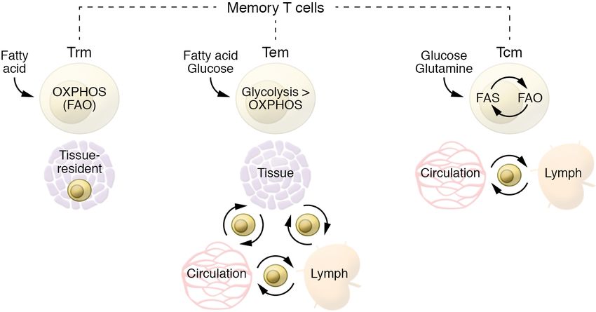
The Journal of Clinical Investigation                                                    R E V I E W S E R I E S : I M M U N O M E TA B O L I S M

petent Tm cells (31). The interaction between the T cell coreceptor       higher mitochondrial reserve capacity in these cells represents a
CD28 and the B7 molecules CD80 and CD86 on activated APCs                 bioenergetic advantage that underlies their rapid recall proper-
prevents T cell anergy and allows development of Tm cells by reg-         ties (67, 68). Distinct from Teff cells, Tm cells are characterized
ulating the cell cycle, cytokine production, and the epigenetic and       by elongated mitochondria with tight cristae structure (Figure 1C)
transcriptional landscape (11, 32). Metabolically, CD28 was origi-        (29), which together support efficient OXPHOS (19, 69). In line
nally characterized to promote higher and more efficient glycolytic       with this observation, T cells lacking the master regulator of inner
flux during activation via engagement of PI3K and Akt, which in           mitochondrial membrane fusion OPA1 or the phosphatase PTP-
turn upregulate mammalian target of rapamycin (mTOR) activity             MT1, responsible for the rate-limiting step in cardiolipin synthesis,
(33, 34). Our group showed that CD28 costimulation during T cell          fail to develop into Tm cells in vivo (29, 70). Conversely, promo-
activation provides important signals to the mitochondria, tran-          tion of mitochondrial elongation, increasing of cardiolipin con-
siently promoting the early expression of carnitine palmitoyltrans-       tent, and inhibition of the repressor of OXPHOS efficiency MCJ1
ferase 1a (CPT1a) before the first cell division and, thus, promoting     are all strategies able to increase long-term survival and function
FAO. Further, CD28 signals restrain mitochondrial cristae loosen-         of Tm cells (29, 70, 71).
ing and endow cells with enhanced spare respiratory capacity after
primary and secondary activation (30), allowing the generation of         Metabolism of different Tm cell subsets
competent Tm cells. Other groups showed a similar function for            CD8+ Tm cells can be categorized broadly into three major subsets
4-1BB costimulation (35). From an immunotherapy perspective it is         according to their functional properties, selectin molecule expres-
of note that the failure of first-generation chimeric antigen receptor    sion, and homing: central memory T (Tcm) cells, tissue-resident
(CAR) T cells in efficiently priming resting T cells and generating       memory T (Trm) cells, and effector memory T (Tem) cells (72).
long-lasting Tm cells was overcome by the engineering of con-             Tcm cells continuously circulate through secondary lymphoid
structs harboring costimulatory signaling domains (e.g., CD28 or          organs, while Trm cells are permanently located in peripheral
4-1BB) together with the CD3ζ domain (36–40). Moreover, it has            tissues, where, with their rapid cytotoxicity, they are the first line
been appreciated that the 4-1BB domain is better for the generation       of defense upon infection, but tend to be shorter-lived than Tcm
of long-lived cells compared with CD28 (41–43), with 4-1BB favor-         cells. The third subset, Tem cells, are a heterogeneous popula-
ing a mitochondrial metabolic signature (44). Thus, costimulatory         tion of T cells able to home to peripheral tissues that retain higher
molecules present during activation are able to modulate metabo-          expression of effector molecules and support Trm cells in tissue
lism and function of Tm cells long after activation signals are gone.     protection by quickly migrating to the site of infection. Tcm cells
Understanding the exact mechanisms underlying this phenome-               largely downregulate effector properties during differentiation.
non may be critical to improving immunotherapy or efficient T cell        Nevertheless, they are able to rapidly recall their function, pro-
priming upon vaccination.                                                 duce a broader spectrum of cytokines, and undergo a more robust
     Cytokine milieu also plays a central role during activation.         proliferation than Tem or Trm cells upon challenge from a previ-
Duration and strength of IL-2 signals control the extent of T cell        ously encountered antigen. Although higher reliance on OXPHOS
proliferation, as well as the pool of Tm cells generated (45–50).         is a common metabolic trait of Tm cells, different Tm cell sub-
IL-2 also supports the proliferative capacity of T cells by modu-         populations have nuanced differences in terms of OXPHOS/gly-
lating T cell metabolism as it stimulates the expression of the           colysis ratios as well as substrate utilization. Tem cells rely less on
transcription factor Myc, the activation of mTOR complex 1                OXPHOS than Tcm or Trm cells. Indeed, Tm cells develop even
(mTORC1), and the stabilization of hypoxia-inducible factor 1α            when glycolysis is genetically enforced in T cells by deletion of the
(HIF1α) to support the uptake of nutrients such as amino acids            von Hippel Lindau (VHL) protein, but they are skewed toward a
and glucose that are necessary to rapidly synthesize nucleotides,         Tem phenotype (73). Tcm and Trm cells, although similar in terms
lipids, and proteins (51). Asymmetric cell division (and thus             of OXPHOS dependency, differ in terms of substrate utilization,
asymmetric activation of mTOR, Myc, and PI3K; refs. 52–55) at             with Trm cells having a unique requirement for acquisition of
the first round of division after activation is also able to dictate      exogenous fatty acids to fuel mitochondrial respiration (74).
future effector/memory phenotypes, with the daughter cell distal
to the APC having an increased propensity to acquire a memory             Different substrates fuel Tm cells
phenotype, and the proximal cell to the APC more prone to be a            Glucose, glutamine, and long-chain and short-chain fatty acids
SLEC (56). Moreover, during the effector phase, T cells with low-         can all be acquired by Tm cells to fuel OXPHOS (75, 76). Nev-
er levels of glycolysis, Akt, or mTOR activation, and mitochon-           ertheless, different Tm subsets preferentially use different sub-
drial membrane potential or reduced cell size, are more likely to         strates (Figure 2) (77). Indeed, although they mainly rely on FAO
acquire Tm cell features (7, 57–61).                                      for their energy demands, Tcm and Trm cells differ in the sub-
     When infection has been resolved (or cancer cells eliminated)        strate of choice (57, 74). Ex vivo Tcm cells and in vitro–generated
and T cells undergo contraction, the anabolic processes that char-        IL-15–cultured Tcm cells engage an apparently futile cycle with
acterize effector T cell response fade (7, 62). During this phase,        the uptake of glucose used to generate fatty acids that are sub-
waning of antigen stimulation and inflammatory signals leads to           sequently burned by FAO (75). These Tcm cells take up a lower
the activation of AMPK and the MAPK-dependent inhibition of               amount of fatty acid compared with Teff or Trm cells and even
mTOR to activate autophagy and allow the generation and surviv-           survive in a lipid-depleted medium (74, 75). Conversely, Trm cells
al of Tm cells (6, 63–65). T cells that are able to engage catabolic      are able to acquire a greater amount of fatty acids directly from
processes to fuel OXPHOS will persist as Tm cells (57, 66), and the       the microenvironment (74, 78). In line with this observation, lipid

                                      J Clin Invest. 2022;132(1):e148546 https://doi.org/10.1172/JCI148546                                  3
R E V I E W S E R I E S : I M M U N O M E TA B O L I S M The Journal of Clinical Investigation
metabolism — fed by serine — also impairs
CD4+ T cell activation by blocking mito-
chondrial biogenesis (86). These observa-
tions point to a specific role for amino acid
availability and utilization during different
phases of T cell development. More nuanced
approaches including inducible knockout
mouse models in the context of infection
(with Cre recombinase activated only at the
peak or after the effector phase) are required
to dissect how specific amino acid require-
ments might impact Tm over Teff cell gen-
eration and function. Notably, in addition
to fueling metabolic programs, amino acids
also bridge metabolism and epigenetics.
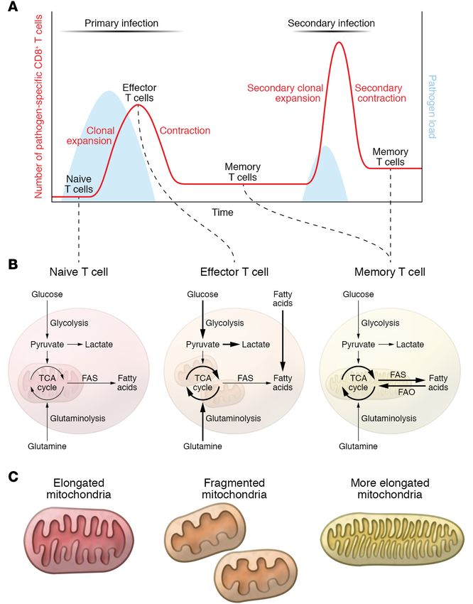
Figure 2. Metabolic features of different memory T cell subsets. Different Tm cell subsets prefer- Methionine availability, for example, regu-
entially use different substrates. Although they mainly rely on FAO for their energy demands, Tcm lates the biosynthesis of the universal methyl
and Trm cells differ in the substrate of choice. Ex vivo Tcm cells and in vitro–generated IL-15–cul- donor S-adenosyl-l-methionine (SAM) and
tured Tcm cells engage an apparently futile cycle with the uptake of glucose used to generate fatty
the H3K4 methylation (H3K4me3) state of
acids that are subsequently burned by FAO. These Tcm cells take up a lower amount of fatty acid
compared with Teff or Trm cells and can even survive in a lipid-depleted medium (75). Conversely, CD4+ T cells, controlling proliferation and
Trm cells are able to acquire a greater amount of fatty acids directly from the microenvironment. cytokine production of Th17 cells (87).
Tem cells are relatively more metabolically active, as they are able to use multiple substrates to fuel
glycolysis and OXPHOS. Epigenetic control of
metabolism in Tm cells
Specific signaling, metabolic, or antigen-
chaperones like FABP4/5 and CD36 are specifically upregulated driven information is imprinted during the primary effector phase
in Trm cells, and ex vivo exogenous supplementation of fatty acids and conserved over time, contributing to immune memory through
increased spare respiratory capacity only in Trm cells, not in Tcm epigenetic modification of histones (9). Interestingly, in addition
cells (74). A recent study challenged the central role for FAO in Tm to directly modulating bioenergetic pathways via substrate avail-
cells (79). However, multiple explanations might reconcile these ability, metabolites can act as signaling molecules, often by modi-
and previous findings, including thymic selection compensation, fying the epigenetic landscape of immune cells (88, 89). High lev-
metabolic adaptation to alternative fuel sources, the increased els of acetate experienced by T cells at the peak of effector phase
utilization of short-chain over long-chain fatty acids, or compen- are responsible for the acetylation of GAPDH, stimulating higher
satory enhanced peroxisomal FAO (23). Catabolic and anabolic glycolysis, while also acetylating histone 3 lysine 9 (H3K9), thus
processes coexist in Tm cells. They have indeed been observed not enhancing chromatin accessibility of specific promoter regions
only for fatty acid synthesis and FAO (75), but also for gluconeo- of Teff cell–associated genes in Tm cells, favoring their rapid
genesis and glycogenolysis (76), and for triacylglycerol synthesis expression upon restimulation (90–92). Another metabolite that
and lipolysis (80). When the metabolic equilibrium between these influences the epigenome is α-ketoglutarate, which reduces the
pathways is genetically or pharmacologically perturbed, Tm cells expression of the DNA methyltransferases Dnmt3a and Dnmt3b
fail to develop. These observations highlight the metabolic flexi- via inhibition of the transcription factor OTX2 (93). DNMT3A-
bility of Tm cells. Nevertheless, additional studies are required to mediated erasure of de novo methylated regions during the Teff
further investigate and clarify the role and crosstalk of multiple cell phase regulates re-expression of Tn cell–associated genes,
anabolic and catabolic processes in these cells. allowing Tm cells to differentiate from a fate-permissive subset of
Multiple observations show that pharmacologic or genetic Teff cells (94); this explains the partially conserved DNA methyl-
interference with mTOR activity promotes Tm cell generation (6, ation profile between Teff and Tm cells in both mice and humans
7). mTOR is a critical hub for sensing amino acid content and the and suggests a lineage relationship between these two populations
metabolic status of the cell. It is therefore not surprising that ami- (94, 95). There are many more examples of how metabolism influ-
no acid availability, transporter expression, and amino acid uptake ences the epigenome, and a more comprehensive overview of the
are all critical for T cell activation and expansion, naturally lim- epigenetic control of T cell fate and differentiation can be found in
iting Tm cell development (81, 82). Glutamine uptake via ASCT2 other recent reviews (9, 96).
coregulates leucine transport and mTOR upon T cell activation,
and genetic ablation of Slc1a5 (the gene encoding ASCT2) results T cell dysfunctions in patients with
in accumulation of CD4+ Tem cells with no apparent differences mitochondrial diseases
in CD8+ cell subsets (83, 84). In a different setting, limiting ser- Mitochondrial diseases (MDs) are the most common group of
ine availability during CD8+ T cell activation and primary expan- inherited metabolic disorders and arise from mutations in mito-
sion impairs proliferation upon secondary infection, resulting in chondrial genes encoded by the nuclear (nDNA) or mitochondri-
limited bacterial clearance (85). Genetic inhibition of one-carbon al (mtDNA) genome (97, 98). They are heterogeneous in etiology
4 J Clin Invest. 2022;132(1):e148546 https://doi.org/10.1172/JCI148546The Journal of Clinical Investigation R E V I E W S E R I E S : I M M U N O M E TA B O L I S M
immune memory and partially explaining the repetitive suscepti-
Table 1. The immune phenotype of patients with mitochondrial bility to bacterial and viral infections of patients with MDs.
diseases Thus, considering that infections can be more deleterious in
Cell-intrinsic defects children with inborn errors of metabolism (IEM), preventing infec-
tions via vaccination is key. Nevertheless, the same mechanisms
Reduced cytokine production
Impaired oxidative metabolism in T cells that negatively affect the immune response to natural infections
Systemic defects might do the same in response to vaccines. Despite this reasonable
Leukopenia concern, multiple studies reported positively on the immunogenic-
Reduced Tm cell frequencies ity, safety, and tolerability of vaccines in children with IEM (111,
Lactic acidosis 112). Vaccination regimens are recommended in IEM patients and
Up to 50% of patients with mitochondrial diseases experience recurrent are not associated with increased risk of serious adverse effects
infections, especially of the respiratory tract, with a high prevalence of during the month after vaccination, although the risk might be
sepsis and pneumonia. The immune impairment can be explained by a more pronounced for the more metabolically unstable patients
combination of cell-intrinsic and systemic immune defects. (113). Administration of live attenuated vaccines should be evalu-
ated carefully in immunocompromised patients (111). While these
considerations are valid and vaccines are highly recommended
for patients with MDs, further MD-specific studies are needed to
(mutations in nDNA or mtDNA) and in inheritance mechanisms establish pathology-specific guidelines and vaccine regimens —
(maternal for mtDNA mutations, autosomal dominant/recessive perhaps by adding additional boost doses at regular intervals, as is
or X-linked for nDNA mutations) (97, 98). Moreover, although all often recommended for immunocompromised patients.
MDs are characterized by dysfunctional OXPHOS, mtDNA integ- Notably, although not of genetic etiology, the progressive
rity, or mitochondrial maintenance, MDs manifest in a pheno- decline in mitochondrial function reported during aging (114, 115)
typically diverse spectrum often affecting muscle, heart, or brain also involves T cells (86, 116, 117), and could be one factor contribut-
physiology with variable penetrance and severity (97, 98). Recent ing to lower T cell responses to vaccines in elderly populations (118).
advances in the immune characterization of patients affected
by (as well as mouse models of) MDs suggest that immune dys- Impact of metabolic health on T cell function
function might be added to the features of MDs (99). Up to half Obesity and metabolic syndrome are major public health issues,
of patients with MD experience recurrent or severe upper respira- with numbers of obese people increasing worldwide (119). Meta-
tory tract infections, often resulting in life-threatening conditions bolic disruptions leading to metabolic syndrome include the com-
(100, 101). This percentage increases to almost 90% of pediatric bination of at least three of the following factors: central adiposity,
MD patients (101), with sepsis (55%) and pneumonia (29%) as the elevated blood glucose and plasma triacylglycerols, high blood
two most common causes of death (102). Immune dysfunction in pressure, and low plasma HDL-cholesterol (120). Moreover, meta-
MDs might be due to multiple factors, including the higher inci- bolic syndrome is often characterized by endothelial cell dysfunc-
dence of leukopenia observed in patients affected by Barth syn- tion, atherogenic dyslipidemia, insulin resistance, and chronic
drome, Pearson syndrome, and Leigh syndrome (103). Defects low-grade inflammation (121). Metabolic alterations and inflam-
have manifested in lower Tm (CD45RO+) cell frequencies in a mation engage in a vicious cycle with T cell activation, senes-
pediatric cohort of MD patients (101), deficient cytokine produc- cence, and proinflammatory cytokine production that worsens
tion in a small cohort of Barth syndrome patients (70), or impaired pathologic conditions and results in higher rates of vaccine failure
antibody production upon vaccination, which was observed in a and complications from infection (122, 123). Mechanistically, in
case of fatal neonatal-onset mitochondrial respiratory chain dis- addition to its effects on innate immune cells, leptin — the levels
ease (104). Interestingly, supporting a role for FAO and CPT1a of which are increased in obese or metabolically impaired individ-
in shaping an efficient long-lasting immune response, a study of uals — also modulates adaptive immunity by inducing expression
Native Alaskan children carrying a hypomorphic variant of CPT1a of activation markers on T cells (124), inhibiting proliferation of
showed a higher incidence of respiratory tract infection and otitis Tm cells (125, 126), and polarizing Th cells toward Th1 proinflam-
in comparison with the control group (105). matory phenotype while simultaneously inhibiting Treg function
More broadly speaking, the immune phenotype in patients (127–129). Hyperinsulinemia as an adaptation to systemic insulin
with MDs could result either from functional defects intrinsic to resistance is a common feature of obesity, fostering type 2 diabe-
T cells (or other immune cells) or from cell-extrinsic mechanisms tes onset and progression. Multiple studies showed that the insulin
(Table 1). Febrile temperature, a physiologic response to infection, receptor (INSR) is also present on T cells, where it modulates glu-
increases basal metabolic rate (10% higher per 1°C increase in body cose and amino acid uptake and is generally upregulated during T
temperature) (106). In patients with MDs the increase in metabolic cell activation (130, 131). Whole-body knockout of INSR showed
rate during an infection coupled with the impairment of OXPHOS reduced cytokine production, proliferation, and migration, as well
might exacerbate lactic acidosis, an already common feature of as increased apoptosis of T cells, although the results from this
MDs (107) that is known to inhibit T cell function (108–110). Genet- model were confounded by the underlying hyperglycemia asso-
ic defects in OXPHOS might also directly impact Tm cell develop- ciated with systemic loss of INSR function (132). T cell–intrinsic
ment given the established role of mitochondrial respiration during defects were confirmed by selective ablation of INSR on T cells
this phase (29, 57, 66, 70), potentially contributing to the impaired and observation of deficiencies in proliferation and cytokine pro-
J Clin Invest. 2022;132(1):e148546 https://doi.org/10.1172/JCI148546 5R E V I E W S E R I E S : I M M U N O M E TA B O L I S M The Journal of Clinical Investigation
Figure 3. Metabolic interventions in cancer immunotherapy. Three main scenarios for metabolic interventions in the context of cancer immunotherapy
can be imagined: (A) In vitro preconditioning to prime T cell metabolism before autologous in vivo transfer. One caveat to consider in this approach is the
loss of the induced preconditioning upon T cell transfer in vivo. (B) Systemic administration of drugs that alter tumor and T cell metabolism. A limitation
of this strategy is the potential for the development of systemic side effects. (C) Targeted delivery of metabolic modulators directly in the TME or adminis-
tration of precursor drugs selectively activated in the TME could potentially overcome the issues described for the first two approaches.
duction that resulted in impaired inflammatory and T cell–specific of implanted tumors from two pancreatic cell lines derived from
responses to influenza virus (133). These observations could well early- and late-stage pancreatic ductal adenocarcinoma (PDAC)
contribute to the higher susceptibility to severe infections and can- showed lower glucose in the interstitial fluid of advanced PDAC
cer as well as the weakened vaccine effectiveness associated with tumors (152). A recent study analyzing human renal cell carci-
systemic insulin resistance observed in obese people (134–137). noma and mouse subcutaneous MC38 tumors compared with
adjacent healthy tissue challenged the idea of glucose restriction
Overcoming metabolic competition in the tumor in the TME (153). Moreover, glucose uptake measured in vivo by
microenvironment to improve Tm cells 18
F-fluorodeoxyglucose PET imaging revealed that T cells are able
Although T cell therapy has shown great preclinical and clinical to acquire glucose in the TME, although they remain functionally
success in treatment of hematologic malignancies (138, 139), its impaired. Overall this study suggested that there is selective nutri-
efficacy in the treatment of solid tumors has been disappointingly ent partitioning among different cells in the TME. Inhibiting the
low (140). Many studies have combined T cell therapy with admin- higher glutamine uptake in cancer cells unleashed glucose uptake
istration of proinflammatory cytokines or checkpoint blockade by TME-resident cells, including T cells, beyond basal levels,
inhibitors to improve success (141), but the severe side effects as restoring their function; this suggests that glutamine metabolism
well as the unsatisfactory results observed prove the necessity of suppresses glucose uptake by T cells without glucose being a lim-
developing new approaches (142–144). Lack of antigen recogni- iting factor in the TME (153). In a different study, divergent met-
tion, chronic activation and exhaustion, and hyporesponsiveness abolic programs upon glutamine metabolism blockade were also
of T cells are common mechanisms of immune evasion in cancer observed between cancer cells and T cells, and they were associat-
(145–147). In recent years, metabolic competition in the tumor ed with increased glucose availability in the tumor and functional
microenvironment (TME) has been increasingly recognized as an and metabolic rescue of T cells (154). It has also been shown that
additional effective immune escape strategy (148). Many cancer checkpoint blockade therapy (149) or inhibition of the N6-meth-
cells rely on glucose through Warburg metabolism and compete yladenosine RNA demethylase FTO (155) can directly impact
with T cells for this substrate, leading to lower concentration of tumor cell metabolism while increasing glucose uptake by T cells.
glucose in the TME compared with plasma (149–151). The paral- While the presence and extent of glucose restriction in TME might
lel use of mouse models of regressing and progressive sarcoma reflect cancer type heterogeneity, competition for metabolites
tumors and melanoma-bearing Braf/Pten mice revealed that the between cancer and immune cells remains a key factor governing
TME of progressive tumors had a lower glucose concentration the balance between cancer progression and regression.
compared with that of regressive ones (149, 150). These two stud- Notably, many interventions to overcome TME inhibitory
ies together formally cemented the idea that nutrient competi- effects on T cells coincide with treatments that either directly
tion occurs in the TME, and that this as a distinct mechanism can stimulate mitochondrial activity or mimic pro-memory metabol-
drive cancer progression. A similar experiment comparing TME ic features. With regard to harnessing metabolism for therapeu-
6 J Clin Invest. 2022;132(1):e148546 https://doi.org/10.1172/JCI148546The Journal of Clinical Investigation R E V I E W S E R I E S : I M M U N O M E TA B O L I S M
tic interventions in cancer immunotherapy, three main scenarios A third strategy is targeted delivery of metabolic modulators
have been envisioned: in vitro metabolic preconditioning, system- directly in the TME. This strategy includes drug delivery using
ic in vivo metabolic treatments, and targeted delivery of metabolic nanoparticles (171), oncolytic viruses (172), use of metabolically
modulators in the TME (156); and multiple strategies have been engineered CAR T cells (173, 174), or precursor drugs selectively
designed to address them (157) (summarized in Figure 3). The first activated in the TME (154). In line with this idea, an intriguing
approach embraces in vitro preconditioning of cells with metabol- strategy based on click chemistry (175) has been used to backpack
ic modulators before adoptive transfer. Examples include inhibit- the ACAT1 inhibitor avasimibe directly on T cell membrane to
ing Akt function to favor a more Tm cell–like metabolic phenotype locally increase cholesterol, improving T cell receptor clustering
in mouse melanoma models (59); using sodium bicarbonate to and, thus, T cell activation and function in mouse models of glio-
reverse the lactic acid–induced interference with T cell glycolysis blastoma and melanoma (176). A similar approach could be envis-
and cytotoxic function in mouse models and patients with acute aged coupling other metabolic modulators directly on T cells,
myeloid leukemia (110); and transiently exposing donor lympho- creating a new generation of combinatorial anticancer therapies.
cytes to 39°C prior to infusion in a myeloid leukemia mouse model Additional strategies exploit other features of the TME to achieve
(158). Similarly, preconditioning T cells with IL-15, which drives a site-specific activation of T cells or drugs. Stemming from the
Tm cell differentiation and metabolically increases spare respi- observation that CAR T cell efficiency can be manipulated by met-
ratory capacity (66), has similar positive results in HER2-positive abolic engineering of these cells (e.g., via CD28 or 4-1BB) (36–40),
tumors, leukemia, and glioma models (159–161). An approach new improved versions of these cells have been generated. Oxy-
based on the transient rest of CAR T cell receptor signaling has gen-sensing CAR T cells activated by the hypoxic TME have been
been suggested to restore functionality of T cells and reverse their developed to reduce possible off-target effects in solid tumors and
exhausted phenotype (162). Interestingly, from a metabolic per- have proven effective in mouse models of hypoxic HN3 tumors
spective, continuous T cell stimulation in a hypoxic microenviron- (174). Alternatively, precursor drugs can also be engineered to
ment drives T cell exhaustion by inducing mitochondrial stress become active only in the tumor. For example, to avoid systemic
(163). Pharmacologic treatments aimed at reducing reactive oxy- toxicity of comprehensive glutamine metabolism inhibitors, pre-
gen species (ROS) or lowering tumor hypoxia improve response to cursor drugs have been designed to be cleaved by specific prote-
immunotherapy in mice (163). Therefore, a rest period could also ases at the tumor site, where they can locally exert their inhibitory
reinvigorate mitochondrial metabolism to sustain long-term T cell function and promote OXPHOS and Tm cell development (154).
persistence and function. It should also be considered that in vitro An emerging area of research aims at investigating how sys-
generation and amplification of tumor-infiltrating lymphocytes temic metabolic interventions like glucose restriction or keto-
for adoptive cell transfer or CAR T cells often require incubation genic diet might be exploited to boost anticancer treatment. This
in supraphysiologic concentrations of nutrients such as glucose, research area stems from pioneering work showing that the sys-
or cytokines like IL-2 (164). Based on preclinical observations (7, temic hyperinsulinemia observed in cancer patients treated with a
30, 165), a transient preincubation in a more physiologic medi- PI3K inhibitor, which is able to reactivate mTOR signaling in can-
um inducing glucose restriction or treatment with rapamycin (or cer cells, can be blocked by a ketogenic diet, allowing more effec-
analogs) is a promising strategy to prime T cells for Tm cell dif- tive control of tumor growth than PI3K inhibitor treatment alone
ferentiation (161, 164, 166, 167). Along the same line, the treat- (177). Despite this promising observation, ketogenic diet interven-
ment of CD8+ T cells with the engineered IL-2 partial agonist H9T tions may only be valid in PI3K-dependent tumors, and could be
improves mitochondrial fitness and promotes a stem cell–like detrimental in other tumors that are able to metabolically adapt
state (168). Boosting mitochondrial elongation or cardiolipin con- their growth to the use of ketone bodies as a fuel source.
tent and blocking the repressor of OXPHOS efficiency MCJ1 are
all metabolic preconditioning strategies to increase long-term sur- Developing vaccines that elicit efficient T cell
vival and function of Tm cells in mouse models (29, 70, 71). responses
This in vitro metabolic preconditioning strategy has the clear A persistent challenge in vaccine development is the genera-
caveat that it might be reversed or lost when cells are transferred tion of vaccines able to elicit efficient T cell responses and Tm
in vivo and approach the TME. To overcome this issue, a sec- cell generation in addition to humoral responses. This could
ond strategy could employ systemic administration of metabolic be particularly important for patients with B cell deficiency or
modulators. Various clinical trials are under way to test clinical functional decline. Many modern vaccines, which are often not
efficacy of vaccination with NY-ESO-1 tumor antigen–based vac- based on live attenuated pathogens, are not highly effective at
cines in combination with rapamycin treatment (ClinicalTrials. priming persistent T cell immunity (4). This might be related
gov NCT01536054, NCT02833506, and NCT01522820) or IL-15 to the fact that, unlike B cells, T cells recognize antigen in the
superagonists (NCT02384954) (169). Preclinical studies in mouse context of MHC on APCs, which requires cross-presentation of
models showed that systemic inhibition of the cholesterol ester- antigen by APCs, higher amounts of the initial antigen dose, and
ification enzyme ACAT1 potentiates the CD8+ T cell antitumor a longer persistence of antigen. Vaccines that efficiently prime
response by increasing cholesterol concentration in the plasma a T cell response are often live attenuated vaccines, like those
membrane and enhancing T cell receptor clustering and signaling used against yellow fever (YF-17D) or smallpox (178, 179). Con-
(170). Possible systemic side effects and complexity of pharmaco- versely, vaccines against influenza virus poorly activate T cells
kinetics of the compounds used are a persistent issue with these (180). In order to improve vaccine-mediated T cell responses,
nevertheless promising approaches. multiple strategies have been deployed, including the use of var-
J Clin Invest. 2022;132(1):e148546 https://doi.org/10.1172/JCI148546 7R E V I E W S E R I E S : I M M U N O M E TA B O L I S M The Journal of Clinical Investigation
ious DNA-based vaccines and viral vectors, the study of specific the transcriptional level (195). It could therefore be envisioned
prime-boost regimens, and adjuvant combinations. Often, these that the implementation of similar systems biology methods, fed
strategies showed limited success (181–183). The mRNA vaccines also by metabolomics data, could be used to identify more prom-
BNT162b1 and mRNA-1273, developed during the COVID-19 ising vaccine candidates earlier in the developmental stage, or
pandemic, were shown to promote high frequencies and per- to identify vaccine nonresponders in particularly fragile popula-
sistence of SARS-CoV-2 receptor-binding domain–specific CD4+ tions and accordingly modulate other pharmacologic and non-
and CD8+ T cells highly effective in IFN-γ production (184–186). pharmacologic interventions.
The mechanisms, which remain elusive, might be related to how
mRNA is expressed by target cells, and to the persistence of anti- Concluding remarks
gen expression. Adenovirus (Ad) vector–based vaccines using The current challenge of the COVID-19 pandemic has made
the backbone of human Ads (huAd5 or huAd26) or chimeric vec- clear that new creative ways to confront long-lasting immunolo-
tors based on chimpanzee viruses (ChAdOX1) are also able to gy questions are key for advancing therapies. Unveiling the met-
elicit a CD8+ T cell response to viral antigens (187–189) owing to abolic circuits regulating immunity and combining discoveries
their ability to promote a niche of persistent antigen presentation from cancer metabolism, vaccine development, and T cell biol-
in fibroblastic stromal cells in the lungs (190). ogy are central strategies to tackle this and future pandemics as
While new vaccines are developed, another line of research well as improving our current treatment of cancer, infections, and
includes exploiting metabolic features of innate and adaptive autoimmune diseases. The subclinical presence of mitochondri-
immune cells to generate more efficient T cell activation during al diseases and metabolic disorders could be a critical factor to
vaccination using metabolic adjuvants targeting the activity of take into consideration when prognosis and therapies for cancer,
mTOR or the amino acid sensor GCN2 (191). Alternatively, in infections, and autoimmune diseases are clinically discussed or
line with their role to protect tissue immediately upon infection, vaccine efficacy is evaluated.
the ability of adjuvants and vaccines to specifically induce anti-
gen-specific Trm cells has also been intensively studied (192). In Acknowledgments
this regard, it is of great interest to integrate metabolic phenotyp- MC is supported by a Barth Syndrome Foundation Idea grant. ELP
ing into vaccinology (193, 194). Changes in plasma metabolites is supported by NIH grant R01AI156274.
upon vaccination have been investigated in pioneering studies on
the live attenuated shingles vaccine Zostavax, showing how ste- Address correspondence to: Mauro Corrado, CECAD Research
rol metabolism integrates cellular and humoral responses (195). Center, Joseph Stelzmannstrasse 26, Cologne 50931, Germa-
In this study alterations in plasma metabolites were observed ny. Email: mcorrado@uni-koeln.de. Or to: Erika L. Pearce,
already at day 1 after vaccination and anticipated the concordant Bloomberg-Kimmel Institute for Cancer Immunotherapy, The
transcriptional changes observed 48 hours later, suggesting that Bunting Blaustein Cancer Research Bldg., 1650 Orleans St., Room
changes in metabolism precede and maybe instruct changes at 344, Baltimore, Maryland 21287, USA. Email: epearce6@jhmi.edu.
1. Farber DL, et al. Immunological memory: lessons 1997;185(2):251–262. 2013;155(1):160–171.
from the past and a look to the future. Nat Rev 11. Borowski AB, et al. Memory CD8+ T cells 20. Quiros PM, et al. Mitonuclear communication
Immunol. 2016;16(2):124–128. require CD28 costimulation. J Immunol. in homeostasis and stress. Nat Rev Mol Cell Biol.
2. Kalia V, et al. Differentiation of memory B and T 2007;179(10):6494–6503. 2016;17(4):213–226.
cells. Curr Opin Immunol. 2006;18(3):255–264. 12. Kaech SM, et al. Selective expression of the inter- 21. Williams MA, Bevan MJ. Effector and mem-
3. Omilusik KD, Goldrath AW. The origins of mem- leukin 7 receptor identifies effector CD8 T cells ory CTL differentiation. Annu Rev Immunol.
ory T cells. Nature. 2017;552(7685):337–339. that give rise to long-lived memory cells. Nat 2007;25:171–192.
4. Gilbert SC. T-cell-inducing vaccines — what’s the Immunol. 2003;4(12):1191–1198. 22. Geltink RIK, et al. Unraveling the complex inter-
future. Immunology. 2012;135(1):19–26. 13. Buck MD, et al. Metabolic instruction of immuni- play between T cell metabolism and function.
5. Testa JS, Philip R. Role of T-cell epitope-based ty. Cell. 2017;169(4):570–586. Annu Rev Immunol. 2018;36:461–488.
vaccine in prophylactic and therapeutic applica- 14. Spinelli JB, Haigis MC. The multifaceted contri- 23. O’Sullivan D. The metabolic spectrum of memory
tions. Future Virol. 2012;7(11):1077–1088. butions of mitochondria to cellular metabolism. T cells. Immunol Cell Biol. 2019;97(7):636–646.
6. Xu X, et al. Autophagy is essential for effector Nat Cell Biol. 2018;20(7):745–754. 24. Wherry EJ, Ahmed R. Memory CD8 T-cell
CD8(+) T cell survival and memory formation. 15. Ploumi C, et al. Mitochondrial biogene- differentiation during viral infection. J Virol.
Nat Immunol. 2014;15(12):1152–1161. sis and clearance: a balancing act. FEBS J. 2004;78(11):5535–5545.
7. Araki K, et al. mTOR regulates memo- 2017;284(2):183–195. 25. Reading JL, et al. The function and dysfunction of
ry CD8 T-cell differentiation. Nature. 16. Ng MYW, et al. Quality control of the mitochon- memory CD8+ T cells in tumor immunity. Immu-
2009;460(7251):108–112. drion. Dev Cell. 2021;56(7):881–905. nol Rev. 2018;283(1):194–212.
8. Salerno F, et al. Dynamic post-transcriptional 17. Pernas L, Scorrano L. Mito-morphosis: mito- 26. Joshi NS, et al. Inflammation directs memory
events governing CD8+ T cell homeosta- chondrial fusion, fission, and cristae remodeling precursor and short-lived effector CD8(+) T cell
sis and effector function. Trends Immunol. as key mediators of cellular function. Annu Rev fates via the graded expression of T-bet transcrip-
2020;41(3):240–254. Physiol. 2016;78:505–531. tion factor. Immunity. 2007;27(2):281–295.
9. Tough DF, et al. Epigenetic regulation of T cell 18. Chan DC. Mitochondrial dynamics and its 27. Sarkar S, et al. Functional and genomic profiling
memory: recalling therapeutic implications. involvement in disease. Annu Rev Pathol. of effector CD8 T cell subsets with distinct mem-
Trends Immunol. 2020;41(1):29–45. 2019;15:235–259. ory fates. J Exp Med. 2008;205(3):625–640.
10. Liu Y, et al. Distinct costimulatory molecules 19. Cogliati S, et al. Mitochondrial cristae shape 28. Kalia V, et al. Prolonged interleukin-2Ralpha
are required for the induction of effector and determines respiratory chain supercomplex- expression on virus-specific CD8+ T cells favors
memory cytotoxic T lymphocytes. J Exp Med. es assembly and respiratory efficiency. Cell. terminal-effector differentiation in vivo. Immu-
8 J Clin Invest. 2022;132(1):e148546 https://doi.org/10.1172/JCI148546The Journal of Clinical Investigation R E V I E W S E R I E S : I M M U N O M E TA B O L I S M
nity. 2010;32(1):91–103. ation and expansion of responding CD8+ T cells ory development. Immunity. 2012;36(1):68–78.
29. Buck MD, et al. Mitochondrial dynamics controls rather than promotion of cell death. Proc Natl 67. van der Windt GJ, et al. CD8 memory T cells
T cell fate through metabolic programming. Cell. Acad Sci U S A. 2002;99(5):3001–3006. have a bioenergetic advantage that underlies
2016;166(1):63–76. 47. Blattman JN, et al. Therapeutic use of IL-2 to their rapid recall ability. Proc Natl Acad Sci U S A.
30. Geltink RIK, et al. Mitochondrial priming by enhance antiviral T-cell responses in vivo. Nat 2013;110(35):14336–14341.
CD28. Cell. 2017;171(2):385–397. Med. 2003;9(5):540–547. 68. Bantug GR, et al. Mitochondria-endoplasmic
31. Villegas EN, et al. Role of CD28 in the generation 48. Kim MT, et al. Manipulating memory CD8 T cell reticulum contact sites function as immunomet-
of effector and memory responses required for numbers by timed enhancement of IL-2 signals. abolic hubs that orchestrate the rapid recall
resistance to Toxoplasma gondii. J Immunol. J Immunol. 2016;197(5):1754–1761. response of memory CD8+ T cells. Immunity.
1999;163(6):3344–3353. 49. Suzuki H, et al. Deregulated T cell activation 2018;48(3):542–555.
32. Esensten JH, et al. CD28 Costimulation: and autoimmunity in mice lacking interleukin-2 69. Gomes LC, et al. During autophagy mitochondria
from mechanism to therapy. Immunity. receptor beta. Science. 1995;268(5216):1472–1476. elongate, are spared from degradation and sustain
2016;44(5):973–988. 50. Kundig TM, et al. Immune responses in cell viability. Nat Cell Biol. 2011;13(5):589–598.
33. Frauwirth KA, et al. The CD28 signaling path- interleukin-2-deficient mice. Science. 70. Corrado M, et al. Dynamic cardiolipin synthesis
way regulates glucose metabolism. Immunity. 1993;262(5136):1059–1061. is required for CD8+ T cell immunity. Cell Metab.
2002;16(6):769–777. 51. Ross SH, Cantrell DA. Signaling and function of 2020;32(6):981–995.
34. Jacobs SR, et al. Glucose uptake is limiting in interleukin-2 in T lymphocytes. Annu Rev Immu- 71. Champagne DP, et al. Fine-tuning of CD8(+) T
T cell activation and requires CD28-mediated nol. 2018;36:411–433. cell mitochondrial metabolism by the respiratory
Akt-dependent and independent pathways. 52. Pollizzi KN, et al. Asymmetric inheritance of chain repressor MCJ dictates protection to influ-
J Immunol. 2008;180(7):4476–4486. mTORC1 kinase activity during division dictates enza virus. Immunity. 2016;44(6):1299–1311.
35. Menk AV, et al. 4-1BB costimulation induces CD8(+) T cell differentiation. Nat Immunol. 72. Martin MD, Badovinac VP. Defining memory
T cell mitochondrial function and biogenesis 2016;17(6):704–711. CD8 T cell. Front Immunol. 2018;9:2692.
enabling cancer immunotherapeutic responses. 53. Verbist KC, et al. Metabolic maintenance of cell 73. Phan AT, et al. Constitutive glycolytic metab-
J Exp Med. 2018;215(4):1091–1100. asymmetry following division in activated T lym- olism supports CD8+ T cell effector memory
36. Subklewe M, et al. Chimeric antigen receptor phocytes. Nature. 2016;532(7599):389–393. differentiation during viral infection. Immunity.
T cells: a race to revolutionize cancer therapy. 54. Chen YH, et al. Asymmetric PI3K activity in lym- 2016;45(5):1024–1037.
Transfus Med Hemother. 2019;46(1):15–24. phocytes organized by a PI3K-mediated polarity 74. Pan Y, et al. Survival of tissue-resident memory T
37. Brocker T, Karjalainen K. Signals through T pathway. Cell Rep. 2018;22(4):860–868. cells requires exogenous lipid uptake and metab-
cell receptor-zeta chain alone are insufficient 55. Borsa M, et al. Modulation of asymmetric cell olism. Nature. 2017;543(7644):252–256.
to prime resting T lymphocytes. J Exp Med. division as a mechanism to boost CD8+ T cell 75. O’Sullivan D, et al. Memory CD8(+) T cells use
1995;181(5):1653–1659. memory. Sci Immunol. 2019;4(34):eaav1730. cell-intrinsic lipolysis to support the metabolic
38. Gong MC, et al. Cancer patient T cells genetically 56. Chang JT, et al. Asymmetric T lymphocyte programming necessary for development. Immu-
targeted to prostate-specific membrane antigen division in the initiation of adaptive immune nity. 2014;41(1):75–88.
specifically lyse prostate cancer cells and release responses. Science. 2007;315(5819):1687–1691. 76. Ma R, et al. A Pck1-directed glycogen metabolic
cytokines in response to prostate-specific mem- 57. Pearce EL, et al. Enhancing CD8 T-cell memory program regulates formation and mainte-
brane antigen. Neoplasia. 1999;1(2):123–127. by modulating fatty acid metabolism. Nature. nance of memory CD8+ T cells. Nat Cell Biol.
39. Krause A, et al. Antigen-dependent CD28 2009;460(7251):103–107. 2018;20(1):21–27.
signaling selectively enhances survival and 58. Sukumar M, et al. Inhibiting glycolytic metabolism 77. Ecker C, et al. Differential reliance on lipid
proliferation in genetically modified activated enhances CD8+ T cell memory and antitumor metabolism as a salvage pathway underlies func-
human primary T lymphocytes. J Exp Med. function. J Clin Invest. 2013;123(10):4479–4488. tional differences of T cell subsets in poor nutri-
1998;188(4):619–626. 59. Crompton JG, et al. Akt inhibition enhances ent environments. Cell Rep. 2018;23(3):741–755.
40. Porter DL, et al. Chimeric antigen receptor-mod- expansion of potent tumor-specific lymphocytes 78. Han SJ, et al. White adipose tissue is a reservoir
ified T cells in chronic lymphoid leukemia. N Engl with memory cell characteristics. Cancer Res. for memory T cells and promotes protective
J Med. 2011;365(8):725–733. 2015;75(2):296–305. memory responses to infection. Immunity.
41. Porter DL, et al. Chimeric antigen receptor T 60. Pollizzi KN, et al. Cellular size as a means of 2017;47(6):1154–1168.
cells persist and induce sustained remissions in tracking mTOR activity and cell fate of CD4+ 79. Raud B, et al. Etomoxir actions on regulato-
relapsed refractory chronic lymphocytic leuke- T cells upon antigen recognition. PLoS One. ry and memory T cells are independent of
mia. Sci Transl Med. 2015;7(303):303ra139. 2015;10(4):e0121710. Cpt1a-mediated fatty acid oxidation. Cell Metab.
42. Ying Z, et al. Parallel comparison of 4-1BB or 61. Sukumar M, et al. Mitochondrial membrane 2018;28(3):504–515.
CD28 co-stimulated CD19-targeted CAR-T cells potential identifies cells with enhanced stemness 80. Cui G, et al. IL-7-Induced glycerol transport and
for B cell non-Hodgkin’s lymphoma. Mol Ther for cellular therapy. Cell Metab. 2016;23(1):63–76. TAG synthesis promotes memory CD8+ T cell
Oncolytics. 2019;15:60–68. 62. Rao RR, et al. The mTOR kinase determines longevity. Cell. 2015;161(4):750–761.
43. Zhao X, et al. Efficacy and safety of CD28- or effector versus memory CD8+ T cell fate by 81. Ren W, et al. Amino-acid transporters in T-cell
4-1BB-based CD19 CAR-T cells in B cell acute regulating the expression of transcription activation and differentiation. Cell Death Dis.
lymphoblastic leukemia. Mol Ther Oncolytics. factors T-bet and Eomesodermin. Immunity. 2017;8(3):e2655.
2020;18:272–281. 2010;32(1):67–78. 82. Marchingo JM, et al. Quantitative analysis of
44. Kawalekar OU, et al. Distinct signaling of core- 63. Rolf J, et al. AMPKα1: a glucose sensor that how Myc controls T cell proteomes and meta-
ceptors regulates specific metabolism pathways controls CD8 T-cell memory. Eur J Immunol. bolic pathways during T cell activation. Elife.
and impacts memory development in CAR T 2013;43(4):889–896. 2020;9:e53725.
cells. Immunity. 2016;44(2):380–390. 64. Puleston DJ, et al. Autophagy is a critical regu- 83. Nakaya M, et al. Inflammatory T cell responses
45. Cheng LE, Greenberg PD. Selective delivery of lator of memory CD8(+) T cell formation. Elife. rely on amino acid transporter ASCT2 facilitation
augmented IL-2 receptor signals to responding 2014;3:e03706. of glutamine uptake and mTORC1 kinase activa-
CD8+ T cells increases the size of the acute anti- 65. Corrado M, et al. Macroautophagy inhibition tion. Immunity. 2014;40(5):692–705.
viral response and of the resulting memory T cell maintains fragmented mitochondria to foster 84. Sinclair LV, et al. Control of amino-acid transport
pool. J Immunol. 2002;169(9):4990–4997. T cell receptor-dependent apoptosis. EMBO J. by antigen receptors coordinates the metabolic
46. Cheng LE, et al. Enhanced signaling through the 2016;35(16):1793–1809. reprogramming essential for T cell differentia-
IL-2 receptor in CD8+ T cells regulated by anti- 66. van der Windt GJ, et al. Mitochondrial respiratory tion. Nat Immunol. 2013;14(5):500–508.
gen recognition results in preferential prolifer- capacity is a critical regulator of CD8+ T cell mem- 85. Ma EH, et al. Serine is an essential metabo-
J Clin Invest. 2022;132(1):e148546 https://doi.org/10.1172/JCI148546 9You can also read