The Gut-Brain Axis in Multiple Sclerosis. Is Its Dysfunction a Pathological Trigger or a Consequence of the Disease?
←
→
Page content transcription
If your browser does not render page correctly, please read the page content below
REVIEW
                                                                                                                                    published: 21 September 2021
                                                                                                                                  doi: 10.3389/fimmu.2021.718220

                                           The Gut-Brain Axis in Multiple
                                           Sclerosis. Is Its Dysfunction a
                                           Pathological Trigger or a
                                           Consequence of the Disease?
                                           Benedetta Parodi 1* and Nicole Kerlero de Rosbo 1,2*
                                           1Department of Neurosciences, Rehabilitation, Ophthalmology and Maternal-Fetal Medicine (DINOGMI), University of Genoa,
                                           Genoa, Italy, 2 TomaLab, Institute of Nanotechnology, Consiglio Nazionale delle Ricerche (CNR), Rome, Italy

                                           A large and expending body of evidence indicates that the gut-brain axis likely plays a crucial
                                           role in neurological diseases, including multiple sclerosis (MS). As a whole, the gut-brain axis
                                           can be considered as a bi-directional multi-crosstalk pathway that governs the interaction
                                           between the gut microbiota and the organism. Perturbation in the commensal microbial
                           Edited by:      population, referred to as dysbiosis, is frequently associated with an increased intestinal
                  Robert O. Watson,
     College of Medicine, Texas A&M
                                           permeability, or “leaky gut”, which allows the entrance of exogeneous molecules, in
             University, United States     particular bacterial products and metabolites, that can disrupt tissue homeostasis and
                       Reviewed by:        induce inflammation, promoting both local and systemic immune responses. An altered gut
                  Francesca Ronchi,
                                           microbiota could therefore have significant repercussions not only on immune responses in
      University of Bern, Switzerland
                        Wakiro Sato,       the gut but also in distal effector immune sites such as the CNS. Indeed, the dysregulation of
    National Center of Neurology and       this bi-directional communication as a consequence of dysbiosis has been implicated as
                    Psychiatry, Japan
                                           playing a possible role in the pathogenesis of neurological diseases. In multiple sclerosis
                  *Correspondence:
                     Benedetta Parodi
                                           (MS), the gut-brain axis is increasingly being considered as playing a crucial role in its
         benedetta.parod@gmail.com         pathogenesis, with a major focus on specific gut microbiota alterations associated with the
             Nicole Kerlero de Rosbo
                                           disease. In both MS and its purported murine model, experimental autoimmune
       nicole.kerleroderosbo@unige.it
                                           encephalomyelitis (EAE), gastrointestinal symptoms and/or an altered gut microbiota have
                   Specialty section:      been reported together with increased intestinal permeability. In both EAE and MS, specific
         This article was submitted to     components of the microbiota have been shown to modulate both effector and regulatory
                      Multiple Sclerosis
              and Neuroimmunology,         T-cell responses and therefore disease progression, and EAE experiments with germ-free
               a section of the journal    and specific pathogen-free mice transferred with microbiota associated or not with disease
              Frontiers in Immunology
                                           have clearly demonstrated the possible role of the microbiota in disease pathogenesis and/
            Received: 31 May 2021
     Accepted: 07 September 2021
                                           or progression. Here, we review the evidence that can point to two possible consequences
     Published: 21 September 2021          of the gut-brain axis dysfunction in MS and EAE: 1. A pro-inflammatory intestinal
                               Citation:   environment and “leaky” gut induced by dysbiosis could lead to an altered
    Parodi B and Kerlero de Rosbo N        communication with the CNS through the cholinergic afferent fibers, thereby contributing
 (2021) The Gut-Brain Axis in Multiple
         Sclerosis. Is Its Dysfunction a   to CNS inflammation and disease pathogenesis; and 2. Neuroinflammation affecting efferent
              Pathological Trigger or a    cholinergic transmission could result in intestinal inflammation as disease progresses.
        Consequence of the Disease?
          Front. Immunol. 12:718220.       Keywords: dysbiosis, intestinal permeability, neuroinflammation, experimental autoimmune encephalomyelitis,
    doi: 10.3389/fimmu.2021.718220         enteric nervous system, vagus nerve, probiotics, short-chain fatty acids (SCFA)

Frontiers in Immunology | www.frontiersin.org                                    1                                  September 2021 | Volume 12 | Article 718220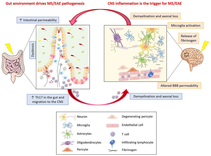
Parodi and Kerlero de Rosbo Gut-Brain Axis in Multiple Sclerosis
INTRODUCTION varying according to age, diet, and other environmental
factors, so an “optimal microbiota composition” does not exist
Multiple sclerosis (MS) is an inflammatory demyelinating disease (3). However, microbiota composition is quite stable at phylum
of the central nervous system (CNS), the etiology of which is still level, and an alteration in the ratio between different phyla, with
unclear, albeit believed to result from an autoimmune attack on the loss of microbiota diversity, has been associated with diseases
CNS components. MS is highly heterogeneous, with clinical (2, 4, 5). In homeostatic conditions, the microbiota not only
patterns characteristic of each individual patient and no single exerts a large number of functions that are related to food
pathognomonic marker known. Infection has long been digestion and vitamin synthesis, but it also has an important
investigated as a possible trigger of MS, albeit without concrete role in the maintenance of intestinal barrier integrity and in the
evidence for a particular agent. More recently, however, the regulation of the immune system (6). The microbiota
concept of a pro-inflammatory gut microbiota as a trigger of communicates with the host mainly through bacterial
autoimmunity has arisen, with the possible implication of metabolites of dietary substrates such as tryptophan
demonstrated dysbiosis in MS, based on pre-clinical studies in metabolites and short chain fatty acids (SCFA), modification of
its purported animal model, experimental autoimmune host molecules such as bile acids, or directly through bacterial
encephalomyelitis (EAE) (1). Here, we review how the possible components such as lipopolysaccharide (LPS) (7).
dysfunction of the gut-brain axis might impact neurological The intestinal barrier is a semipermeable mucosa which
diseases, with particular emphasis on MS. consists of the mucus, protecting the underlying layers; the
intestinal epithelium, mostly composed of enterocytes and
minor secretory cell subsets; and the lamina propria, a thin
WHAT IS THE GUT-BRAIN AXIS? layer of connective tissue that is densely populated by cells of the
innate and the adaptive immune system. The intestinal barrier
The gut-brain axis acts as a link between the external environment allows the entry of essential nutrients and beneficial molecules
and the CNS. Its main components are the microbiota for its role from the gastrointestinal tract, while preventing the entrance of
in gastrointestinal homeostasis, the intestinal barrier that regulates pathogens and harmful antigens (8).
the entry of food and microbial metabolites into the organism, and The ENS is the largest part of the peripheral nervous system
the sympathetic and parasympathetic arms of the autonomic that innervates the gastrointestinal tract, with nerve fiber endings
nervous system, namely the enteric nervous system (ENS) and of the myenteric plexus terminating in close proximity to intestinal
the vagus nerve, which transmit signals to the brain. epithelial cells (IEC) (Figure 1); it is connected to the CNS
The human gut microbiota comprises 500-1000 bacterial through the parasympathetic (via the vagus nerve) and
species and an undetermined number of viruses, fungi, and sympathetic (via the prevertebral ganglia) nervous systems. The
others (2); it is highly heterogeneous between individuals, ENS is composed of an extensive network of neurons and enteric
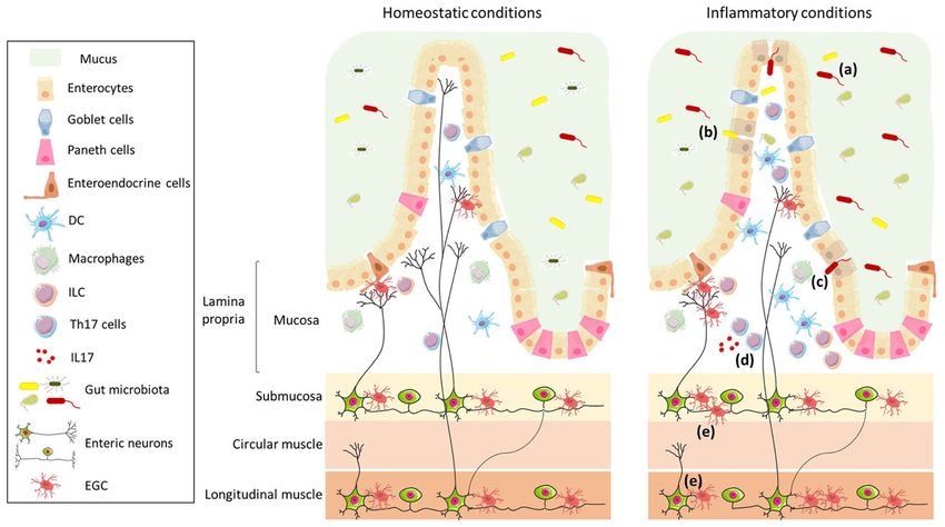
FIGURE 1 | Schematic representation of the intestinal wall in homeostatic and inflammatory conditions. The intestinal barrier is composed of mucus, which acts as
physical and biochemical barrier, of a continuous monolayer of intestinal epithelial cells (including enterocytes, goblet cells, Paneth cells, and enteroendocrine cells), and
of the lamina propria, which is densely populated by immune cells (such as DC, macrophages, ILC, and T cells). The intestinal barrier is directly exposed to the gut
microbiota that has an important role in the maintenance of barrier integrity and in the regulation of the immune system. The lamina propria is innervated by enteric
nerve fibres, which originate in the myenteric and submucosal plexi and are in contact with EGC. The left panel shows the intestinal wall under homeostatic conditions;
in the right panel, under inflammatory conditions, the composition of the gut microbiota is altered (a), the intestinal epithelial barrier is impaired (b), enabling translocation
of microbes (or their products) (c), and resulting in an increase in Th17 cells (d) and enteric axonal loss and gliosis (e).
Frontiers in Immunology | www.frontiersin.org 2 September 2021 | Volume 12 | Article 718220Parodi and Kerlero de Rosbo Gut-Brain Axis in Multiple Sclerosis
glial cells (EGC), which play an important role in the maintenance acid receptors at CNS level in a vagus nerve-dependent manner
of intestinal barrier function (8, 9). ENS functions include the (21). Similarly, the administration of another Lactobacillus strain
regulation not only of gastrointestinal motility, but also of (L. reuteri) improves social behavior in mouse models of autism
secretion, nutrient absorption, immune regulation, and defence spectrum disorder (ASD), which are associated with alterations
(10). In particular, glial cell-derived neurotrophic factor secreted of the gut microbiota, possibly through stimulation of the
by EGC is involved in the regulation of ILC3, which are a vagus nerve. Indeed, treatment of ASD mice with L. reuteri led
component of the glial–ILC3–epithelial cell unit that surveys gut to an increase in the number and fluorescence intensity of
environment and control its defence (11). In a recent study, Yan oxytocin-positive neurons in the periventricular nucleus of the
and co-authors demonstrated that enteric neurons themselves can hypothalamus where vagal fibers project, with release of oxytocin
exert a key role in the regulation of immune cells, by preventing (19) that is known to reverse social deficits in ASD mice (22).
microbial-induced differentiation of regulatory T (Treg) cells Contrasting data were however obtained in another study where
through the release of IL-6 (12). administration of L. reuteri induces a depression-like behavior via
the vagus nerve in mice treated with an antibiotic cocktail,
suggesting that the effect of L. reuteri likely depends on complex
synergistic interaction between intestinal microorganisms (23).
HOW THE GUT INTERACTS WITH Altogether, these studies suggest an anti-inflammatory role for
THE BRAIN vagus nerve stimulation, which was shown to be responsible for
modulating microglia activation in aged APP/PS1 mice, a genetic
The bidirectional communication between the gut and the brain
model of Alzheimer’s disease (AD) (24) and to have a positive effect
occurs in different ways.
on cognition in AD patients (25). In this context, it has been
demonstrated that vagal afferent fibers in the gastrointestinal tract
Through the ENS are involved in hippocampal-mediated memory function (26).
The ENS is directly connected to the brain through the
sympathetic and parasympathetic innervation of the gut. The Through Cross-Talk With the Immune
sympathetic pathway is composed of approximately 50% afferent System
and 50% efferent nerve fibres, while the parasympathetic The immune system represents an important gatekeeper in the
innervation of the gut is mainly provided by the vagus nerve, gut-brain axis, as it closely interacts with nerves and epithelial
composed of 80% afferent and 20% efferent fibers (13, 14). Many cells, and can sense microbiota metabolites (27). The crosstalk
studies have focused on the role of the vagus nerve, as part of the between the immune and nervous systems occurs because
cholinergic anti-inflammatory pathway and for its capacity to immune cells express a wide variety of receptors for
sense the gut stimuli and transmit them to the brain (14). neurotransmitters/neuropeptides and, in turn, neurons are
Efferent fiber signals from the brain to the gut, in turn, can responsive to cytokines. Besides the anti-inflammatory role of
affect gut motility, secretion, and epithelial permeability, and, the cholinergic (parasympathetic) system, the sympathetic
through the ensuing change in physical environment, alter the nervous system also affects intestinal immune cells differently.
composition of the intestinal microbiota. Of note, vagal efferent Indeed, its effects depends on several factors, including the type
fibers also regulate immune responses and cytokine production and the concentration of the neurotransmitter (norepinephrine,
through the activation of the acetylcholine/a7nicotinic- adenosine), the affinity of the receptor subtypes on immune cells,
acetylcholine receptor signaling (15). These fibers innervate the and the different functionality of the sympathetic nervous system
intestinal wall indirectly through synapses contacting the ENS in during different phases of inflammation (28). In this context,
the myenteric plexus (14), suggesting that central stimulation of macrophages present in the lamina propria or the muscularis
the vagus nerve leads to the activation of enteric neurons that express opposite phenotypes, pro-inflammatory vs tissue-
subsequently release factors, such as acetylcholine, that might protective, respectively, at steady state, a divergence that is in
affect the local immune system (16). Vagal afferent fibers can part related to norepinephrine signaling via b2 adrenergic
sense a wide spectrum of pro-inflammatory and neuroprotective receptors highly expressed on muscularis macrophages (29).
molecules produced by the microbiota or by the host (cytokines,
SCFA, LPS, neurotransmitters, etc…) and thereby transmit gut
signals to the CNS. Increasing evidence has shown that oral Through Sensing Microbial Metabolites
administration of probiotics, defined as “live microbial strains Microbiota metabolites can directly and indirectly influence CNS
that beneficially affect the host when ingested in sufficient doses” cells. In particular, they regulate microglia development (30, 31)
(17), have a high impact on vagus nerve activity (18, 19). The and functions (32). In this context, microglia from germ-free
precise mechanism underlying probiotics-vagus nerve interaction (GF) mice have a dysregulated expression profile of maturation
is not fully elucidated. Nevertheless, an increase in the spontaneous and activation markers, an altered/immature morphology, and a
firing frequency of the vagal afferent fibers following application of defective response to pathogens. Microbial metabolites, in
the anxiolytic probiotic Lactobacillus rhamnosus has been particular SCFA that include acetate, propionate and butyrate,
demonstrated, which was abolished upon vagotomy (20). Thus, are involved in the regulation of blood-brain barrier (BBB)
oral administration of L. rhamnosus reduces anxiety and permeability. In this context, GF mice have an increased BBB
depression in mice by reverting the expression of g-aminobutyric permeability with an altered expression of tight junction, and
Frontiers in Immunology | www.frontiersin.org 3 September 2021 | Volume 12 | Article 718220Parodi and Kerlero de Rosbo Gut-Brain Axis in Multiple Sclerosis
colonization with specific bacterial strain or the administration pathophysiology (54). The influence of the microbiota in AD
of SCFA restores BBB integrity (33). appears related not only to dysbiosis, but also to the ability of
amyloids to traverse the gut wall. Gut microbiota produces a high
quantity of amyloids in the biofilm that covers the gastrointestinal
tract, among which curli is the most studied; it is produced by
NEUROLOGICAL CONSEQUENCES OF A Escherichia coli in stressful conditions (55, 56) and promotes
“LEAKY” GUT intestinal inflammation (56–58). Interestingly, bacterial amyloids
may act as prion proteins, eliciting cross-seeding, through
Pathological or environmental factors directly or indirectly
molecular mimicry with amyloid-b (Ab), in vitro and in vivo,
impair intestinal barrier integrity. This can occur through
suggesting that they could induce the formation of Ab aggregates
changes in microbiota (34) or upon an increased activation of
in the CNS (59, 60). In this context, injection of brain extract
mucosal immune cells, such as occurs in inflammatory bowel
containing Ab in the stomach and gut wall (in the serosa) of the
disease (IBD) (35, 36). Dysfunction of this barrier allows the
outbred wild-type ICR mice induces CNS amyloidosis and AD‐
translocation of microbes or their products (such as LPS) from
like dementia by spreading of Ab through the ENS and vagus
the gut lumen into the lamina propria and from there into the
nerve (61).
blood, thereby promoting inflammation/an aberrant immune
The idea that neuropathology could be triggered by a leaky
response, not only in the gut, but also at systemic level. Such an
gut is best exemplified by recent studies in the neurodegenerative
increase in intestinal permeability, also known as “leaky gut”,
Parkinson’s disease (PD) and its experimental model. It stems
together with an alteration in the commensal population
from studies of Braak et al. in the early 2000’s who suggested that
composition, has been associated not only with gastrointestinal
Lewy body pathology could develop first in extra-CNS sites,
disorders, but also with extra-intestinal diseases (37, 38).
showing the presence of a-synuclein aggregates both in the
In the context of the gut-brain axis, a leaky gut has become an
olfactory bulb and in the dorsal motor nucleus of the vagus
important focus of interest for neurological disorders in the past
nerve (in the brainstem) (62), as well as in the myenteric and
decade, with the demonstration of intestinal microbiota dysbiosis
submucosal plexus of PD patients (63). These data led to the
and/or an impaired intestinal barrier in neurological diseases
hypothesis that a-synuclein aggregates could reach the brain
with different aetiologies, such as stroke (39), Huntington’s
from the ENS, as shown by EGC activation (64), via an
disease (40, 41), and amyotrophic lateral sclerosis (42, 43).
uninterrupted series of projection neurons, extending from the
Intestinal barrier disruption has also been linked to cognitive
dorsal motor nucleus of the vagal nerve to the cerebral cortex
disorders associated or not with neurodegeneration. In
(63). More recent studies in experimental models support this
particular, a key role for the gut in the pathogenesis of ASD
hypothesis. Thus, the injection of a-synuclein fibrils in the
has been hypothesized with gastrointestinal inflammation
muscle layer of pylorus and duodenum (that are densely
reported in as many as 84% of the cases (44). In particular,
innervated by vagus nerve) of wild-type mice leads to
altered expression of intestinal tight junctions (45), together with
pathologic propagation of a-synuclein to the CNS via the
an altered composition of the gut microbiota and their metabolic
vagus nerve, resulting in loss of dopaminergic neurons and
products, have been observed in ASD patients and/or in a mouse
PD-like clinical symptoms (65). Hence the hypothesis that a-
model with ASD features (45–47). Interestingly, an increased use
synuclein may originate in the ENS and go to the brain via
of antibiotics during pregnancy seems to be correlated with a
retrograde axonal transport through the vagus nerve (66, 67).
higher risk for the development of ASD (47). In this context,
Interestingly, in PD patients, gastrointestinal manifestations start
administration of an emerging probiotic, Bacteroides fragilis,
years (decades) before the clinical onset of disease and are
ameliorated gut alterations, dysbiosis, and ASD symptoms in
associated with increased intestinal permeability and dysbiosis
the mouse model (48). While gut dysbiosis is now well
(68). Also corroborating this hypothesis is the observation that
established as a possible contributor of AD, both in patients
intestinal infection with Gram-negative bacteria triggers PD-like
and in several animal models (49–51), there is, to our knowledge,
symptoms in the Pink-/- mouse model of PD (69).
no direct evidence for a dysfunctional intestinal barrier in
That a leaky gut could indeed lead to neurologic manifestations
patients, although it should be noted that intestinal permeability
is also clearly corroborated by the noted observation that IBD is
increases with age in the human (52). Interestingly, age-dependent
often associated with CNS demyelination, the hallmark of MS (70,
alterations in the gut microbiota associated with gut leakiness are
71); conversely, neurological disorders are often associated with
found in the 5xFAD mouse model of AD, with a significant
gastrointestinal symptoms (72).
decrease in microbial diversity; transplantation of microbiota from
aged AD, but not wild-type, mice accelerated AD pathology in
young mice, effects that were mitigated by probiotic bacteria-
enriching treatment (50). Similarly, in the ADLPAPT mouse AD THE GUT-BRAIN AXIS IN MS AND EAE
model, reducing gut dysbiosis through transplantation of faecal
microbiota from WT mice improved AD pathogenesis and Gut Dysbiosis
cognitive impairment (53). In contrast, in another mouse AD The possible implication of gut dysbiosis in MS was first studied
model, AppNL-G-F mice, administration of probiotics ameliorated in EAE through the use of GF mice or antibiotic treatment,
intestinal inflammation and leakiness, but not neurological which demonstrated that the microbiota plays a crucial role in
Frontiers in Immunology | www.frontiersin.org 4 September 2021 | Volume 12 | Article 718220Parodi and Kerlero de Rosbo Gut-Brain Axis in Multiple Sclerosis
directing both pro- and anti-inflammatory immune responses in highly relevant to MS that dysbiosis in patients is frequently
the CNS (73). The importance of microbiota in the pathogenesis associated with an increased intestinal permeability (84) and
of EAE is supported by studies in which the prophylactic (but not both alterations have been also demonstrated in EAE (85–87).
therapeutic) antibiotic treatment protects against the disease, That such conditions might promote a systemic inflammatory
possibly by increasing Treg- and Th2-cell responses (74–76). condition is supported by the increased plasma levels of LPS
Importantly, antibiotic treatment impaired the development of observed in both MS patients and EAE- affected mice (88, 89), as
EAE if given per os specifically, rather than intravenously (76). well as by the increased anti-microbiota systemic IgG responses
EAE experiments with mice treated with specific antibiotics or observed in MS patients, which suggest an enhanced bacterial
GF mice monocolonized with particular bacteria have clearly translocation from the gut lumen to systemic circulation in
demonstrated that signals from an altered gut microbiota can MS (90).
induce inflammation in extra-intestinal tissues (74, 75, 77, 78).
This concept has been further elaborated through experiments in
which microbiota from MS patients were tested for their ability
The ENS Link
In view of the close proximity of ENS neuronal endings to gut
to modulate EAE. Several studies have demonstrated a dysbiotic
luminal contents, it is likely that the gut microbiota impacts enteric
gut microbiota in MS, albeit with variables findings (73). A more
neurons, as demonstrated by the changes in electrophysiological
recent analysis showed significant association with MS of several
properties of myenteric neurons (91) and intestinal motility (92)
Acinetobacter species, which are rare in healthy human gut,
in response to alterations of the microbiota, providing a potential
together with the decreased presence of Parabacteroides, more
link between the microbiota and the CNS. Although the ENS has
particularly P. distasonis. Interestingly, exposure of lymphocytes
been poorly studied in MS, gastrointestinal motility disorder is an
from healthy individuals to MS microbiota or extract from MS-
additional burden in MS (93), and two recent studies suggest that
associated Acinetobacter calcoaceticus increased their differentiation
the ENS is involved. Thus, signs of ENS degeneration, including
to Th1 type cells and reduced the proportion of CD25+FoxP3+
gliosis and axonal loss in the myenteric plexus, which were
Treg cells, whereas exposure to P. distasonis extract could skew
antibody-mediated and caused significant decrease in intestinal
their T-cell phenotype to regulatory (79).
motility, were observed in a B cell- and antibody-dependent
mouse EAE model before the onset of CNS disease; myenteric
Gut-Brain Cross-Talk plexus alterations with nerve fiber loss and enteric glia activation
EAE experiments further corroborated the pro-inflammatory
were also detected in colon resectates from two of three MS
environment imparted by MS microbiota. Thus, EAE severity in
patients analysed (94). Serum autoantibodies against potential
mice pre-colonized with faecal MS microbiota was significantly
target antigens derived from enteric glia and/or neurons were
greater than in mice colonized with control microbiota, and was
identified in both EAE-affected mice and MS patients (94). Similar
associated with decreased IL-10+ Treg-cell induction in mesenteric
observations were made in three other mouse EAE models where
lymph nodes (79). Similarly, colonization with MS microbiota of
autoantibodies targeting ENS components were implicated in
transgenic mice bearing the T-cell receptor (TCR) for an
altered gastrointestinal motility (95).
encephalitogenic epitope significantly increased the spontaneous
incidence of EAE in these mice (80). However, the gut environment
can also favor the generation of Treg cells that can improve EAE. Restoring Gut Homeostasis: Probiotics
Indeed, not only effector T cells (81), but also Treg cells are and Fecal Microbiota Transplantation
activated within the gut mucosa in response to commensal The clear involvement of the gut-brain axis in MS pathogenesis
dysbiosis. In this context, a population of autoreactive CD4+ suggests that it could be a potential therapeutic target.
intraepithelial lymphocytes shown to proliferate in mouse GALT Accordingly, in view of the profound impact dysbiosis could
and gut lamina propria in response to non-self-antigens derived have on MS, the use of probiotics has been proposed as a
from the intestinal contents, infiltrated the CNS upon adoptive potential therapy. Probiotics are thought to contribute to
transfer to regulate inflammation, decrease demyelination, and maintaining or restoring a balanced and diverse microbiota
ameliorate disease severity (82). Not only T cells, but also B cells (96). Although their mode of action is not fully understood,
reactive to gut commensals could be involved in the cross-talk increasing evidence indicates that they have a high impact on
between gut and brain. Thus, in MS patients, IgA+ B cells specific vagus nerve activity (18). Several studies have shown that both
for MS-associated immune-stimulatory bacterial strains traffic to the preventive and the therapeutic administration of Bacteroides
the inflamed CNS where they colocalize with active lesions (83). As fragilis can suppress EAE by promoting Treg-cell function (97,
these CNS-infiltrating IgA+ B cells recognizing gut microbiota did 98). This anti-inflammatory effect is apparently mediated by
not cross-react with brain antigens and were associated with IL10 polysaccharide A (PSA), a component of B. fragilis capsula,
expression, the authors suggest that these gut-originating IgA+ B which, as a ligand for TLR2, induces the differentiation of Treg
cells represent regulatory cells actively recruited to the inflamed cells (99). Oral treatment with PSA induced an increase in the
CNS through inflammation-dependent factors, independently of number of CD103+ dendritic cells (DC), a subset of DC involved
their reactivity to microbial antigens (83). in the differentiation of Treg cells, in cervical lymph nodes of
Altogether these studies clearly demonstrate the importance treated mice (98). Similarly, the preventive per os administration
of the microbiota composition in determining the inflammatory of Escherichia coli strain Nissle 1917, a probiotic used for the
environment in the gut, and its consequences on the CNS. It is treatment of many gastrointestinal disorders (100), ameliorates
Frontiers in Immunology | www.frontiersin.org 5 September 2021 | Volume 12 | Article 718220Parodi and Kerlero de Rosbo Gut-Brain Axis in Multiple Sclerosis
EAE outcome by modulating cytokine production and migration induced for EAE, daily gavage with fecal supernatant from naïve
of autoreactive CD4+ T cells and preventing EAE-induced mice delayed disease onset and reduced disease expression at
intestinal alterations (101). The combination of different both clinical and neuropathological levels, preventing BBB
strains of probiotics could exert synergistic therapeutic effects; impairment and reducing neuroinflammation, demyelination
thus, three strains of Lactobacillus, each singly shown to be and axonal loss; the beneficial effect was associated with a
effective in preventing the development of EAE when trend towards a restored gut microbiota diversity in the treated
administered before disease onset, were shown to inhibit mice (110). In MS, however, studies to assess the possible effect of
established disease when administered as a therapeutic mixture FMT are scarce. Serendipitous findings from three MS patients
(102). In MS patients, the administration of a mixture of treated with 5-10 FMT infusions for severe chronic constipation
probiotics (enriched with Lactobacillus, Streptococcus and demonstrated not only resolution of the condition, but also
Bifidobacterium) induced a switch in the peripheral immune progressive neurological improvement (111). A recent proof-
response to anti-inflammatory, and reverted the alteration in of-concept single-subject longitudinal study was conducted to
microbiota composition associated with MS (103, 104). Whether evaluate the potential impact of FMT on relapsing-remitting MS.
or not these changes were associated with clinical improvement The patient received FMT infusions from five healthy donors
was not assessed in this short-term study, but the data from and was followed over 12 months for clinical assessment, fecal
randomized double-blind placebo-controlled clinical trials of microbiome composition, fecal SCFA concentrations, and serum
three- and four-month duration with similar probiotic levels of inflammatory and neuroprotective biomarkers (112).
mixtures (Lactobacilli and Bifidobacteria) suggest that daily The treatment resulted in an improved microbiome with
probiotic supplementation could ameliorate clinical symptoms increased bacterial diversity partly due to increased relative
of MS (as judged by a small but significant improvement in the abundance of butyrate-producing bacterial species, that was
expanded disability status scale) (105). However, recent studies accompanied by an increased concentration of the anti-
in EAE suggest that, in the design of probiotic treatment, it is inflammatory SCFA, butyrate. The modified microbiota was
important to consider that probiotic strains could exert different, associated with decreased levels of inflammatory cytokines and
even opposite, effects depending on the timing of exposure, the increased levels of the neuroprotective molecule, brain-derived
microbiome composition of the recipient, and the recipient’s growth factor, in the serum. At clinical level, the patient showed
genetic background. This is best exemplified by the divergent progressive improvement in gait and walking and balance
effects of treatment of EAE with L. reuteri. Thus, despite its metrics over the course of the study (112). Similarly, a case
beneficial effect in suppressing the development of the disease report indicated that treatment with FMT for Clostridum difficile
when administered before the onset of the symptoms (106), enterocolitis in a patient with secondary progressive MS was
L. reuteri within a probiotic mixture could instead contribute associated with disease stability (113). Albeit limited, these
to CNS autoimmunity, through potential mimicry with an findings argue for further investigations of the potential benefit
encephalitogenic protein leading to activation of encephalitogenic of rebalancing the microbiota through FMT as an adjunct
T cells in the small intestine (74). In studies of EAE in consomic approach to MS therapy, and clinical trials to this effect are
mice where transfer of the most divergent microbiota from these ongoing (114).
mice into genetically identical recipients showed that genetically
determined gut microbiota modulates EAE severity, abundance of
L. reuteri in the transferred microbiota, or through its stable Modulation of the Gut Microbiota Through
introduction in the microbiota associated with the less severe the Diet
disease, was seen to co-segregate with exacerbated disease (107). Another strategy to modulate the gut microbiota is through the
In another approach to therapy targeting the microbiota, diet, since it is considered the main factor that shapes its composition.
humanized HLA-transgenic mice induced for EAE were A vegetarian diet promotes an increase in the number of SCFA-
gavaged before disease onset with Prevotella histolica, a producing bacteria, leading to immunomodulatory effects not only in
commensal decreased in the microbiota of MS patients as the gut but also at systemic level (See above) (115). In this context,
compared to that of healthy subjects, but increased in microbiota preventive, but not therapeutic, treatment by daily gavage with
of MS patients receiving disease-modifying therapies. The treatment propionic acid ameliorated EAE at both clinical and pathological
restored the gut microbiota to that preceding the encephalitogenic levels by inducing the differentiation of Treg cells in the small
challenge and resulted in reduced gut permeability and BBB intestine (116). Interestingly, propionic acid is reduced in serum
dysfunction, with reduced CNS inflammation; disease and faeces of MS patients in association with an altered microbiota,
amelioration was associated with an immunomodulatory effect at and dietary supplementation for three years with this SCFA resulted
systemic level, with a decrease in Th1- and Th17-cell levels and in clinical and pathological amelioration in the treated patients. This
increased frequencies of Treg cells (108). was associated with a rebalancing of the Treg/Th17 cell ratio towards
Restoring gut homeostasis thus appears as a potentially a more regulatory profile and an upregulation of genes related to
important aspect of the therapeutic approach to MS. In this induction of Treg cells in the intestine (117). The induction of Treg
context, fecal microbiota transplantation (FMT), which has given cells by SCFA can occur directly or indirectly by enhancing the
promising results in IBD (109), is being considered as a possible production of retinoic acid by CD103+ DC, which drives the
approach to reestablish gut microbiota composition. In mice differentiation of FoxP3+Treg cells (118, 119). In particular, it has
Frontiers in Immunology | www.frontiersin.org 6 September 2021 | Volume 12 | Article 718220Parodi and Kerlero de Rosbo Gut-Brain Axis in Multiple Sclerosis
been shown that the anti-inflammatory effect of the commensal yet impossible to ascertain which of gut or brain inflammation
butyrate occurs through activation of its receptor, hydroxycarboxylic comes first in MS. In EAE, intestinal barrier permeability, together
acid receptor 2 (HCAR2), which triggers an increased production of with morphological alterations, was observed already at 7 days post-
retinoic acid and IL-10 in intestinal DC (120), modulates the over- immunization (85), prior to clinical disease onset, and was associated
activation of ILC3 (121), and decreases pro-inflammatory gene with an increase in potentially pathogenic T cells infiltrating the gut
expression in IEC (122, 123). SCFA can also affect B cells, as lamina propria. However, it has not been assessed at earlier post-
recently demonstrated in a mouse model of arthritis, in which gut- immunization times, nor correlated concomitantly with BBB
derived acetate was shown to promote the differentiation of anti- integrity, which is already compromised at this pre-clinical stage
inflammatory IL-10-producing B-cells (124). Of note in this context, (Kerlero de Rosbo and Cedola, unpublished data).
serum levels of butyrate correlated positively with the percentage of
IL-10-producing B cells and levels of acetate correlated negatively The Gut Environment Drives MS
with TNF production by IgM+ B cells in patients with MS or Pathogenesis
clinically isolated syndrome preceding MS (125). Highly relevant in this context is the observation that T cells get
Other dietary modes that can impact CNS integrity through activated in the gut (74, 131) and intestinal CD4+ T cells can play
the microbiota, and thereby through the Th17/IL-17 pathway in a role in the initiation of CNS autoimmunity. A recent study
the gut, include intermittent fasting. Thus, intermittent fasting linked autoimmunity in a spontaneous mouse model of MS to
exerted a beneficial effect on EAE, ameliorating the clinical course the expression of the transforming growth factor beta (TGF-b)
and pathology of the fasting mice through an effect partially inhibitor, Smad7, in intestinal CD4+ T cells (132). TGF- b and
dependent on their microbiota that led to a decrease in IL-17- Smad7 are involved in the regulation of naïve CD4+ T-cell
producing T cells together with an increase of Treg cells in GALT differentiation and Smad7 in particular was shown to drive
(126). In this context, it should be noted that long-term calorie Th1-cell responses in MS and EAE (133). Overexpression of
restriction seemingly has a health-promoting effect in mice by Smad7 in the T cells of these mice led to highly increased disease
inducing a balanced gut microbiota, with increased proportion of parameters at all levels, with disease dependent upon induction
beneficial bacterial strains involved in the maintenance of of the CD4+ T cells at the intestinal mucosal barrier before their
intestinal homeostasis (such as Lactobacillus) (127). Moreover, migration to the CNS (132). Adoptive transfer EAE experiments
the reduction in protein intake, and in particular the omission of showed that only gut-derived T cells or T cells treated to induce
the amino acid tryptophan, have been shown to prevent EAE expression of gut-homing receptors were able to trigger disease
induction by affecting encephalitogenic T cell responses (128). upon transfer in wild-type recipients. Of note, EAE induction
This effect is dependent on the presence of gut microbiota, as was dependent on the presence of a full gut microbiota (132). At
dietary tryptophan restriction leads to an enrichment of the human level, the analysis of intestinal tissue biopsies from 27
tryptophan-synthesizing bacterial genera (128). MS patients and 27 control individuals revealed an altered TGF-
The influence of the diet on the gut-brain axis in MS and EAE b-dependent Th-cell differentiation pattern in the lamina propria
is also exemplified by a recent EAE study based on the observation with increased frequencies of inflammatory T cells and reduced
that MS is less prevalent in countries where the diet includes high FoxP3 expression (132), a process akin to the defective TGF-b1/
amounts of isoflavones, a class of phytoestrogens known for their Smad signaling in IBD (134).
antioxidant and anti-inflammatory health benefits. The authors of Interestingly, it has been suggested that the levels of
the study observed a more severe EAE course in mice fed a diet circulating mucosal-associated invariant T cells, which are
devoid of isoflavones whereas a diet rich in isoflavone was abundant in gut lamina propria, may reflect disease activity in
associated with an ameliorated EAE course as compared to the MS, with reduced numbers observed during exacerbation
standard mouse chow. The beneficial effect was associated with correlating with increased MRI activity in some patients, while
decreased activation and proliferation of the encephalitogenic T at remission increased numbers coincided with decreased MRI
cells and with reduced immune cell infiltrates in the CNS. As activity (135). As recently demonstrated, T cells specific for an
shown by the difference in gut microbiome composition between encephalitogenic epitope might be expanded and activated by the
mice fed an isoflavone-rich diet and those fed an isoflavone-free small intestine microbiota, albeit without evidence that these
diet, disease amelioration was dependent on the presence of cells could migrate to the CNS (74). Another recent study in
isoflavone-metabolizing bacteria and their metabolites, which, adoptive transfer EAE, which showed the infiltration of
interestingly, are reduced in MS patients (129). encephalitogenic Th17 cells in the lamina propria at a time
when no such cells were detected in the CNS, demonstrated that
the encephalitogenic cells could later be traced to the CNS of
WHAT COMES FIRST IN MS, GUT OR CNS mice with clinical symptoms while they decreased in the gut;
INFLAMMATION? blocking the initial transit of the autoreactive T cells to the gut
resulted in reduced disease severity (131). In the context of gut
Although gastrointestinal manifestations are common in MS inflammation, an interesting study showed that the lack of
(130) and possible indication can be obtained from retrospective disease expression in mice lacking IL-17 was not related to the
studies in MS patients, such as when comorbidity with IBD encephalitogenic potential of the T cells, which were fully active
occurs or demyelination consecutive to IBD is observed, it is as upon transfer in wild-type mice, but rather to the absence of
Frontiers in Immunology | www.frontiersin.org 7 September 2021 | Volume 12 | Article 718220Parodi and Kerlero de Rosbo Gut-Brain Axis in Multiple Sclerosis
IL-17 in the gut. Restoring the microbiota of IL-17-deficient mice Th17 cells is possibly due to a diet-dependent shift in microbiome
to that of IL-17-sufficient mice resulted in full-blown EAE, composition (140) and is induced by the activation of serum
indicating that the reduced susceptibility to EAE in IL-17- glucocorticoid kinase 1 that regulates Na+ intake (141). Studies in
deficient mice was strongly associated with the altered MS patients demonstrated that a high salt diet induces an increase
microbiota observed in these mice, suggesting that IL-17 in the in Th17 cells in the blood and a decrease in intestinal Lactobacillus
gut can modulate the intestinal microbiota (136). This study strain, and is associated with worsened clinical symptoms and
demonstrated the crucial impact of intestinal IL-17 on enhanced radiological activity (140, 142). However, contrasting
microbiota composition and thereby on EAE expression, as data have been reported in MS, where in a five-year clinical trial
reintroducing IL-17 expression specifically in IEC of IL-17- high levels of urinary sodium in patients with clinically isolated
deficient mice resulted in a restored microbiota (136). Highly syndrome did not correlate with increased rates of conversion to
relevant in this context are the observations that, in MS, there is definite MS or with clinical worsening and/or enhanced MRI
an expansion of Th17 cells in the intestine, which is associated activity (143). Similarly, in EAE a high salt diet suppressed the
with microbiota alterations and correlates with high disease opticospinal encephalomyelitis mouse model by promoting the
activity (137). tightening of the BBB, possibly via increasing the serum level of
It therefore seems that inflammation in the gut can indeed corticosterone (144).
result in activation of encephalitogenic T cells that can travel to Although EAE experiments in mice transferred with
the CNS where they can induce inflammatory damage with microbiota from EAE-affected mice or MS patients have shown
subsequent demyelination and axonal loss (Figure 2). In that transfer with a dysbiotic gut microbiota increases disease
addition, inflammation in the gut can also influence microbiota severity and conversely, there is as yet no concrete evidence that
diversity and can lead to dysbiosis, which can itself propagate microbiota dysbiosis and its potential inflammatory consequences
intestinal inflammation. A possible trigger for intestinal on the gut-driven immune response are by themselves sufficient
inflammation, through dysbiosis, could be the diet, which can to cause EAE or MS. However, in addition to its “non-specific”
alter both the number and the diversity of microbiota species. effect related to inflammation, the microbiota could be involved in
Studies have shown that a diet rich in salt could be associated with another route leading to brain inflammation. Thus, a recent EAE
a higher risk for development of EAE and dementia, via the study showed that T cells specific for an encephalitogenic peptide
induction of Th17 cell differentiation; in particular, mice fed a can be activated through mimicry with microorganisms in the
high-salt diet developed neurovascular and cognitive impairments small intestine. Lactobacilli produce many peptides that have
through the expansion of Th17 cells in the small intestine, leading molecular mimicry with the TCR- binding residues of the
to an increase in IL-17 in the plasma (138, 139). This increase in encephalitogenic peptide, myelin oligodendrocyte glycoprotein
FIGURE 2 | What comes first in MS, gut or CNS inflammation? Gut environment drives MS/EAE pathogenesis. In MS/EAE, dysbiosis together with an increased
intestinal permeability, possibly due to alterations in intestinal epithelial barrier morphology, have been demonstrated. These alterations allow the translocation of
microbes or their products from the gut lumen into the lamina propria, thereby promoting inflammation. Inflammation in the gut can result in activation of
encephalitogenic T cells that can travel to the CNS where they can induce inflammatory damage with subsequent demyelination and axonal loss. CNS inflammation
is the trigger for MS/EAE. Neuroinflammation in MS/EAE could be the consequence of an altered vascular permeability of BBB. The leakage of plasma components,
such as fibrinogen, leads to rapid activation of microglia that might be the trigger of the disease, with ensuing infiltration of homing inflammatory cells and signal
transmission to the gut resulting in intestinal barrier permeability thereby propagating the brain-gut inflammation loop.
Frontiers in Immunology | www.frontiersin.org 8 September 2021 | Volume 12 | Article 718220Parodi and Kerlero de Rosbo Gut-Brain Axis in Multiple Sclerosis
(MOG)35-55; in particular, a specific peptide produced by in cervical lymph nodes. Interestingly, disruption of the
L. reuteri induces proliferation of MOG-specific CD4 T cells meningeal lymphatics or removal of the cervical lymph nodes
(74). Although the L. reuteri peptide was not by itself resulted in delayed EAE onset (161).
encephalitogenic, these findings suggest that molecular mimicry CNS inflammation can also be triggered by leakage from CNS
with gut microbiota elements can amplify a pathogenic vessels. More particularly, dysfunction of the BBB is a hallmark
autoimmune T-cell response. of MS. Although not performed concomitantly with assessment
of the gut barrier, a two-photon microscopy monitoring study of
CNS Inflammation Is the Trigger for MS blood vessel integrity in EAE clearly demonstrated that focal
Molecular mimicry with microbial proteins is in fact one of the transient vessel leaks occur in the cortical gray matter within the
most common etiological hypotheses for pathogenic first three days post-encephalitogenic challenge (163). Such an
autoimmunity, including MS. Indeed, there are many examples altered vascular permeability at this early stage of EAE induction
of autoimmune disease development following an infection and appeared to be an initiating event, rather than a consequence of
epidemiological data indicate that viral infections are a risk factor effector T-cell aggregation together with tissue damage at
for MS (145). Molecular mimicry is dependent on TCR perivascular lesions, as it was not accompanied with an
degeneracy, a common feature of antigen recognition, whereby infiltration of inflammatory cells. However, leakage of plasma
a single TCR is able to recognise a multiplicity of peptides (146). components such as fibrinogen leads to rapid inflammatory
Such degeneracy has been documented for self-reactive T cells activation of microglia (Figure 2) and has been observed in
(145, 146) and an early seminal study (147), later confirmed (148, MS and in EAE induced in marmosets where microglial
149), demonstrated that viral peptides can activate human T cells activation was detected in conjunction with early fibrinogen
specific for myelin protein epitopes derived from patients with deposits prior to tissue damage or gadolinium enhancement on
MS. Moreover, cloned MS-derived T cells specific for a disease- magnetic resonance imaging (164). Interestingly, depletion of
relevant human myelin epitope could be activated by microbial fibrinogen ameliorates EAE (165). In both EAE and MS, there is
peptides subsequently shown to induce EAE in humanized mice evidence that microglia activation might be a trigger of disease.
transgenic for the relevant human TCR (150). Autoreactive Thus, in EAE it was shown to precede infiltration of inflammatory
T cells that express a low-avidity TCR bypass clonal deletion cells (166) and impairment of microglia reduces EAE severity (167);
(151) and T cells with specificity to myelin epitopes are present in in MS, the presence of frequent, macroscopically invisible, pre-active
healthy humans (152, 153) and naive wild-type mice (154). Such lesions, not associated with BBB disruption, suggests CNS-intrinsic
self-reactive T cells have been shown to respond to their self- factors leading to innate immune activation (168). However, there is
antigen or its variants during infection, where inflammation and no indication that focal vessel leakage such as seen shortly after EAE
high levels of antigen presentation prevail (155, 156). It should be induction, occurs in the absence of specific barrier breaching events
noted that active EAE does not develop in the absence of a strong and the nature of possible CNS-intrinsic stimuli of innate immune
inflammatory Th1 response induced by Mycobacterium activation is unclear. In this context, however, dysfunction or
tuberculosis. These findings suggest that infection could trigger degeneration of pericytes, resulting in BBB permeability and
a strong autoimmune response by degenerate myelin-reactive accumulation of blood-derived fibrinogen in brain parenchyma
T cells through mimicry with the infectious agent. Interestingly, could play a crucial role in mediating neuroinflammation (169).
T cells expressing degenerate TCRs are present at higher Interestingly, in early MS, vascular permeability in white matter
frequency in patients with MS (157). Such activated myelin- tracts is associated with pericyte dysfunction (170), and soluble
reactive T cells could then home to the brain to induce fibrinogen can trigger pericyte death by autophagy (171). In
inflammation and tissue damage. Indeed, T cells primed in the addition, in view of their close association with the brain vessels,
periphery can bypass the BBB (158), and it is clear that in both activated pericytes, which express receptors for inflammatory
MS and EAE, peripherally activated T cells home to the CNS, molecules, could propagate inflammation generated elsewhere in
provided that they recognise an antigen expressed, either the body and release pro-inflammatory factors deleterious to BBB
inherently or through transgenic manipulation, in that organ integrity and function (169).
(159, 160). In addition, in the last few years, the demonstration of On the other hand, signals from the brain, in particular
meningeal lymphatic vessels (161) has provided a possible link through the sympathetic and parasympathetic nervous systems
between the CNS and the peripheral immune system that could and the ENS, can trigger intestinal inflammation and increase
be highly relevant to the development of an inflammatory CNS intestinal barrier permeability following CNS injury (172). This
disease such as MS. Thus, CNS antigens collected through the in turn will lead to gastrointestinal dysfunction, immune cell
glymphatic system from brain parenchyma into the CSF, could be activation in the gut, and gut dysbiosis, which in turn can result
transported by meningeal lymphatics (162) into cervical lymph in an increased CNS inflammation.
nodes with the potential to induce an autoimmune response These different hypotheses, however, are not mutually exclusive.
(161). In this context, DC and T cells, which are found in healthy
meninges, can migrate to cervical lymph nodes via meningeal
lymphatics under both normal and pathological conditions (161). DISCUSSION
It is therefore possible that, under inflammatory conditions,
potentially autoreactive degenerate T cells could initiate an At this stage, the primary trigger for the dysfunctional gut-brain
immune response to a relevant CNS antigen presented by DC axis in MS still hides within a chicken-egg causality dilemma,
Frontiers in Immunology | www.frontiersin.org 9 September 2021 | Volume 12 | Article 718220Parodi and Kerlero de Rosbo Gut-Brain Axis in Multiple Sclerosis
with an apparent self-feeding loop of brain-gut inflammation. mechanisms that underlie the deleterious T-cell response might
Thus, innate immune activation in the brain might itself be totally differ, with gut events promoting MS, while CNS events
triggered by signals from the gut. Metabolites of the gut promote EAE, or vice versa. Regardless, it is clear that the gut-
microbiota affect CNS cells (30–32, 173) and the innate brain interaction is of utmost importance in the progression of
immune response in the CNS was shown to be affected by the the disease and therapeutic approaches which target gut
microbiota. Indeed, an increased production of SCFA, in dysbiosis and intestinal barrier dysfunction must be seriously
particular butyrate, caused by an alteration of the microbiota considered as drug adjuncts in the future pharmacopeia of MS.
upon feeding mice a high-fiber diet, resulted in a decrease in
inflammatory microglia gene expression (174). Similarly,
microbial metabolites of dietary tryptophan controlled by the AUTHOR CONTRIBUTIONS
commensal microbiota act directly on microglia and regulate
microglia control of astrocyte-mediated neuroinflammation, via All authors listed have made a substantial, direct, and intellectual
the aryl hydrocarbon receptor (173), while gut microbiota- contribution to the work and approved it for publication.
modulated production of IFNg by meningeal NK cells drives
the differentiation of a new subset of astrocytes (characterized by
the expression of LAMP1 + TRAIL + ) that have an anti- FUNDING
inflammatory function in EAE (175). To shed light on the
implication of gut dysbiosis in diseases like MS, it will be We gratefully acknowledge the funding by FISR-Tecnopolo per
necessary to clearly define the sequence of events that lead to la Medicina di Precisione. D.G.R. n. 2117 of 21.11.2018.
intestinal barrier dysfunction vs brain barrier dysfunction and
delineate the timing at which these first occur. This could be
done in EAE, but is nearly impossible in MS where disease ACKNOWLEDGMENTS
initiation is unclear and could occur many years before actual
symptoms drive the diagnosis. Obviously, EAE is not MS and the We thank N. Rigoli for help in preparing the figures.
Cells and Gut Defence. Nature (2016) 535(7612):440–3. doi: 10.1038/
REFERENCES nature18644
1. Wekerle H. Nature, Nurture, and Microbes: The Development of Multiple 12. Yan Y, Ramanan D, Rozenberg M, McGovern K, Rastelli D, Vijaykumar B,
Sclerosis. Acta Neurol Scand (2017) 136 Suppl 201:22–5. doi: 10.1111/ et al. Interleukin-6 Produced by Enteric Neurons Regulates the Number and
ane.12843 Phenotype of Microbe-Responsive Regulatory T Cells in the Gut. Immunity
2. Sekirov I, Russell SL, Antunes LC, Finlay BB. Gut Microbiota in Health and (2021) 54(3):499–513.e495. doi: 10.1016/j.immuni.2021.02.002
Disease. Physiol Rev (2010) 90(3):859–904. doi: 10.1152/physrev.00045.2009 13. Matteoli G, Boeckxstaens GE. The Vagal Innervation of the Gut and
3. Rinninella E, Raoul P, Cintoni M, Franceschi F, Miggiano GAD, Gasbarrini Immune Homeostasis. Gut (2013) 62(8):1214–22. doi: 10.1136/gutjnl-
A, et al. What Is the Healthy Gut Microbiota Composition? A Changing 2012-302550
Ecosystem Across Age, Environment, Diet, and Diseases. Microorganisms 14. Goverse G, Stakenborg M, Matteoli G. The Intestinal Cholinergic Anti-
(2019) 7(1):14. doi: 10.3390/microorganisms7010014 Inflammatory Pathway. J Physiol (2016) 594(20):5771–80. doi: 10.1113/
4. McBurney MI, Davis C, Fraser CM, Schneeman BO, Huttenhower C, JP271537
Verbeke K, et al. Establishing What Constitutes a Healthy Human Gut 15. Rosas-Ballina M, Olofsson PS, Ochani M, Valdé s-Ferrer SI, Levine YA,
Microbiome: State of the Science, Regulatory Considerations, and Future Reardon C, et al. Acetylcholine-Synthesizing T Cells Relay Neural Signals in
Directions. J Nutr (2019) 149(11):1882–95. doi: 10.1093/jn/nxz154 a Vagus Nerve Circuit. Science (2011) 334(6052):98–101. doi: 10.1126/
5. Mosca A, Leclerc M, Hugot JP. Gut Microbiota Diversity and Human science.1209985
Diseases: Should We Reintroduce Key Predators in Our Ecosystem? Front 16. Bonaz B, Bazin T, Pellissier S. The Vagus Nerve at the Interface of the
Microbiol (2016) 7:455. doi: 10.3389/fmicb.2016.00455 Microbiota-Gut-Brain Axis. Front Neurosci (2018) 12:49. doi: 10.3389/
6. Fung TC, Olson CA, Hsiao EY. Interactions Between the Microbiota, fnins.2018.00049
Immune and Nervous Systems in Health and Disease. Nat Neurosci 17. Hill C, Guarner F, Reid G, Gibson GR, Merenstein DJ, Pot B, et al. Expert
(2017) 20(2):145–55. doi: 10.1038/nn.4476 Consensus Document. The International Scientific Association for
7. Lavelle A, Sokol H. Gut Microbiota-Derived Metabolites as Key Actors in Probiotics and Prebiotics Consensus Statement on the Scope and
Inflammatory Bowel Disease. Nat Rev Gastroenterol Hepatol (2020) 17 Appropriate Use of the Term Probiotic. Nat Rev Gastroenterol Hepatol
(4):223–37. doi: 10.1038/s41575-019-0258-z (2014) 11(8):506–14. doi: 10.1038/nrgastro.2014.66
8. Aubé AC, Cabarrocas J, Bauer J, Philippe D, Aubert P, Doulay F, et al. 18. Fülling C, Dinan TG, Cryan JF. Gut Microbe to Brain Signaling: What Happens
Changes in Enteric Neurone Phenotype and Intestinal Functions in a in Vagus. Neuron (2019) 101(6):998–1002. doi: 10.1016/j.neuron.2019.02.008
Transgenic Mouse Model of Enteric Glia Disruption. Gut (2006) 55 19. Sgritta M, Dooling SW, Buffington SA, Momin EN, Francis MB, Britton RA,
(5):630–7. doi: 10.1136/gut.2005.067595 et al. Mechanisms Underlying Microbial-Mediated Changes in Social
9. Bush TG, Savidge TC, Freeman TC, Cox J, Campbell EA, Mucke L, et al. Behavior in Mouse Models of Autism Spectrum Disorder. Neuron (2019)
Fulminant Jejuno-Ileitis Following Ablation of Enteric Glia in Adult 101(2):246–59.e246. doi: 10.1016/j.neuron.2018.11.018
Transgenic Mice. Cell (1998) 93(2):189–201. doi: 10.1016/s0092-8674(00) 20. Perez-Burgos A, Wang B, Mao YK, Mistry B, McVey Neufeld KA,
81571-8 Bienenstock J, et al. Psychoactive Bacteria Lactobacillus Rhamnosus (JB-1)
10. Niesler B, Kuerten S, Demir IE, Schäfer KH. Disorders of the Enteric Elicits Rapid Frequency Facilitation in Vagal Afferents. Am J Physiol
Nervous System - A Holistic View. Nat Rev Gastroenterol Hepatol (2021) Gastrointest Liver Physiol (2013) 304(2):G211–20. doi: 10.1152/ajpgi.
18(6):393–410. doi: 10.1038/s41575-020-00385-2 00128.2012
11. Ibiza S, Garcı́a-Cassani B, Ribeiro H, Carvalho T, Almeida L, Marques R, 21. Bravo JA, Forsythe P, Chew MV, Escaravage E, Savignac HM, Dinan TG,
et al. Glial-Cell-Derived Neuroregulators Control Type 3 Innate Lymphoid et al. Ingestion of Lactobacillus Strain Regulates Emotional Behavior and
Frontiers in Immunology | www.frontiersin.org 10 September 2021 | Volume 12 | Article 718220You can also read