NnFormer: Interleaved Transformer for Volumetric Segmentation
←
→
Page content transcription
If your browser does not render page correctly, please read the page content below
nnFormer: Interleaved Transformer for
 Volumetric Segmentation

 Hong-Yu Zhou?1,2 , Jiansen Guo?1 , Yinghao Zhang?1 , Lequan Yu3 ,
 Liansheng Wang1 , and Yizhou Yu2
 1
 Department of Computer Science, Xiamen University
 2
 Department of Computer Science, The University of Hong Kong
arXiv:2109.03201v5 [cs.CV] 1 Oct 2021

 3
 Department of Statistics and Actuarial Science, The University of Hong Kong
 whuzhouhongyu@gmail.com, {jsguo,zhangyinghao}@stu.xmu.edu.cn,
 lqyu@hku.hk, lswang@xmu.edu.cn, yizhouy@acm.org

 Abstract. Transformers, the default model of choices in natural language
 processing, have drawn scant attention from the medical imaging commu-
 nity. Given the ability to exploit long-term dependencies, transformers
 are promising to help atypical convolutional neural networks (convnets)
 to overcome its inherent shortcomings of spatial inductive bias. However,
 most of recently proposed transformer-based segmentation approaches
 simply treated transformers as assisted modules to help encode global
 context into convolutional representations without investigating how to
 optimally combine self-attention (i.e., the core of transformers) with
 convolution. To address this issue, in this paper, we introduce nnFormer
 (i.e., not-another transFormer), a powerful segmentation model with an
 interleaved architecture based on empirical combination of self-attention
 and convolution. In practice, nnFormer learns volumetric representations
 from 3D local volumes. Compared to the naive voxel-level self-attention
 implementation, such volume-based operations help to reduce the com-
 putational complexity by approximate 98% and 99.5% on Synapse and
 ACDC datasets, respectively. In comparison to prior-art network con-
 figurations, nnFormer achieves tremendous improvements over previous
 transformer-based methods on two commonly used datasets Synapse
 and ACDC. For instance, nnFormer outperforms Swin-UNet by over 7
 percents on Synapse. Even when compared to nnUNet, currently the best
 performing fully-convolutional medical segmentation network, nnFormer
 still provides slightly better performance on Synapse and ACDC. Codes
 and models are available at https://github.com/282857341/nnFormer.

 Keywords: First keyword · Second keyword · Another keyword.

 1 Introduction
 Transformers [33], which are the de-facto choices for natural language processing
 (NLP) problems, have recently been widely exploited in vision-based applica-
 tions [8, 20]. The core idea behind is to apply the self-attention mechanism
 ?
 This work is done when Hong-Yu Zhou was visiting Xiamen University.
 First three authors contributed equally.
2 Authors Suppressed Due to Excessive Length

to capture long-range dependencies. Compared to convnets (i.e., convolutional
neural networks [14]), transformers relax the inductive bias of locality, making
them more capable of dealing with non-local interactions [42]. It has also been
investigated that the prediction errors of transformers are more consistent with
those of humans than convnets [31].
 Given the fact that transformers are naturally more advantageous than
convnets, there are a number of approaches trying to apply transformers to the
field of medical image analysis. [6] first time proposed TransUNet to explore
the potential of transformers in the context of medical image segmentation. The
overall architecture of TransUNet is similar to that of U-Net [27], where convnets
act as feature extractors and transformers help encode the global context. In
fact, a major feature of TransUNet and most of its followers [4, 5, 32, 41] is to
treat convnets as main bodies, on top of which transformers are further applied
to capture long-term dependencies. However, such characteristic may cause a
problem: advantages of transformers are not fully exploited. In other words, we
believe one- or two-layer transformers are not enough to entangle long-term
dependencies with convolutional representations that often contain precise spatial
information and provide hierarchical concepts.
 To address the above issue, some researchers [3, 12, 18] started to use trans-
formers as the main stem of segmentation models. [12] first time introduced a
convolution-free segmentation model by forwarding flattened image representa-
tions to transformers, whose outputs are then reorganized into 3D tensors to
align with segmentation masks. Recently, Swin Transformer [20] showed that
by referring to the feature pyramids used in convnets, transformers can learn
hierarchical object concepts at different scales by applying appropriate down-
sampling to feature maps. Inspired by this idea, Swin-UNet [3] utilized hierarchical
transformer blocks to construct the encoder and decoder within a U-Net like
architecture, based on which DS-TransUNet [18] added one more encoder to
accept different-sized inputs. Both Swin-UNet and DS-TransUNet have achieved
consistent improvements over TransUNet. Nonetheless, they did not explore how
to appropriately combine convolution and self-attention for building an optimal
medical segmentation network.
 The main contribution of nnFormer (i.e., not-another transFormer) is its
hybrid stem where convolution and self-attention are interleaved to give full play
to their strengths. Figure 1 presents the effects of different components used in
the encoder of nnFormer. Firstly, we put a light-weight convolutional embedding
layer ahead of transformer blocks. In comparison to directly flattening raw
pixels and applying 1D pre-processing in [12], the convolutional embedding layer
encodes precise (i.e., pixel-level) spatial information and provide low-level yet high-
resolution 3D features. After the embedding block, transformer and convolutional
down-sampling blocks are interleaved to fully entangle long-term dependencies
with high-level and hierarchical object concepts at various scales, which helps
improve the generalization ability and robustness of learned representations.
 The other contribution of nnFormer lies in proposing a computational-efficient
way to capture inter-slice dependencies. To be specific, nnFormer introduces

nnFormer: Interleaved Transformer for Volumetric Segmentation 3

 Incorporating long-term
 1. Precise spatial encoding. Modeling object concepts from high-
 dependencies into high-
 2. High-resolution low-level features. level features at multiple scales.
 level features.

 Convolutional Transformer Convolutional Transformer
 embedding blocks down-sampling blocks

 Fig. 1: Overview of the interleaved stem used in the encoder of nnFormer.

volume-based multi-head self-attention (V-MSA) to learn representations on 3D
local volumes, which are then aggregated to produce whole-volumetric predictions.
Compared to the naive multi-head self-attention (MSA) [33], V-MSA is able to
reduce the computational complexity by about 98% and 99.5% in transformer
blocks on Synapse and ACDC datasets, respectively.
 In the experiment section, we compare nnFormer with a wide range of baseline
segmentation approaches. The proposed nnFormer surpasses Swin-UNet by over
7 percents in the task of multi-organ segmentation on Synapse. When performing
automated cardiac diagnosis on ACDC dataset, nnFormer outperforms Swin-
UNet by nearly 2 percents in average. Considering the average dice score on
ACDC is over 90 percents, we believe 2-percent improvements on ACDC are as
impressive as the 7-percent improvements on Synapse.

2 Related work
In this section, we mainly review methodologies that resort to transformers to
improve segmentation results of medical images. Since most of them employ
hybrid architectures of convolution and self-attention [33], we divide them into
two categories based on whether the majority of the stem is convolutional or
transformer-based.

Convolution-based stem. TransUNet [6] first time applied transformer to im-
prove the segmentation results of medical images. TransUNet treats the convnet as
a feature extractor to generate a feature map for the input slice. Patch embedding
is then applied to patches of feature maps in the bottleneck instead of raw images
in ViT [8]. Concurrently, similar to TransUNet, Li et al. [16] proposed to use a
squeezed attention block to regularize the self-attention modules of transformers
and an expansion block to learn diversified representations for fundus images,
which are all implemented in the bottleneck within convnets. TransFuse [41]
introduced a BiFusion module to fuse features from the shallow convnet-based
encoder and transformer-based segmentation network to make final predictions on
2D images. Compared to TransUNet, TransFuse mainly applied the self-attention
mechanism to the input embedding layer to improve segmentation models on 2D
images. Yun et al. [39] employed transformers to incorporate spectral information,
4 Authors Suppressed Due to Excessive Length

which are entangled with spectral information encoded by convolutional features
to address the problem of hyperspectral pathology. Xu et al. [38] extensively
studied the trade-off between transformers and convnets and proposed a more
efficient encoder named LeViT-UNet. Li et al. [17] presented a new up-sampling
approach and incorporated it into the decoder of UNet to model long-term
dependencies and global information for better reconstruction results. TransClaw
U-Net [4] utilized transformers in UNet with more convolutional feature pyramids.
TransAttUNet [5] explored the feasibility of applying transformer self attention
with convolutional global spatial attention. Xie et al. [37] adopted transformers
to capture long-term dependencies of multi-scale convolutional features from
different layers of convnets. TransBTS [34] first utilized 3D convnets to extract
the volumetric spatial features and down-sample the input 3D images to produce
hierarchical representations. The outputs of the encoder in TransBTS are then
reshaped into a vector (i.e. token) and fed into transformers for global feature
modeling, after which an ordinary convolutional decoder is appended to up-sample
feature maps for the goal of reconstruction. Different from these approaches that
directly employ convnets as feature extractors, our nnFormer functionally relies
on convolutional and transformer-based blocks, which are interleaved to take
advantages of each other.

Transformer-based stem. Valanarasu et al. [32] proposed a gated axial-attention
model (i.e., MedT) which extends the existing convnet architectures by intro-
ducing an additional control mechanism in the self-attention. Karimi et al. [12]
removed the convolutional operations and built a 3D segmentation model based
on transformers. The main idea is to first split the local volume block into 3D
patches, which are then flattened and embedded to 1D sequences and passed to
a ViT-like backbone to extract representations. Swin-UNet [3] built a U-shape
transformer-based segmentation model on top of transformer blocks in [20], where
observable improvements were achieved. DS-TransUNet [18] further extended
Swin-UNet by adding one more encoder to handle multi-scale inputs and in-
troduced a fusion module to effectively establish global dependencies between
features of different scales through the self-attention mechanism. Compared to
these transformer-based stems, nnFormer inherits the superiority of convolution
in encoding precise spatial information and producing hierarchical representations
that help model object concepts at various scales.

3 Method
3.1 Overview
The overall architecture of nnFormer is presented in Figure 2, which maintains a
similar U shape as that of U-Net [27] and mainly consists of two branches, i.e.,
the encoder and decoder. Concretely, the encoder involves one embedding block,
seven transformer blocks and three down-sampling blocks. Symmetrically, the
decoder branch includes seven transformer blocks, three up-sampling blocks and
one patch expanding block for making final predictions. Inspired by U-Net [27],
nnFormer: Interleaved Transformer for Volumetric Segmentation 5

 a.
 Output size Output size

 × × ×
 × × × Embedding Expanding
 4 4 2

 Transformer Transformer
 block ×2 block ×2

 Residual connection

 × × ×2
 8 8 4
 Down-sampling + Up-sampling × × ×
 4 4 2

 Transformer Transformer
 block ×2 block ×2

 Residual connection

 × × ×4
 16 16 8
 Down-sampling + Up-sampling × × ×2
 8 8 4

 Transformer Transformer
 block ×2 block ×2

 Residual connection

 × × ×8
 32 32 16
 Down-sampling + Up-sampling × × ×4
 16 16 8

 Transformer Transformer
 block ×1 block ×1

 Encoder Decoder
 b. GELU + LayerNorm
 3 × 3 × 3 Conv 2 × 2 × 2 Conv
 Down-sampling 2 × 2 × 2 stride
 2 × 2 × 2 Stride
 GELU + LayerNorm
 3 × 3 × 3 Conv
 1 × 1 × 1 Stride 2 × 2 × 2 DeConv
 Up-sampling 2 × 2 × 2 stride
 Embedding GELU + LayerNorm
 3 × 3 × 3 Conv LayerNorm
 2 × 2 × 1 Stride
 GELU + LayerNorm 4 × 4 × 2 DeConv
 3 × 3 × 3 Conv
 Expanding 4 × 4 × 2 stride
 1 × 1 × 1 Stride

Fig. 2: Architecture of nnFormer. In (a), we show the overall architecture of
nnFormer. In (b), we present more details of the embedding, down-sampling, up-
sampling and the last expanding blocks. M denotes the number of segmentation
classes. Note that the displayed architecture is applied to images from Synapse
dataset. In practice, the architecture may slightly vary depending on the input
patch size.
6 Authors Suppressed Due to Excessive Length

we add long residual connections between corresponding feature pyramids of the
encoder and decoder in a symmetrical manner, which helps to recover fine-grained
details in the prediction.

3.2 Encoder
The input of nnFormer is a 3D patch X ∈ RH×W ×D (usually randomly cropped
from the original image), where H, W and D denote the height, width and depth
of each input patch, respectively.

Embedding block. The embedding block is responsible for transforming each in-
 H W D
put scan X into a high-dimensional tensor Xe ∈ R 4 × 4 × 2 ×C , where H W
 4 × 4 × 2
 D

represents the number of the patch tokens and C represents the sequence length.
In practice, we set C to 192 and 96 on Synapse and ACDC datasets, respectively.
Different from ViT [8] and Swin Transformer [20] that use large convolutional
kernels in the embedding block to extract features, we found that applying
successive convolutional layers with small convolutional kernels bring more
benefits in the initial stage, which could be explained from two perspectives,
i.e., i) why applying successive convolutional layers and ii) why using small-sized
kernels. For i), we use convolutional layers in the embedding block because they
encode pixel-level spatial information, more precisely than patch-wise positional
encoding used in transformers. For ii), compared to large-sized kernels, small
kernel sizes help reduce computational complexity while providing equal-sized
receptive field. As shown in Figure 2b, the embedding block consists of four
convolutional layers whose kernel size is 3. After each convolutional layer (except
the last one), one GELU [10] and one layer normalization (i.e., LayerNorm) [1]
layers are appended. In practice, depending on the size of input patch, strides of
convolution in the embedding block may accordingly vary.

Transformer block. After the embedding block, we pass the high-dimensional
tensor Xe to interleaved transformer blocks. The main point behind is to fully
entangle the captured long-term dependencies with hierarchical object concepts
at various scales provided by following down-sampling convolution and high-
resolution spatial information encoded by the initial embedding block. Compared
to Swin Transformer [20], we employ a hierarchical way to conduct self-attention
but compute self-attention within 3D local volumes (i.e., V-MSA, volume-based
multi-head self-attention) instead of 2D local windows.
 Supposing Xt ∈ RL×C being the input of transformer blocks, it would be
first reshaped to X̂t ∈ RNV ×NT ×C , where NV is the number of 3D local volumes
and NT = SH × SW × SD denotes the number of patch tokens in each volume.
{SH , SW , SD } stand for the size of local volume. In nnFormer, to adapt to
various shape of MRI/CT scans, we design {SH , SW , SD } to make it cover all
patch tokens of the output of the last transformer block in the encoder. The
intuition behind is that it may not be desirable to brute-forcely pad the data
in order to satisfy fixed {SH , SW , SD }. Thus, the size of the input cropped
patch X needs to adaptively adjusted in order to accord with the size of local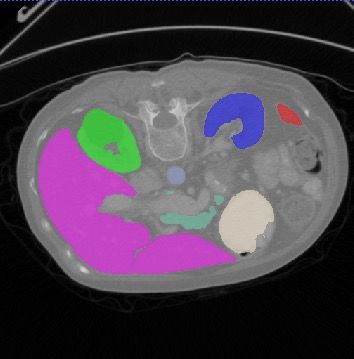
nnFormer: Interleaved Transformer for Volumetric Segmentation 7

volumes. In practice, we set {SH , SW , SD } on Synapse and ACDC to {4, 4, 4}
and {5, 5, 3}, respectively.
 We follow [20] to conduct two successive transformer blocks, where the main
difference lies in that our computation is built on top of 3D volumes instead of
2D windows. The computational procedure can be summarized as follows:

 X̂tl = V-MSA LayerNorm Xtl−1 + Xtl−1 ,

 Xtl = MLP LayerNorm X̂tl + X̂tl ,
    (1)
 X̂tl+1 = SV-MSA LayerNorm Xtl + Xtl ,

 Xtl+1 = MLP LayerNorm X̂tl+1 + X̂tl+1 .

Here, l stands for the layer index. MLP is an abbreviation for multi-layer percep-
tron. V-MSA and SV-MSA denote the volume-based multi-head self-attention
and its shifted version. The computational complexity of V-MSA on a volume of
h × w × d patches is:

 Ω(V-MSA) = 4hwdC 2 + 2SH SW SD hwdC. (2)

Compared to the complexity of the naive multi-head self-attention (MSA) [33]
used in ViT [8], i.e.,

 Ω(MSA) = 4hwdC 2 + 2(hwd)2 C. (3)

V-MSA reduces the computational complexity by approximate 98% and 99.5%
on Synapse and ACDC datasets, respectively.
 SV-MSA displaces the 3D local volume used in V-MSA by S2H , S2W , S2D


to introduce more interactions between different local volumes.
 The query-key-value (QKV) attention [33] in each 3D local volume can be
computed as follows:

 QK T

 Attention(Q, K, V) = softmax √ + B V, (4)
 dk

where Q, K, V ∈ RNT ×dk denote the query, key and value matrices of dimension
dk . B ∈ RNT is the relative position encoding. In practice, we first initialize
a smaller-sized position matrix B̂ ∈ R(2SH −1)×(2SW −1)×(2SD −1) and take corre-
sponding values from B̂ to build a larger position matrix B.
 It is worth noting that the last transformer block of the encoder (the down-
most block in Figure 2) only employs V-MSA as we found introducing SV-MSA
would deteriorate the overall segmentation results.

Down-sampling block. We found that by replacing the neighboring concate-
nation operation in [20] with direct strided convolution, nnFormer provides more
improvements to volumetric segmentation. The intuition behind is that convo-
lutional down-sampling produces hierarchical representations that help model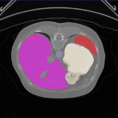
8 Authors Suppressed Due to Excessive Length

object concepts at multiple scales. As displayed in Figure 2b, in most cases, the
down-sampling block involves a strided convolution operation where the stride is
set to 2 in all dimensions. However, in practice, the stride with respect to specific
dimension (refer to Table 1b) is set to 1 as the number of slices is limited in this
dimension and over-down-sampling (i.e., a large stride) might be harmful.

3.3 Decoder

Architectures of transformer blocks of the decoder are highly symmetrical to
those of the encoder. In contrast to the down-sampling blocks, we employ strided
deconvolution to up-sample low-resolution feature maps to high-resolution ones,
which in turn are merged with representations from the encoder via long-range
residual connections to capture both semantic and fine-grained information.
Similar to up-sampling blocks, the last patch expanding block also takes the
deconvolutional operation to produce final predictions.

4 Experiments

To fairly compare nnFormer with previous Transformer-based architectures, we
conduct experiments on Synapse [13] and Automatic Cardiac Diagnosis Challenge
(ACDC) [2] datasets. For each experiment, we repeat it for three times and report
their average results.

Synapse for multi-organ CT segmentation. This dataset includes 30 cases
of abdominal CT scans. Following the split used in [6], 18 cases are extracted to
build the training set while the rest 12 cases are used for testing. We report the
model performance evaluated with the average Dice Similarity Coefficient (DSC)
on 8 abdominal organs, which are aorta, gallbladder, spleen, left kidney, right
kidney, liver, pancreas and stomach.

ACDC for automated cardiac diagnosis. ACDC involves 100 patients, with
the cavity of the right ventricle, the myocardium of the left ventricle and the
cavity of the left ventricle to be segmented. Each case’s labels involve left ventricle
(LV), right ventricle (RV) and myocardium (MYO). The dataset is split into 70
training samples, 10 validation samples and 20 testing samples.

4.1 Implementation details

We run all experiments based on Python 3.6, PyTorch 1.8.1 and Ubuntu 18.04.
All training procedures have been performed on a single NVIDIA 2080 GPU with
11GB memory. The initial learning rate is set to 0.01 and we employ a “poly”
decay strategy as described in Equation 5. The default optimizer is SGD where
we set the momentum to 0.99. The weight decay is set to 3e-5. We utilize both
cross entropy loss and dice loss by simply summing them up. The number of

nnFormer: Interleaved Transformer for Volumetric Segmentation 9

Table 1: Network configurations of our nnFormer and nnUNet on Synapse and
ACDC. We only report the stride of down-sampling (i.e. str. of down-samp.) as
the stride of up-sampling can be easily inferred according to symmetrical down-
sampling operations. Note that the network configuration of nnUNet is automat-
ically determined based on pre-defined hand-crafted rules (for self-adaptation).
 (a) Synapse

 nnFormer nnUNet
 Target spacing [0.76, 0.76, 3] [0.76, 0.76, 3]
 Median image shape 512 × 512 × 148 512 × 512 × 148
 Crop size 128 × 128 × 64 192 × 192 × 48
 Batch size 2 2
 [2, 2, 2], [2, 2, 1], [2, 2, 2], [2, 2, 1], [2, 2, 2], [2, 2, 2],
 Str. of Down-samp.
 [2, 2, 2], [2, 2, 2] [2, 2, 2], [2, 2, 1]

 (b) ACDC

 nnFormer nnUNet
 Target spacing [1.52, 1.52, 6.35] [1.52, 1.52, 6.35]
 Median image shape 246 × 213 × 13 246 × 213 × 13
 Crop size 160 × 160 × 14 256 × 224 × 14
 Batch size 4 4
 [2, 2, 1], [2, 2, 1], [2, 2, 1], [2, 2, 1], [2, 2, 1], [2, 2, 2],
 Str. of Down-samp.
 [2, 2, 2], [2, 2, 2] [2, 2, 1], [2, 2, 1]

training epochs (i.e., max_epoch in Equation 5) is 1000 and one epoch contains
250 iterations. The numbers of heads of multi-head self-attention used in different
encoder stages are [6, 12, 24, 48] and [3, 6, 12, 24] on Synapse and ACDC,
respectively.

 epoch_id 0.9
 lr = initial_lr × (1 − ) . (5)
 max_epoch

Pre-processing and augmentation strategies. All images will be first re-
sampled to the same target spacing. Augmentations such as rotation, scaling,
gaussian noise, gaussian blur, brightness and contrast adjust, simulation of low
resolution, gamma augmentation and mirroring are applied in the given order
during the training process.

Deep supervision. We also add deep supervision during the training stage.
Specifically, the output of each stage in the decoder is passed to the final expanding
block, where cross entropy loss and dice loss would be applied. In practice, given
the prediction of one typical stage, we down-sample the ground truth segmentation
mask to match the prediction’s resolution. Thus, the final training objective
10 Authors Suppressed Due to Excessive Length

function is the sum of all losses at three resolutions:

 Lall = α1 L{H, W, D} + α2 L{ H , W D + α3 L{ H , W D . (6)
 4 4 , 2 } 8 8 , 4 }

Here, α{1, 2, 3} denote the magnitude factors for losses in different resolutions.
In practice, α{1, 2, 3} halve with each decrease in resolution, leading to α2 = α21
and α3 = α41 . Finally, all weight factors are normalized to 1.

Pre-trained model weights. Pre-training can be vastly important to provide
generalized and transferable representations for downstream tasks. Given the
fact that most operations in nnFormer operate on 1D sequences, we explore the
possibility of transfering pre-trained weights on natural images to the medical
imaging field. More concretely, we aim to reap the benefit of pre-trained weights
of MLP layers and QKV attention on ImageNet pre-training. To this goal, we
align channel numbers of transformer blocks to those of pre-trained models so
that we load the weights of MLP layers and QKV attention. Besides, considering
architectures of the encoder and decoder are highly symmetrical, we propose
symmetrical initialization to reuse the pre-trained weights of the encoder in
the decoder. Specifically, transformer blocks with the same input and output
resolution are initialized using the same set of model weights (i.e., symmetrical
transformer blocks of the encoder and decoder in Figure 2).

Network configurations. In Table 1, we display network configurations of
experiments on Synapse and ACDC. Compared to nnUNet, in nnFormer, better
segmentation results can be achieved with smaller-sized input patches.

4.2 Experiments on Synapse
As shown in Table 2, we make experiments on Synapse and to compare our
nnFormer against a variety of both transformer- and convnet-based baselines.
The major evaluation metric is dice score.
 Apart from nnUNet, the best performing convnet-based method is DualNorm-
UNet [36] that achieves an average dice score of 80.37. In comparison, WAD
reports the best transformer-based results whose average is 80.30, slightly lower
than DualNorm-UNet. Our nnFormer is able to outperform WAD and DualNorm-
UNet by over 8 percents and 7 percents in average, respectively, which are quite
impressive improvements on Synapse. Besides, we found that the performance
of nnUNet are severely underestimated. When being carefully tuned, nnUNet
reaches an average dice score of 86.99, which is much better than DualNorm-UNet
and WAD but still worse than proposed nnFormer.

4.3 Experiments on ACDC
Table 3 presents the experimental results on ACDC, where the overall performance
of transformer-based baselines are better than those of convnet-based ones. The
underlying reason is that images from ACDC have many fewer slices on z-axis
nnFormer: Interleaved Transformer for Volumetric Segmentation 11 Table 2: Experiments on Synapse (dice score in %). Best results are bolded. Methods Average Aotra Gallbladder Kidnery(L) Kidnery(R) Liver Pancreas Spleen Stomach VNet [22] 68.81 75.34 51.87 77.10 80.75 87.84 40.04 80.56 56.98 DARR [9] 69.77 74.74 53.77 72.31 73.24 94.08 54.18 89.90 45.96 R50 U-Net [27] 74.68 87.74 63.66 80.60 78.19 93.74 56.90 85.87 74.16 U-Net [27] 76.85 89.07 69.72 77.77 68.60 93.43 53.98 86.67 75.58 R50 Att-UNet [28] 75.57 55.92 63.91 79.20 72.71 93.56 49.37 87.19 74.95 Att-UNet [23] 77.77 89.55 68.88 77.98 71.11 93.57 58.04 87.30 75.75 VIT None [8] 61.50 44.38 39.59 67.46 62.94 89.21 43.14 75.45 68.78 VIT CUP [8] 67.86 70.19 45.10 74.70 67.40 91.32 42.00 81.75 70.44 R50 VIT CUP [8] 71.29 73.73 55.13 75.80 72.20 91.51 45.99 81.99 73.95 R50-Deeplabv3+ [7] 75.73 86.18 60.42 81.18 75.27 92.86 51.06 88.69 70.19 DualNorm-UNet [36] 80.37 86.52 55.51 88.64 86.29 95.64 55.91 94.62 79.8 CGNET [35] 75.08 83.48 65.32 77.91 72.04 91.92 57.37 85.47 67.15 ContextNet [25] 71.17 79.92 51.17 77.58 72.04 91.74 43.78 86.65 66.51 DABNet [15] 74.91 85.01 56.89 77.84 72.45 93.05 54.39 88.23 71.45 EDANet [21] 75.43 84.35 62.31 76.16 71.65 93.20 53.19 85.47 77.12 ENet [24] 77.63 85.13 64.91 81.10 77.26 93.37 57.83 87.03 74.41 FPENet [19] 68.67 78.98 56.35 74.54 64.36 90.86 40.60 78.30 65.35 FSSNet [40] 74.59 82.87 64.06 78.03 69.63 92.52 53.10 85.65 70.86 SQNet [30] 73.76 83.55 61.17 76.87 69.40 91.53 56.55 85.82 65.24 FastSCNN [26] 70.53 77.79 55.96 73.61 67.38 91.68 44.54 84.51 68.76 TransUNet [6] 77.48 87.23 63.16 81.87 77.02 94.08 55.86 85.08 75.62 SwinUNet [3] 79.13 85.47 66.53 83.28 79.61 94.29 56.58 90.66 76.6 TransClaw U-Net [4] 78.09 85.87 61.38 84.83 79.36 94.28 57.65 87.74 73.55 LeVit-UNet-384s [38] 78.53 87.33 62.23 84.61 80.25 93.11 59.07 88.86 72.76 WAD [17] 80.30 87.73 69.93 83.95 79.78 93.95 61.02 88.86 77.16 nnUNet (3D) [11] 86.99 93.01 71.77 85.57 88.18 97.23 83.01 91.86 85.25 nnFormer 87.40 92.04 71.09 87.64 87.34 96.53 82.49 92.91 89.17 (i.e., the spacing on z-axis is quite large in Figure 1b), which is the exactly the case where transformer has more advantages as they are designed to deal with 2D inputs with less interaction on z-axis. From Table 3, we can see that the best transformer model is LeViT-UNet-384s, which average dice is slightly higher than SwinUNet but much higher than convnet-based Dual-Attn. In contrast, nnFormer surpasses LeViT-UNet-384s by nearly 1.5 percents on average, again displaying its advantages over transformer-based baselines. 4.4 Ablation study In this section, we present the significance of the embedding block and convolu- tional down-sampling blocks. Besides, we also investigate the influences of adding more transformer blocks to the encoder and employing natural images based pre-training. Significance of the embedding block. To investigate the influence of our embedding block, we replace it with patch-wise convolution, as shown in Table 4. Patch-wise convolution [8, 20] only contains one convolutional layer that has large kernel size and stride. For instance, on Synapse, both the kernel size and convolutional stride are set to [4,4,2]. We can see that the successive convolutional
12 Authors Suppressed Due to Excessive Length

 Table 3: Experiments on ACDC (dice score in %). Best results are bolded.
 Methods Average RV Myo LV
 R50-U-Net [27] 87.55 87.10 80.63 94.92
 R50-Attn UNet [28] 86.75 87.58 79.20 93.47
 VIT-CUP [8] 81.45 81.46 70.71 92.18
 R50-VIT-CUP [8] 87.57 86.07 81.88 94.75
 TransUNet [6] 89.71 88.86 84.54 95.73
 SwinUNet [3] 90.00 88.55 85.62 95.83
 LeViT-UNet-384s [38] 90.32 89.55 87.64 93.76
 nnUNet (3D) [11] 91.59 90.25 89.10 95.41
 nnFormer 91.78 90.22 89.53 95.59

Table 4: Investigation of the embedding block on Synapse. Patch-wise con-
volution consists of only one convolutional layer with large kernel size and
stride.
 Average Aotra Gallbladder Kidnery(L) Kidnery(R) Liver Pancreas Spleen Stomach
Patch-wise convolution 84.63 88.84 65.33 86.97 85.98 95.58 77.30 91.83 85.15
 Ours 87.40 92.04 71.09 87.64 87.34 96.53 82.49 92.91 89.17

layers of the embedding block surpasses the atypical patch-wise convolution by
nearly 3 percents in average. Actually, similar phenomena were first time observed
in [29], where small-sized kernels are found to be more effective than large ones.

Influences of convolutional down-sampling blocks. In Table 5, we report
the results of using neighboring concatenation [20] in nnFormer to replace convo-
lutional down-sampling blocks. The main operation of neighboring concatenation
is to concatenate neighboring patches of features maps in the channel dimension,
after which a fully-connected layer is added to reduce the number of channels.
From Table 5, we can see that convolutional down-sampling blocks provide over 3
percents improvements over neighboring concatenation in average, demonstrating
that applying interleaved convolutional down-sampling blocks are more helpful
to build hierarchical object concepts at various scales.

Investigation of adding more transformer blocks. We add one more SV-
MSA block to the last encoder stage and report its results in Table 6. Generally
speaking, more transformer blocks help nnFormer to encode more long-term
dependencies into representations. Somewhat interestingly, from Table 6, we
can find that capturing more long-term dependencies may not be an optimal
choice for nnFormer. For instance, although introducing more transformer blocks
achieve a higher segmentation dice score on Gallbladder (the most difficult organ
on Synapse), it deteriorates the segmentation performance on other organs. We
will explore the reason behind in the future.
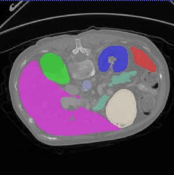
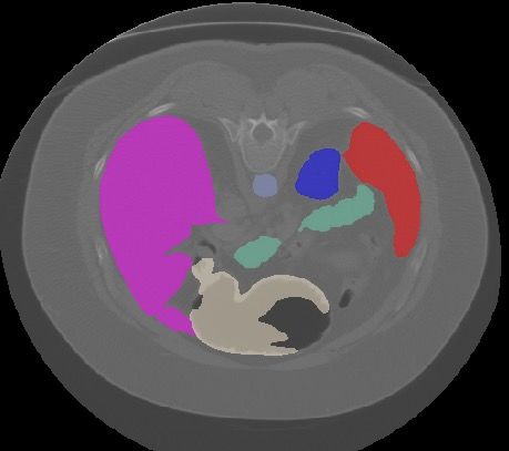
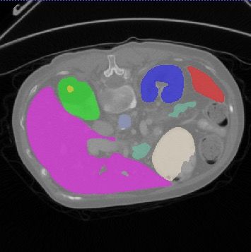
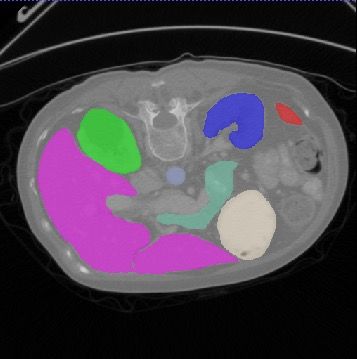
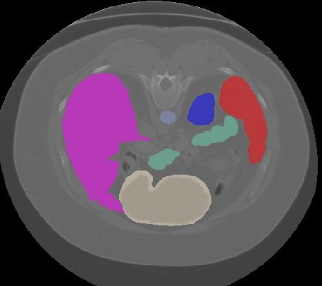
nnFormer: Interleaved Transformer for Volumetric Segmentation 13

 Spleen Pancreas Right kidney Left kidney Gallbladder Liver Stomach Aorta

 GT

 nnUNet

nnFormer

 GT

 nnUNet

nnFormer

 Fig. 3: Segmentation results of some hard samples on Synapse.
14 Authors Suppressed Due to Excessive Length

 RV Myo LV

 GT

 nnUNet

nnFormer

 GT

 nnUNet

nnFormer

 Fig. 4: Segmentation results of some hard samples on ACDC.
nnFormer: Interleaved Transformer for Volumetric Segmentation 15

 Table 5: Investigation of the convolutional down-sampling blocks.
 Average Aotra Gallbladder Kidnery(L) Kidnery(R) Liver Pancreas Spleen Stomach
 Neighboring concatenation 84.30 88.00 67.60 87.52 87.38 95.31 80.63 85.29 82.69
 Ours 87.40 92.04 71.09 87.64 87.34 96.53 82.49 92.91 89.17

 Table 6: Investigation of adding more transformer blocks.
 Average Aotra Gallbladder Kidnery(L) Kidnery(R) Liver Pancreas Spleen Stomach
 More transformer blocks 85.98 89.02 71.74 86.76 87.06 96.37 82.30 89.04 85.51
 Ours 87.40 92.04 71.09 87.64 87.34 96.53 82.49 92.91 89.17

 Table 7: Benefits of using pre-trained weights on natural images.
 Average Aotra Gallbladder Kidnery(L) Kidnery(R) Liver Pancreas Spleen Stomach
 No pre-training 84.34 90.15 69.00 86.34 87.48 95.93 80.97 85.23 79.67
 Ours 87.40 92.04 71.09 87.64 87.34 96.53 82.49 92.91 89.17

Influences of using pre-trained models on natural images. In Table 7,
we show that it is crucial to make use of pre-trained weights on natural images,
where removing pre-training deteriorates the overall segmentation performance
by over 3 percents. The underlying reason is that Synapse does not have enough
labeled 3D scans to fully realize the potential of nnFormer.

4.5 Visualization
In Figures 3 and 4, we compare the segmentation results of nnUNet and nnFormer
in some hard samples. On Synapse, it seems that nnFormer has quite apparent
advantages on stomach where nnUNet often fails to generate an integrated
delineation mask. Meanwhile, compared nnUNet, nnFormer has the ability to
reduce false positive predictions of spleen, which are also consistent with the
performance reported in Table 2.
 Similar phenomena can also be observed in Figure 4, where nnFormer can
greatly reduce false positive predictions of right ventricle (RV) and myocardium
(MYO), especially myocardium. These segmentation results help to verify the
fact that nnFormer can produce more robust and discriminative representations
than nnUNet.

5 Conclusion
In this paper, we present a new medical image segmentation network named
nnFormer. nnFormer is constructed on top of an interleaved stem of convolution
and self-attention, where convolution helps encode precise spatial information
into high-resolution low-level features and build hierarchical object concepts at
multiple scales. On the other hand, self-attention in transformer blocks entangles
long-term dependencies with convolutional representations to capture global
16 Authors Suppressed Due to Excessive Length

context. Based on such hybrid architecture, nnFormer achieves tremendous
progress over previous transformer-based segmentation methodologies. Even
when compared to nnUNet, currently the best performing segmentation network,
nnFormer still provides consistent yet observable improvements. In the future, we
hope nnFormer could draw more attention from the medical imaging community
to make efforts on developing more efficient segmentation models.

References

 1. Ba, J.L., Kiros, J.R., Hinton, G.E.: Layer normalization. arXiv preprint
 arXiv:1607.06450 (2016)
 2. Bernard, O., Lalande, A., Zotti, C., Cervenansky, F., Yang, X., Heng, P.A., Cetin,
 I., Lekadir, K., Camara, O., Ballester, M.A.G., et al.: Deep learning techniques for
 automatic mri cardiac multi-structures segmentation and diagnosis: Is the problem
 solved? IEEE transactions on medical imaging 37(11), 2514–2525 (2018)
 3. Cao, H., Wang, Y., Chen, J., Jiang, D., Zhang, X., Tian, Q., Wang, M.: Swin-
 Unet: Unet-like pure transformer for medical image segmentation. arXiv preprint
 arXiv:2105.05537 (2021)
 4. Chang, Y., Menghan, H., Guangtao, Z., Xiao-Ping, Z.: TransClaw U-Net: Claw u-net
 with transformers for medical image segmentation. arXiv preprint arXiv:2107.05188
 (2021)
 5. Chen, B., Liu, Y., Zhang, Z., Lu, G., Zhang, D.: TransAttUnet: Multi-level attention-
 guided u-net with transformer for medical image segmentation. arXiv preprint
 arXiv:2107.05274 (2021)
 6. Chen, J., Lu, Y., Yu, Q., Luo, X., Adeli, E., Wang, Y., et al.: TransUNet: Trans-
 formers make strong encoders for medical image segmentation. arXiv preprint
 arXiv:2102.04306 (2021)
 7. Chen, L.C., Papandreou, G., Kokkinos, I., Murphy, K., Yuille, A.L.: Deeplab:
 Semantic image segmentation with deep convolutional nets, atrous convolution,
 and fully connected crfs. IEEE Transactions on Pattern Analysis and Machine
 Intelligence 40(4), 834–848 (2018)
 8. Dosovitskiy, A., Beyer, L., Kolesnikov, A., Weissenborn, D., Zhai, X., Unterthiner,
 T., et al.: An image is worth 16x16 words: Transformers for image recognition at
 scale. arXiv preprint arXiv:2010.11929 (2020)
 9. Fu, S., Lu, Y., Wang, Y., Zhou, Y., Shen, W., Fishman, E., Yuille, A.: Domain
 adaptive relational reasoning for 3d multi-organ segmentation. In: International
 Conference on Medical Image Computing and Computer-Assisted Intervention. pp.
 656–666. Springer (2020)
10. Hendrycks, D., Gimpel, K.: Gaussian error linear units (gelus). arXiv preprint
 arXiv:1606.08415 (2016)
11. Isensee, F., Jäger, P.F., Kohl, S.A., Petersen, J., Maier-Hein, K.H.: Automated
 design of deep learning methods for biomedical image segmentation. arXiv preprint
 arXiv:1904.08128 (2019)
12. Karimi, D., Vasylechko, S., Gholipour, A.: Convolution-free medical image segmen-
 tation using transformers. arXiv preprint arXiv:2102.13645 (2021)
13. Landman, B., Xu, Z., Igelsias, J.E., Styner, M., Langerak, T., Klein, A.: Miccai
 multi-atlas labeling beyond the cranial vault–workshop and challenge. In: Proc.
 MICCAI: Multi-Atlas Labeling Beyond Cranial Vault-Workshop Challenge (2015)
nnFormer: Interleaved Transformer for Volumetric Segmentation 17

14. LeCun, Y., Bottou, L., Bengio, Y., Haffner, P.: Gradient-based learning applied to
 document recognition. Proceedings of the IEEE 86(11), 2278–2324 (1998)
15. Li, G., Yun, I., Kim, J., Kim, J.: Dabnet: Depth-wise asymmetric bottleneck for
 real-time semantic segmentation. arXiv preprint arXiv:1907.11357 (2019)
16. Li, S., Sui, X., Luo, X., Xu, X., Liu, Y., Goh, R.S.M.: Medical image segmentation
 using squeeze-and-expansion transformers. arXiv preprint arXiv:2105.09511 (2021)
17. Li, Y., Cai, W., Gao, Y., Hu, X.: More than encoder: Introducing transformer
 decoder to upsample. arXiv preprint arXiv:2106.10637 (2021)
18. Lin, A., Chen, B., Xu, J., Zhang, Z., Lu, G.: DS-TransUNet: Dual swin transformer
 u-net for medical image segmentation. arXiv preprint arXiv:2106.06716 (2021)
19. Liu, M., Yin, H.: Feature pyramid encoding network for real-time semantic segmen-
 tation. arXiv preprint arXiv:1909.08599 (2019)
20. Liu, Z., Lin, Y., Cao, Y., Hu, H., Wei, Y., Zhang, Z., Lin, S., Guo, B.: Swin
 transformer: Hierarchical vision transformer using shifted windows. arXiv preprint
 arXiv:2103.14030 (2021)
21. Lo, S.Y., Hang, H.M., Chan, S.W., Lin, J.J.: Efficient dense modules of asymmetric
 convolution for real-time semantic segmentation. In: MMAsia ’19: ACM Multimedia
 Asia (2019)
22. Milletari, F., Navab, N., Ahmadi, S.A.: V-net: Fully convolutional neural networks
 for volumetric medical image segmentation. In: 2016 fourth international conference
 on 3D vision (3DV). pp. 565–571. IEEE (2016)
23. Oktay, O., Schlemper, J., Folgoc, L.L., Lee, M., Heinrich, M., Misawa, K., Mori,
 K., McDonagh, S., Hammerla, N.Y., Kainz, B., et al.: Attention u-net: Learning
 where to look for the pancreas. arXiv preprint arXiv:1804.03999 (2018)
24. Paszke, A., Chaurasia, A., Kim, S., Culurciello, E.: Enet: A deep neural network
 architecture for real-time semantic segmentation. arXiv preprint arXiv:1606.02147
 (2016)
25. Poudel, R.P., Bonde, U., Liwicki, S., Zach, C.: Contextnet: Exploring context and
 detail for semantic segmentation in real-time. arXiv preprint arXiv:1805.04554
 (2018)
26. Poudel, R.P., Liwicki, S., Cipolla, R.: Fast-scnn: Fast semantic segmentation network.
 arXiv preprint arXiv:1902.04502 (2019)
27. Ronneberger, O., Fischer, P., Brox, T.: U-Net: Convolutional networks for biomedical
 image segmentation. In: International Conference on Medical image computing and
 computer-assisted intervention. pp. 234–241. Springer (2015)
28. Schlemper, J., Oktay, O., Schaap, M., Heinrich, M., Kainz, B., Glocker,
 B., Rueckert, D.: Attention gated networks: Learning to leverage
 salient regions in medical images. Medical Image Analysis 53, 197–
 207 (2019). https://doi.org/https://doi.org/10.1016/j.media.2019.01.012,
 https://www.sciencedirect.com/science/article/pii/S1361841518306133
29. Simonyan, K., Zisserman, A.: Very deep convolutional networks for large-scale
 image recognition. arXiv preprint arXiv:1409.1556 (2014)
30. Treml, M., Arjona-Medina, J., Unterthiner, T., Durgesh, R., Hochreiter, S.: Speeding
 up semantic segmentation for autonomous driving. In: NIPS 2016 Workshop - MLITS
 (2016)
31. Tuli, S., Dasgupta, I., Grant, E., Griffiths, T.L.: Are convolutional neural networks
 or transformers more like human vision? arXiv preprint arXiv:2105.07197 (2021)
32. Valanarasu, J.M.J., Oza, P., Hacihaliloglu, I., Patel, V.M.: Medical Trans-
 former: Gated axial-attention for medical image segmentation. arXiv preprint
 arXiv:2102.10662 (2021)
18 Authors Suppressed Due to Excessive Length

33. Vaswani, A., Shazeer, N., Parmar, N., Uszkoreit, J., Jones, L., Gomez, A.N., et al.:
 Attention is all you need. In: Adv. in Neur. Info. Process. Sys. pp. 5998–6008 (2017)
34. Wang, W., Chen, C., Ding, M., Li, J., Yu, H., Zha, S.: TransBTS: Multimodal brain
 tumor segmentation using transformer. arXiv preprint arXiv:2103.04430 (2021)
35. Wu, T., Tang, S., Zhang, R., Cao, J., Zhang, Y.: Cgnet: A light-weight context
 guided network for semantic segmentation. IEEE Transactions on Image Processing
 30, 1169–1179 (2021)
36. Xiao, J., Yu, L., Xing, L., Yuille, A., Zhou, Y.: Dualnorm-unet: Incorporating
 global and local statistics for robust medical image segmentation. arXiv preprint
 arXiv:2103.15858 (2021)
37. Xie, Y., Zhang, J., Shen, C., Xia, Y.: CoTr: Efficiently bridging cnn and transformer
 for 3d medical image segmentation. arXiv preprint arXiv:2103.03024 (2021)
38. Xu, G., Wu, X., Zhang, X., He, X.: LeViT-UNet: Make faster encoders with
 transformer for medical image segmentation. arXiv preprint arXiv:2107.08623
 (2021)
39. Yun, B., Wang, Y., Chen, J., Wang, H., Shen, W., Li, Q.: SpecTr: Spectral
 transformer for hyperspectral pathology image segmentation. arXiv preprint
 arXiv:2103.03604 (2021)
40. Zhang, X., Chen, Z., Wu, Q., Cai, L., Lu, D., Li, X.: Fast semantic segmentation
 for scene perception. IEEE Transactions on Industrial Informatics (2018)
41. Zhang, Y., Liu, H., Hu, Q.: TransFuse: Fusing transformers and cnns for medical
 image segmentation. arXiv preprint arXiv:2102.08005 (2021)
42. Zhou, H.Y., Lu, C., Yang, S., Yu, Y.: ConvNets vs. Transformers: Whose visual
 representations are more transferable? arXiv preprint arXiv:2108.05305 (2021)
You can also read