Non-functional thyroid cystadenoma in three boxer dogs
←
→
Page content transcription
If your browser does not render page correctly, please read the page content below
Maurin et al. BMC Veterinary Research     (2019) 15:228
https://doi.org/10.1186/s12917-019-1948-z

 CASE REPORT                                                                                                                                      Open Access

Non-functional thyroid cystadenoma in
three boxer dogs
Marie-Pauline Maurin* , Dan Davies, Hanne Jahns, Robert E. Shiel and Carmel T. Mooney

  Abstract
  Background: Thyroid neoplasia is a common endocrine neoplasm in dogs. The boxer is one of the reported
  breeds predisposed to malignant thyroid neoplasia. However, the association between thyroid neoplasia,
  malignancy and breed should be considered with caution.
  Cases presentation: This article describes the presentation, clinical pathological findings, computed tomographic
  (CT) imaging findings and histopathological features of benign cystic thyroid tumour (cystadenoma) diagnosed in
  three boxers. These three dogs were presented for investigation of unilateral (n = 2) or bilateral (n = 1) cervical
  masses with no associated clinical signs of thyroid dysfunction. In each case, post-contrast CT scan identified a
  large, lateralised, non-invasive, well-defined homogeneous cystic structure with a hyperattenuating contrast-
  enhancing capsule of suspected thyroid origin displacing the surrounding cervical tissues. Ultrasound-guided fine
  needle aspiration of the cysts yielded fluid with a high thyroxine concentration in each case. Histopathology was
  consistent with thyroid cystadenoma in all cases. One dog was concurrently diagnosed with oral melanoma and
  euthanased. Two dogs underwent surgical excision with one lost to follow-up after 36 months and the other
  euthanased after 16 months following diagnosis of mast cell tumour.
  Conclusions: To the authors’ knowledge, this is the first detailed report of non-functional benign thyroid
  cystadenoma in dogs and provides relevant information about case management for this type of tumour. The
  presence of a large cystic structure associated with benign non-functional thyroid neoplasia may be a condition to
  which boxer dogs are predisposed.
  Keywords: Thyroid neoplasm, Boxer, Thyroid cystic adenoma, Thyroid cyst, Cervical mass

Background                                                                             aggressive biological behaviour. They can extend into
Thyroid tumours account for 1.1 to 3.8% of all neo-                                    or around the wall of the trachea, cervical muscles,
plasms and approximately 10–15% of all head neoplasms                                  oesophagus, larynx, nerves and vessels [12]. Early inva-
in dogs [1–5]. In some studies, boxers, beagles, golden                                sion of the cranial and caudal thyroid veins with forma-
retrievers and Siberian huskies were more commonly                                     tion of tumour cell thrombi can lead to pulmonary
affected compared to other breeds [2, 4–8]. However,                                   metastases even before involvement of the retropharyn-
another recent study found no association between                                      geal and caudal cervical lymph nodes [7, 11]. The ma-
breed and development of thyroid neoplasia [1]. Thyroid                                jority of dogs with thyroid tumours are euthyroid,
carcinomas are more frequently diagnosed antemortem                                    although both hyperthyroidism and hypothyroidism
than thyroid adenomas [1, 4–6, 15], which are often                                    have been described [9–11]. In a recent large study of
discovered serendipitously during physical examination                                 canine thyroid carcinoma, 12 of 57 (21%) cases where
or at necropsy [5, 8]. Dogs with thyroid carcinomas have                               thyroid function was investigated were hyperthyroid
evidence of metastasis at the time of diagnosis in from                                [10]. Up to 30% of dogs are described as hypothyroid
16 to approximately 60% of case [11, 14, 16]. Carcin-                                  secondary to destruction of the normal thyroid paren-
omas are generally large in size with rapid growth and                                 chyma [12]. In accord with this, one experimental study
                                                                                       of Beagles suggested that hypothyroidism may be a pre-
                                                                                       existing condition [13]. However, in many older studies
* Correspondence: marie-pauline.maurin@ucdconnect.ie
School of Veterinary Medicine, University College Dublin, Dublin, Ireland              the diagnosis of hypothyroidism is questionable. In a

                                         © The Author(s). 2019 Open Access This article is distributed under the terms of the Creative Commons Attribution 4.0
                                         International License (http://creativecommons.org/licenses/by/4.0/), which permits unrestricted use, distribution, and
                                         reproduction in any medium, provided you give appropriate credit to the original author(s) and the source, provide a link to
                                         the Creative Commons license, and indicate if changes were made. The Creative Commons Public Domain Dedication waiver
                                         (http://creativecommons.org/publicdomain/zero/1.0/) applies to the data made available in this article, unless otherwise stated.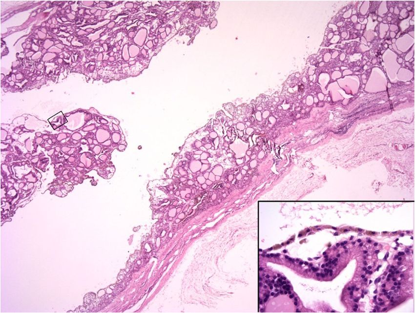
Maurin et al. BMC Veterinary Research   (2019) 15:228                                                                  Page 2 of 9

recent study, only 2 of 57 (3.5%) of cases were truly              There were no haematological abnormalities in any
hypothyroid, although such a diagnosis could not be             case (Table 2). Serum biochemistry identified hyperchol-
excluded in approximately 30% of cases [10].                    esterolaemia in case 3. Otherwise only mildly increased
   Marked rapid enlargement of the thyroid gland could          liver enzyme activities of no clinical significance were
also be caused by cysts. Thyroid cysts are fluid filled cav-    noted (Table 2). Total T4 and canine thyroid-stimulating
ities, in dogs mainly as thyroid cystadenomas [17]. Cysts       hormone (cTSH) concentrations were measured in cases
can also be observed with malignant thyroid tumours,            2 and 3 (Table 2). Results were most consistent with
namely papillary thyroid carcinomas, which are rarely           non-thyroidal illness although additional testing (e.g.
reported in dogs [15, 17]. Although cystadenomas are oc-        measurement of free T4 concentration or thyroglobulin
casionally mentioned in large case series of thyroid neopla-    autoantibody status) was not performed. In case 2, the
sia in dogs, or as incidental post-mortem findings,             cTSH concentration approached the upper limit of the
detailed clinical features of these cases have not been         reference interval at the time of initial presentation. Re-
described [1, 5, 7, 18]. Two individual case reports            peat measurements of T4 and cTSH were recommended
described thyroid cystadenoma with concurrent hyper-            and performed six weeks after surgical treatment.
thyroidism in a German shepherd dog and an English                 Cervical ultrasonography confirmed an ovoid, cystic
springer spaniel [19, 20].                                      lesion in the region of the right thyroid lobe in case 1
   The current case report describes the clinical signs,        and of the left lobe in case 2. In case 3, a large cystic
diagnosis and treatment of thyroid cystadenomas in              structure was localised in the left thyroid lobe and sev-
three boxer dogs that presented with enlarging masses           eral small cystic structures in the right thyroid lobe.
on the ventral neck. The presence of hyperthyroidism            Ultrasound-guided aspiration of the cystic lesions was
was not identified in any case. Furthermore, the meas-          performed in all cases. In cases 1 and 2, cytologic ana-
urement of the thyroxine (T4) concentration in the cys-         lysis of the fluid contained in the large cystic lesions was
tic fluid allowed rapid confirmation of the thyroidal           non-diagnostic, revealing proteinaceous fluid with mild
origin of the mass in each case.                                cell necrosis. In case 3, fine needle aspirate of the het-
                                                                erogeneous part of the left thyroid lobe was performed
Case presentations                                              and cytological analysis revealed abundant cellular deb-
Between 2012 and 2018, three boxer dogs were inde-              ris, small numbers of red blood cells (RBC) and very
pendently evaluated at University College Dublin (UCD)          large cells up to 10 times RBC diameter with eccentric-
Veterinary Hospital for investigation of ventral cervical       ally displaced large nuclei with predominantly cytoplas-
masses (Table 1). The masses were present for a period          mic area filled with light-to-dark blue material with
ranging from six weeks to six months prior to referral.         frequent coarse dark-blue granules. Occasionally, these
Episodes of regurgitation after excitement or strenuous         cells occured in small clusters with indistinct cell bor-
exercise were also reported in case 1, and hypersalivation      ders. No significant criteria of malignancy were found.
in case 3. In cases 1 and 2, the primary veterinary sur-        These features were considered consistent with thyroid
geon drained fluid from the mass by percutaneous fine           gland adenoma with marked cell necrosis; however, well-
needle aspiration prior to referral; the swelling recurred      differentiated carcinoma could not be excluded. Some of
within a few weeks in both cases.                               the aspirated fluid was placed in plain tubes for T4
  At presentation, palpation of the ventral neck revealed a     measurement using the same assay as for serum samples
3 to 5 cm diameter, mobile, well-circumscribed, non-            (Immulite 2000 Canine Total T4, Siemens Healthcare
painful, ovoid, subcutaneous mass in close proximity to         Diagnostics using an Immulite 2000 Analyzer). Concen-
the trachea in each case. Mandibular and pre-scapular           trations were markedly increased (compared to the
lymph node palpation revealed no abnormalities. Cardio-         serum reference interval) in each case, confirming thy-
thoracic auscultation and abdominal palpation were unre-        roidal origin (Table 2).
markable in all cases. No dermatological or neurological           Plain computed tomography (CT) of the neck and
abnormalities were noted in any case.                           chest were performed for staging purpose in all cases

Table 1 Signalment, histopathologic diagnosis and outcome in three dogs with thyroid cystadenoma
Cases            Gender                    Age (years)     Diagnosis                               Outcome
1                Male castrated            8               Thyroid cystadenoma (Right lobe)        Last follow-up
                                                                                                   36 months after surgical removal
2                Male castrated            8               Thyroid cystadenoma (Left lobe)         Euthanased
                                                                                                   16 months after surgical removal
3                Female neutered           10              Thyroid cystadenoma (Left lobe)         Euthanased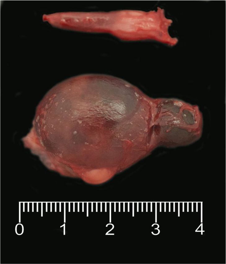
Maurin et al. BMC Veterinary Research       (2019) 15:228                                              Page 3 of 9

Table 2 Haematological and biochemical findings in three dogs with thyroid cystadenoma
                                          Case 1                       Case 2                  Case 3
                                          (Reference intervals)        (Reference intervals)   (Reference intervals)
Haematocrit                               0.48 (0.37–0.55 L/L)         0.49 (0.37–0.55 L/L)    0.46 (0.37–0.55 L/L)
Haemoglobin                               170 (120–180 g/L)            164 (120–180 g/L)       156 (120–180 g/L)
Red Blood Cells                           7.10                         7.09                    6.24
                                          (5.5–8.5 × 10¹²/L)           (5.5–8.5 × 10¹²/L)      (5.5–8.5 × 10¹²/L)
MCHC                                      352 (310–362 g/L)            333 (315–370 g/L)       340 (310–362 g/L)
MCV                                       68.1 (60–77 fL)              68.2 (60–80 fL)         73.5 (60–77 fL)
Platelet count                            261                          216                     339
                                          (150–500 × 109/L)            (160–500 × 109/L)       (150–500 × 109/L)
White Blood Cells                         7.31 (6–17 × 109/L)          7.40 (6–15 × 109/L)     6.81 (6–17 × 109/L)
                                                             9                            9
Neutrophil count                          4.56 (3–11.5 × 10 /L)        5.18 (3–11.5 × 10 /L)   3.13 (3–11.5 × 10⁹/L)
Lymphocyte count                          2.07 (1–3.6 × 10⁹/L)         1.55 (1–4.8 × 10⁹/L)    2.66 (1–3.6 × 10⁹/L)
Monocyte count                            0.37 (0–1.35 × 10⁹/L)        0.52 (0–1.30 × 10⁹/L)   0.61 (0–1.35 × 10⁹/L)
Eosinophil count                          0.26 (0–1.47 × 10⁹/L)        0.07 (0–1.25 × 10⁹/L)   0.41 (0–1.47 × 10⁹/L)
Smear                                     NAD                          NAD                     NAD
Total protein                             69.7 (54–71 g/L)             62.0 (54–77 g/L)        69.7 (54–71 g/L)
Urea                                      4.5 (3.6–8.6 mmol/L)         4.9 (2.0–9.0 mmol/L)    7.2 (3.6–8.6 mmol/L)
ALT                                       50 (0–36 U/L)                67 (0–25 U/L)           61 (0–36 U/L)
GGT                                       0 (0–16 U/L)                 n/p                     2 (0–16 U/L)
CK                                        106 (0–122 U/L)              77 (0–190 U/L)          76 (0–122 U/L)
Amylase                                   974 (400–1300 U/L)           1063 (0–1800 U/L)       1039 (400–1300 U/L)
Cholesterol                               5.97                         6.50                    8.87
                                          (3.2–6.5 mmol/L)             (3.8–7.0 mmol/L)        (3.2–6.5 mmol/L)
Phosphate                                 1.37                         1.10                    1.30
                                          (0.8–1.8 mmol/L)             (0.8–1.6 mmol/L)        (0.8–1.8 mmol/L)
Sodium                                    150.4                        148.8                   151.2
                                          (137–151 mmol/L)             (139–154 mmol/L)        (137–151 mmol/L)
Chloride                                  109.6                        109.0                   111.3
                                          (105–117 mmol/L)             (99–125 mmol/L)         (105–117 mmol/L)
Triglycerides                             0.39                         0.42                    0.73
                                          (0.11–1.69 mmol/L)           (0.45–1.90 mmol/L)      (0.11–1.69 mmol/L)
Albumin                                   31.8 (25–38 g/L)             32.0 (26–40 g/L)        33.0 (25–38 g/L)
Creatinine                                94 (20–120 umol/L)           95 (40–106 umol/L)      75 (20–120 umol/L)
ALP                                       92 (0–82 U/L)                44 (0–25 U/L)           129 (0–82 U/L)
GLDH                                      3 (0–16 U/L)                 4 (0–10 U/L)            6 (0–16 U/L)
Lipase                                    28 (0–130 U/L)               36 (0–150 U/L)          32 (0–130 U/L)
Glucose                                   5.4 (3–6.5 mmol/L)           4.8 (2.0–5.5 mmol/L)    6.6 (3–6.5 mmol/L)
Total Bilirubin                           5.2 (0.9–10 umol/L)          1 (0–9.0 umol/L)        3.8 (0.9–10 umol/L)
Calcium (total)                           2.60 (2.3–3 mmol/L)          2.64 (2–3 mmol/L)       2.60 (2.3–3 mmol/L)
Potassium                                 4.44                         4.73                    3.79
                                          (3.7–5.8 mmol/L)             (3.5–6.0 mmol/L)        (3.7–5.8 mmol/L)
Globulin                                  37.9 (28–42 g/L)             30.0 (20–47 g/L)        36.7 (28–42 g/L)
AST                                       25 (0–37 U/L)                n/p                     21 (0–37 U/L)
Total T4                                  n/p                          14.9 (15–60 nmol/L)     12.1 (15–60 nmol/L)
TSH                                       n/p                          0.643 (< 0.68 ng/mL)    0.275 (< 0.68 ng/mL)
T4 in cystic fluid                        > 193.0 nmol/L               111.0 nmol/L            > 193.0 nmol/L
Total T4/TSH serum concentration and T4 fluid concentration
n/p: not performed
NAD: no abnormalities detected
T4, thyroxine
TSH, thyroid stimulating hormone
Maurin et al. BMC Veterinary Research (2019) 15:228 Page 4 of 9
and demonstrated ovoid, well-defined, homogeneous
fluid attenuating structures within the ventral neck at
the level of the third cervical vertebra on the right side
in case 1, and left side in cases 2 and 3. The structures
measured 3.7 × 2.7 cm in case 1, 3.1 × 4.2 cm in case 2,
and 4.8 × 2.7 cm in case 3. In all cases, the mass partially
displaced the trachea and surrounding structures to the
contralateral side. Post-contrast CT identified a hyperat-
tenuating contrast-enhancing margin in all cases with no
evidence of invasion into surrounding tissues. No other
structure compatible with thyroid tissue could be identi-
fied on the ipsilateral side of the trachea in case 1 and 2.
The contralateral thyroid lobe showed no enlargement
or other abnormality in case 1 or 2 (Fig. 1). In case 3,
the right thyroid lobe was enlarged and lobulated with
multiple ovoid, poorly-defined, hypoattenuating nodules
separated by thin bands of hyperattenuating contrast-
enhancing tissue. The large cystic structure was at the
level of the cranial pole of the left lobe, and the caudal
pole was heterogeneously contrast enhancing similar to
Fig. 2 Case 3: Dorsal plane CT reconstruction at the level of the
the right lobe (Fig. 2). There was no evidence of en- thyroid glands in soft tissue window, post intravenous contrast
larged lymph nodes or pulmonary metastasis identified administration. Cranial is at the top of the image. The right lobe of the
on the CT images. thyroid gland (white arrows) is heterogeneously contrast enhancing
Surgical excision of the thyroid lobe containing the cys- and contains multiple poorly defined fluid attenuating structures
surrounded by contrast enhancing tissue. The caudal aspect of the left
tic structure was performed in cases 1 and 2 via routine
lobe of the thyroid gland (white arrowheads) is similarly
ventral midline incision of the neck. In both cases similar heterogeneously contrast enhancing. The cranial aspect of the left lobe
intraoperative findings were observed and comparable of the thyroid gland is enlarged (3.6 cm length × 2.2 cm height × 2.5
techniques were performed. The cystic structure was cm width), and contains an ovoid fluid attenuating structure
moderately adherent and gently isolated and separated surrounded by a thin margin of peripheral contrast enhancement. An
endotracheal tube is located within the tracheal lumen
from the surrounding tissues by blunt dissection. It was
detached caudally, then reflected rostrally and the main
vascularisation, which was found cranially, was ligated and
the cystic structure removed. In both cases the cystic
structure was removed intact and no perforation occurred
during dissection. Surgery and anaesthetic recovery were
uneventful. Excised thyroid tissue of case 1 and 2 was sub-
mitted for histopathology and in case 3, the entire thyroid
gland was submitted following necropsy. Histopathology
revealed similar morphological features in all three cases,
which consisted of large cystic cavities containing protein-
aceous fluid, erythrocytes and cell debris lined by cuboidal
cells which were often flattened and attenuated, or occa-
sionally lost (Fig. 3). When neoplastic cuboidal lining cells
were lost, the cyst was lined by thick collagen-rich fibrous
tissue with a few trapped follicles (Fig. 4). In all samples
examined including both thyroid lobes of case 3, the solid
Fig. 1 Case 2: Transverse plane CT reconstruction at the level of the
mid-body of the third cervical vertebra in soft tissue window, post thyroid tissue adjoining the cysts was composed of
intravenous contrast administration. A well-defined large (4.3 cm cuboidal neoplastic cells with round-to-oval, normo-
length × 3.5 cm height × 3.3 cm width), ovoid and fluid attenuating chromatic nuclei forming variably-sized follicles or
structure with a thin peripherally contrast-enhancing margin is small, solid nests. The adjacent thyroid tissue was com-
located to the left lateral aspect of the trachea (white arrowheads).
pressed. Based on the above features, a diagnosis of
An endotracheal tube is located within the tracheal lumen
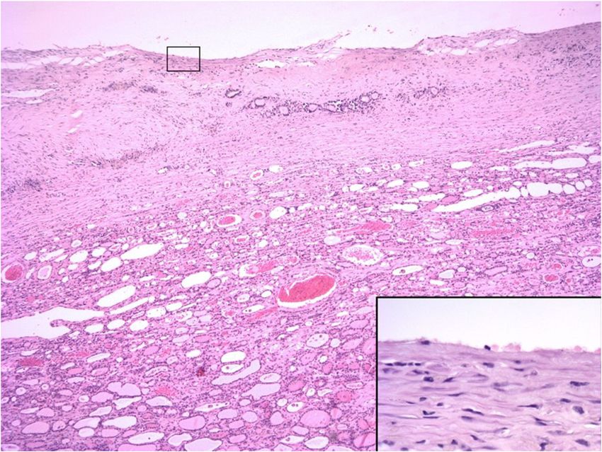
thyroid cystadenoma was made in all three cases.Maurin et al. BMC Veterinary Research (2019) 15:228 Page 5 of 9 Fig. 3 Case 1: Thyroid cystadenoma. The large cystic cavity is lined by cuboidal epithelial cells and contains scant proteinaceous fluid and cell debris. Tumour cells forming either follicles or solid nests surround the cyst and project into the lumen. H&E 20x and inset 400x magnification Cases 1 and 2 were discharged 48 and 72 h postoper- scrotal mast cell tumour was diagnosed. The tumour atively, with follow-up times of 36 and 16 months, was surgically excised completely and adjunctive masi- respectively. No surgical complications were reported. tinib (Masivet, AB Science) was provided. Metastasis of In case 2, as recommended, thyroid function was re- the mast cell tumour to the abdominal lymph nodes oc- assessed six weeks after surgery. The results indicated curred a year later while the dog was still receiving decreased total T4 (8.22 (15–60) nmol/L) and increased chemotherapy, and euthanasia was elected. A rapidly cTSH (2.38 (< 0.68) ng/ml) concentrations. Primary progressive oral amelanotic melanoma was also diag- hypothyroidism was diagnosed and levothyroxine (Solox- nosed in case 3 at the same time as presentation for the ine; Virbac) prescribed. Four months after surgery, an cervical mass. The dog was euthanased and post- intermediate grade Patnaik, high-grade Kiupel dermal mortem examination performed. Fig. 4 Case 2: Thyroid cystadenoma. The cyst is lined by a thick wall composed of collagen rich fibrous tissue with a few trapped follicles. The remaining thyroid tissue is compressed. H&E 40x and inset 400x magnification
Maurin et al. BMC Veterinary Research   (2019) 15:228                                                                 Page 6 of 9

   In order to further investigate a possible predisposition        None of the dogs had overt clinical signs of
for thyroid cystadenoma in the boxer breed, the database         hypothyroidism at the time of diagnosis. This is not
of all the samples (internal and external) submitted to the      unexpected with unilateral lesions. However, information
Veterinary Diagnostic Laboratory at UCD was searched             on thyroid function was not available for Case 1 and thy-
for pathology reports from dogs that included the words          roid dysfunction could not be definitively ruled out. Case
“thyroid” from January 2007 to January 2019. In addition         2 had a low total T4 with upper reference interval cTSH
to the three cases described above, thyroid cystadenomas         concentration. This may suggest subclinical thyroid dys-
were diagnosed in three further dogs that died from unre-        function given that primary hypothyroidism was subse-
lated two of which were boxer dogs diseases, two of which        quently definitively diagnosed six weeks after removal of
were boxer dogs (Table 3). Clinicopathological informa-          the thyroid cystadenoma. Neoplasia within, and subse-
tion was limited for these external cases. One of the            quent removal of, the first lobe may have contributed to
boxers (U351634) was an entire male diagnosed post-              decreased functional thyroid reserve and the development
mortem with unilateral right cystadenoma with the cyst           of overt hypothyroidism. In this case, the T4 concentra-
measuring approximately 1 cm in diameter. The dog died           tion of the cystic fluid was lower compared to cases 1 and
because of a large aortic body carcinoma with marked in-         3. This raises the possibility that the T4 concentration in
filtration of the right atrium. The other boxer (U243968)        the cystic fluid may provide some information on thyroid
was also an entire male and was diagnosed post-mortem            function that warrants further investigation. In humans,
with two large cysts occurring in the left thyroid gland         post-operative hypothyroidism has been reported in up to
(Fig. 5). The dog died as a result of severe diffuse bilateral   one third of patients following hemithyroidectomy be-
renal fibrosis. Unfortunately, further information regard-       cause of suspected neoplasia [21–23]. In these cases,
ing thyroid function of these two external cases could not       lymphocytic infiltration was identified within the resected
be retrieved. In addition, a cyst (2.5 cm × 1.3 cm × 1 cm)       tissue, but it is unclear if the tumour itself acts as a trigger
was reported in association with a bilateral follicular          for autoimmune thyroiditis [21] or if this represents con-
thyroid carcinoma in a 12-year-old bichon frise.                 current but unrelated immune-mediated disease [22].
                                                                 Lymphocytic infiltration was not apparent histologically in
Discussion                                                       the resected tissue of any of the current cases, but thyro-
The current case report describes the presence of an un-         globulin autoantibodies were not assessed. Therefore, the
common tumour, thyroid cystadenoma, in three boxer               cause of hypothyroidism in case 2 remains unclear as the
dogs. Although this tumour type has been previously re-          contralateral thyroid lobe was not removed or sampled.
ported in dogs, detailed clinical features are lacking [1, 5,       It has been proven that fine needle aspiration (FNA)
7, 18]. Greater detail is provided in two case reports, but      of solid cervical masses has a good diagnostic accuracy
the clinical appearance differed substantially from the          in humans [24]. In dogs, one study showed that correl-
current cases because of the presence of hyperthyroid-           ation between cytological results and histopathological
ism [19, 20]. In the current study, investigation was            findings for thyroid carcinomas was only fair and the as-
prompted by identification of a mass, or clinical signs sec-     piration technique resulted in excessive contamination
ondary to a mass, rather than the consequences of hyper-         with blood in one third of cases [25]. Cytology was per-
thyroidism. In addition, the presence of this tumour type        formed in all cases and only the FNA results in case 3
in three dogs of the same breed may suggest a possible           were consistent with the histopathologic diagnosis of ad-
breed predisposition; a finding that is further supported        enoma. It is believed that this sample included tissue
by the identification of this tumour type mainly in boxer        from the solid part of the thyroid tumour, as the enlarge-
dogs in the database review.                                     ment was not confined to a single cyst in this case. In
  Non-functional benign thyroid masses can cause clinical        cases 1 and 2, cytologic evaluation was made from the
signs because of their size and localisation. Dysphagia, dys-    aspirated fluid and was not diagnostic. This supports
pnoea, cough or regurgitation can occur because of com-          previous reports in which cytology of cystic fluid has
pression of surrounding tissues such as the trachea and          rarely offered evidence of a particular tumour type or
oesophagus. In case 1, regurgitation and increased respira-      origin due to the paucity of cellular material and lack of
tory effort after strenuous exercise and excitement were re-     pathognomonic cytological features, revealing only cell
ported. In this case, the CT of the neck and chest showed        necrosis and blood cells [19, 20].
that the mass was causing intermittent partial obstruction          In adult humans presenting with a solitary cystic mass
of the tracheal lumen by compression of the tracheal mem-        in the lateral neck, measurement of thyroglobulin con-
brane. Case 3 was reported to drool excessively. Although        centration in the cystic fluid is advised as an initial step
potentially related to the thyroid masses, the subsequent        to confirm the thyroidal origin [27, 28]. Canine thyro-
discovery of a rapidly growing oral neoplasm was consid-         globulin assays are not commercially available. However,
ered a more likely cause.                                        a similar approach was taken by measurement of cystic
Maurin et al. BMC Veterinary Research (2019) 15:228 Page 7 of 9 Table 3 Cases of thyroid tumours in dogs identified through a database search of the UCD Veterinary Diagnostic Laboratory records from January 2007 to January 2019. The highlighted cases are the thyroid cystadenoma cases, with in bold the three cases described in this report NC: Not communicated; (E): External sample; *Diagnosis at post-mortem; LR Labrador retriever; JRT Jack Russell terrier; CKC Cavalier King Charles; WHWT West Highland White terrier; SBT Staffordshire bull terrier
Maurin et al. BMC Veterinary Research       (2019) 15:228                                                                                   Page 8 of 9

                                                                          Further enlargement occurs due to frequent haemorrhage
                                                                          and desquamation of the follicular cells into the cystic
                                                                          lumen [15]. Follicular cysts have also been described in
                                                                          which the fluid filled cavities are lined by a dense fibrous
                                                                          capsule from which fronds of uniform cells arranged in
                                                                          follicular and/or compact cellular patterns project [8].
                                                                          In these cases, neoplastic tissue is not identified. How-
                                                                          ever, follicular cysts show similar morphological fea-
                                                                          tures to cystadenomas, and the differentiation can be
                                                                          somewhat arbitrary and dependent upon whether or
                                                                          not solid adenomatous tissue is present within the section
                                                                          examined [17, 24, 32]. Indeed, adenomatous change
                                                                          was not initially observed in case 2, and was only
                                                                          identified after preparation and examination of add-
                                                                          itional histopathologic sections.
                                                                             Interestingly, all three dogs in the present study were
                                                                          boxers; a breed in which thyroid tumours have been re-
                                                                          ported with increased frequency in some [2, 7, 8] but not
                                                                          all [1] previous studies. The UCD Veterinary Diagnostic
                                                                          Laboratory database search of all internal and external
                                                                          samples over a 12-year period revealed the presence of
                                                                          this tumour type in only six cases (including the three
 Fig. 5 Boxer (U243968); thyroid glands; The left thyroid was 1 × 0.5     dogs included in this case report), and five of these were
 cm in size, the right was enlarged, with two soft cystic lesions (blue   boxer dogs. Whether boxers are truly predisposed to this
 arrow =1.5 × 1 cm, green arrow = 3.5 × 2.5 cm). Marked parathyroid
 hyperplasia is also observed (grey arrow), likely due to concurrent      particular condition warrants further investigation.
 advanced chronic renal disease diagnosed in this case                       To the authors’ knowledge, this is the first report of
                                                                          non-functional thyroid cystadenomas in dogs. The cystic
                                                                          thyroid structures were detectable clinically because of
T4 concentrations, and the increased values rapidly con-                  their large volume but were not associated with clinical
firmed thyroidal origin in all three cases. This appears to               signs related to thyroid dysfunction. Measurement of
be a useful diagnostic procedure in dogs but has only                     T4 concentration in the cystic fluid was supportive of
been reported once before [19].                                           thyroidal origin and should be considered as a simple,
   Although long-term effects of thyroid cysts are not                    relatively inexpensive first line diagnostic tool. Despite
known, drainage was only temporarily beneficial. With-                    their apparent rarity, thyroid cystadenoma should be
out treatment, thyroid cysts could continue to grow with                  considered a differential for dogs presenting with cer-
progressive compression of adjacent structures, or ma-                    vical masses. Surgical excision is recommended as it
lignant transformation from adenoma to carcinoma                          is curative and because it provides tissue samples to
could occur. Although only suspected in dogs, this has                    investigate the underlying thyroid lesion.
been suggested to occur in cats with hyperthyroidism
[29]. Also, one study suggested that in theory, TSH may                   Abbreviations
                                                                          CT: Computed tomographycTSH: canine thyroid-stimulating hormone;
contribute to further growth of primary thyroid carcin-                   FNA: Fine needle aspiration; RBC: Red blood cells; T4: Thyroxine;
omas in dogs [30]. Surgical excision was therefore rec-                   UCD: University College Dublin
ommended and curative in the two cases in which it was
                                                                          Acknowledgements
performed. In humans, thyroidectomy is recommended                        Not applicable.
for all patients with thyroid cysts who have a history of
prior neck irradiation, abnormal FNA cytology results,                    Authors’ contributions
recurrence of the cyst despite two or more needle aspi-                   M-PM, RS and CM interpreted the three cases haematology and
                                                                          biochemistry results. DD performed the images descriptions. HJ performed
rations and compressive clinical signs despite draining                   the histological examination. All authors read and approved the final
the cystic fluid [31].                                                    manuscript.
   In small animals, thyroid cystadenomas are described as
follicular cell adenomas with one or two large cavities                   Funding
                                                                          Not applicable.
filled with proteinaceous fluid, necrotic debris and eryth-
rocytes [7, 15, 17]. Cysts develop due to follicular disten-              Availability of data and materials
sion and degeneration within thyroid tumours [17].                        All data generated during this study are included in this published article.
Maurin et al. BMC Veterinary Research (2019) 15:228 Page 9 of 9
Ethics approval and consent to participate 23. Chotigavanich C, Sureepong P, Ongard S, Eiamkulvorapong A, Boonyaarunnate
Not applicable. T, Chongkolwatana C, et al. Hypothyroidism after hemithyroidectomy: the
incidence and risk factors. J Med Assoc Thail. 2016;99(1):77–83.
Consent for publication 24. Durante C, Grani G, Lamartina L, Filetti S, Mandel SJ, Cooper DS. The
Not applicable. diagnosis and management of thyroid nodules, a review. JAMA. 2018;319(9):
914–24.
25. Thompson EJ, Stirtzinger T, Lumsden JH, Little PB. Fine needle aspiration
Competing interests
cytology in the diagnosis of canine thyroid carcinoma. Can Vet J. 1980;21:
The authors declare that they have no competing interests.
186–8.
26. Lee M-J, Kim E-K, Kwak JY, Kim MJ. Partially cystic thyroid nodules on
Received: 21 March 2019 Accepted: 4 June 2019
ultrasound: probability of malignancy and sonographic differentiation.
Thyroid. 2009;19(4):341–7.
27. Benmoussa JA, Chen K, Nijjar S, Applewhite M, Warshaw J. Lateral neck
References cystic mass: the role of thyroglobulin measurement in fine needle
1. Bertolini G, Drigo M, Angeloni L, et al. Incidental and nonincidental canine aspiration. Endocrine Pract 2018. Rapid Electronic Article in Press.
thyroid tumors assessed by multidetector row computed tomography: a 28. Yehuda M, Schechter ME, Abu-Ghanem N, Golan G, Horowitz G, Fliss DM, et
single-Centre cross sectional study in 4520 dogs. Vet Radiol Ultrasound. al. The incidence of malignancy in clinically benign cystic lesions of the
2017;58:304–14. lateral neck: our experience and propose diagnostic algorithm. Eur Arch
2. Scarlett JM. Epidemiology of thyroid disease in dogs and cats. Vet Clin Otorhinolaryngol. 2018;275:767–73.
North Am Small Anim Pract. 1994;24:477–86. 29. Peterson ME, Broome MR, Rishniw M. Prevalence and degree of thyroid
3. Harari J, Patterson JS, Rosenthal RC. Clinical and pathological features of pathology in hyperthyroid cats increases with disease duration: a cross-
thyroid tumors in 26 dogs. J Am Anim Hosp Assoc. 1986;188:1160–4. sectional analysis of 2096 cats referred for radioiodine therapy. J Feline Med
4. Birchard SJ, OF R. Neoplasia of the thyroid gland in the dog: a retrospective Surg. 2016;18(2):92–103.
study of 16 cases. J Am Anim Hosp Assoc. 1981;17:369–72. 30. Verschueren CP, Rutteman GR, Vos JH, Van Dijk JE, de Bruin TW.
5. Wucherer KL, Wilke V. Thyroid cancer in dogs: an update based on 638 Thyrotrophin receptors in normal and neoplastic (primary and metastatic)
cases (1995-2005). J Am Anim Hosp Assoc. 2010;46:249–54. canine thyroid tissue. J Endocrinol. 1992;132(2):461–8.
6. Hayes HM, Fraumeni JF. Canine thyroid neoplasms: epidemiologic features. 31. McHenry CR, Slusarczyk SJ, Khiyami A. Recommendations for management
J Natl Cancer Inst. 1975;55:931–4. of cystic thyroid disease. Surgery. 1999;126(6):1167–72.
7. Brodey RS, Kelly DF. Thyroid neoplasm in the dog, a clinicopathologic study 32. Miller ML, Peterson ME, Randolph JF, Broome GD, Norsworthy GD, Rishniw
of fifty-seven cases. Cancer. 1968;22:406–15. M. Thyroid cysts in cats: a retrospective study of 40 cases. J Vet Intern Med.
8. Leav I, Schiller MD, Rijnberk A, Legg MA, der Kinderen PJ. Adenomas and 2017;31:723–9.
carcinomas of the canine and feline thyroid. Am J Path. 1976;83:61–93.
9. Rijnberk A, der Kinderen PJ. Toxic thyroid carcinoma in the dog. Acta
Endocrinol 1969;[Suppl]:138–177. Publisher’s Note
10. Campos M, Ducatelle R, Rutteman G, Kooistra HS, Duchateau L, de Rooster Springer Nature remains neutral with regard to jurisdictional claims in
H, et al. Clinical, pathologic, and Immunohistochemical prognostic factors in published maps and institutional affiliations.
dogs with thyroid carcinoma. J Vet Intern Med. 2014;28:1805–13.
11. Rosol TJ, Meuten DJ. Tumors of the endocrine glands. In: Tumors in domestic
animals. Meuten DJ. Eds. 5th edn. John Wiley & Sons Inc, Iowa, 2017. pp. 791–797.
12. Scott-Moncrieff JC. Canine thyroid tumours and hyperthyroidism. In: Canine
and Feline Endocrinology. Feldman E, Nelson R, Reusch C, Scott-Moncrieff J.
Eds. 4th edn. WB Saunders, Missouri; 2014. pp. 196–212.
13. Benjamin SA, Stephens LC, Hamilton BF, Saunders WJ, Lee AC, Angleton GM,
Mallinckrodt CH. Associations between lymphocytic thyroiditis,
hypothyroidism, and thyroid neoplasia in beagles. Vet Pathol. 1996;33:486–94.
14. Barber LG. Thyroid tumors in dogs and cats. Vet Clin North Am Small Anim
Pract. 2007;37:755–73.
15. Capen CC. Tumors of the endocrine glands. In: Tumors in domestic animals.
Moulton JE. Eds. 2nd ed. Besheley LA, London: University of California Press;
1978. pp. 392–406.
16. Miles KG, Lattimer JC, Jergens AE, Krause GF. A retrospective evaluation of
the radiographic evidence of pulmonary metastatic disease on initial
presentation in the dog. Vet Radiol Ultrasound. 1990;60(3):300–5.
17. Kiupel M, Capen C, Miller M, Smedley R. World Health Organization
histological classification of the tumors of the endocrine system of
domestic animals. Armed forces Institute of Pathology Washington, 2nd
Series Volume XII, D; 2008. p. 27–36.
18. Pollard RE, Bohannon LK, Feldman EC. Prevalence of incidental thyroid
nodules in ultrasound studies of dogs with hypercalcemia (2008-2013). Vet
Radiol Ultrasound. 2015;56:63–7.
19. Mackay B. Hyperthyroidism associated with functional cystic thyroid tumor in a
dog: comparison to feline hyperthyroidism. Aust Vet Practit. 1999;29:54–8.
20. Lawrence D, Thompson J, Layton AW, Calderwood-Mays M, Ellison G,
Mannella C. Hyperthyroidism associated with a thyroid adenoma in a dog. J
Am Vet Med Assoc. 1991;199:81–3.
21. Seiberling KA, Dutra JC, Bafaramovic S. Hypothyroidism following
hemithyroidectomy for benign nontoxic thyroid disease. Ear Nose Throat J.
2007;86(5):295–9.
22. De Carlucci D Jr, Tavares MR, Obara MT, Liporoni Martins LA, Carneiro Hojaij
F, Cernea CR. Thyroid function after unilateral total lobectomy: risk factors
for postoperative hypothyroidism. Arch Otolaryngol Head Neck Surg. 2008;
134:1076–9.You can also read