GAN-based Virtual Re-Staining: A Promising Solution for Whole Slide Image Analysis
←
→
Page content transcription
If your browser does not render page correctly, please read the page content below
GAN-based Virtual Re-Staining: A Promising
arXiv:1901.04059v1 [cs.CV] 13 Jan 2019
                                           Solution for Whole Slide Image Analysis

                                         Zhaoyang Xu? , Carlos Fernández Moro† ,Béla Bozóky† , Qianni Zhang?

                                                               email: zhaoyang.xu@qmul.ac.uk.
                                             ?
                                                 Queen Mary University of London, † Karolinska University Hospital

                                           Abstract. Histopathological cancer diagnosis is based on visual exam-
                                           ination of stained tissue slides. Hematoxylin and eosin (H&E) is a stan-
                                           dard stain routinely employed worldwide. It is easy to acquire and cost
                                           effective, but cells and tissue components show low-contrast with vary-
                                           ing tones of dark blue and pink, which makes difficult visual assessments,
                                           digital image analysis, and quantifications. These limitations can be over-
                                           come by IHC staining of target proteins of the tissue slide. IHC provides
                                           a selective, high-contrast imaging of cells and tissue components, but
                                           their use is largely limited by a significantly more complex laboratory
                                           processing and high cost. We proposed a conditional CycleGAN (cC-
                                           GAN) network to transform the H&E stained images into IHC stained
                                           images, facilitating virtual IHC staining on the same slide. This data-
                                           driven method requires only a limited amount of labelled data but will
                                           generate pixel level segmentation results. The proposed cCGAN model
                                           improves the original network [1] by adding category conditions and in-
                                           troducing two structural loss functions, which realize a multi-subdomain
                                           translation and improve the translation accuracy as well. Experiments
                                           demonstrate that the proposed model outperforms the original method
                                           in unpaired image translation with multi-subdomains. We also explore
                                           the potential of unpaired images to image translation method applied on
                                           other histology images related tasks with different staining techniques.

                                 1        Introduction
                                 Virtual staining, by its literal meaning, is to use computerized algorithms to cre-
                                 ate an artificial effect of staining without physically tampering the slide. With
                                 well-designed applications, this novel approach will introduce a revolutionary im-
                                 pact on histopathology analysis of digitized whole slide images (WSIs). Based on
                                 the digital scan of a real slide stained using a traditional dyeing method, virtual
                                 staining is, in fact, a post-processing step that can generate other stained versions
                                 of the same slide using dedicated computer algorithms. The aim is to provide
                                 different staining effects that can to highlight different relevant histological and
                                 cell features on the same slide. From this point of view, image normalization
                                 can be also regarded as a basic form of virtual staining, which mainly focuses
                                 on reducing the staining variance among slides caused by different staining pro-
                                 tocols, scanners or scanning conditions, and ensuring a coherent appearance of
                                 components.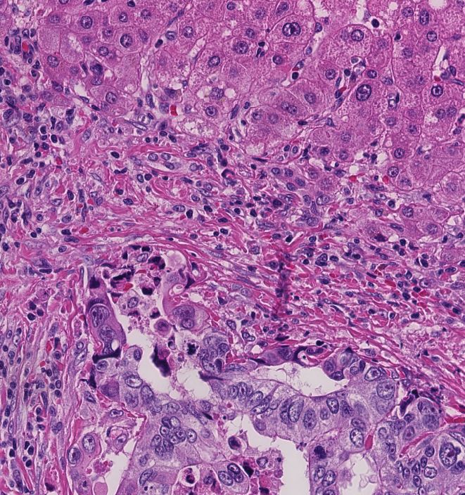
2       Zhaoyang Xu? , Carlos Fernández Moro† ,Béla Bozóky† , Qianni Zhang?

          (a) H&E stained image                  (b) Immuno stained images

      Fig. 1: Matched H&E and IHC stained images of a similar WSI region

    Virtual staining can be considered in two main types: low-level virtual stain-
ing and semantic virtual staining. In low-level virtual staining, the output image
is a weighted liner combination of different channels of the original image in its
colour space. There are not semantic factors considered in producing the result-
ing version of the image. In comparison, the semantic virtual staining methods
further take into account semantic information in the transform process to ensure
the semantic correctness or truthfulness of the virtually stained images.

1.1    Challenges and Objective
The large size of unannotated histology image data has posed critical challenges
for its understanding and analysis. Motivated by the great success of deep learn-
ing models applied in different tasks in natural image analysis, more and more
deep learning algorithms and systems are being designed for histopathology im-
age analysis. Dozens of fruitful outcomes have been achieved based on deep
convolutional neural networks (DCNN), especially using patch-based methods.
With enough number of annotated patches, a DCNN will be trained to detect
cancerous tissue patches against other benign tissue components.
    However, there are intrinsic limitations in the current methods. A major
issue is networks’ high dependence on annotated training sets that are often in-
adequate both in terms quantity, due to the expensive manpower consumption
required for the job, and in terms of quality, because of inter- and intra-observer
subjectivity. Moreover, when multiple tissue types are present in a region in-
tertwined together, it becomes impossible even for experienced pathologists to
create accurate annotation masks, such as the H&E stained image region shown
in Fig. 1 (a). The lack of adequate and accurate training data results in unsat-
isfactory detection accuracy.
    In clinical practice, there are other methods, mainly IHC staining, for the
pathologist to highlight the cancerous or other cell and tissue components of
interest. IHC staining allows the visualization of specific proteins on the tissue
slide using targeted antibodies and subsequently detecting the bound antibodies
using chromogens of different colours. In such a way, by using protein-binded an-
tibodies that are selectively expressed in the cancer cells, these become readily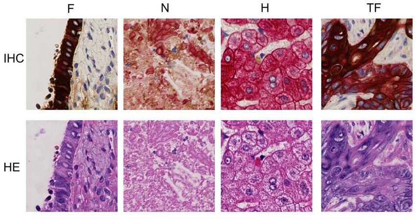
Title Suppressed Due to Excessive Length       3

distinguishable from the other tissues by the high-contrast chromogen staining,
as shown in shown in Fig. 1 (b). Despite its superior features compared to H&E,
there are obstacles that preclude the wide application of IHC staining in clinical
practice - the high cost of antibodies, autostainer machine equipment, and com-
plex laboratory process. Unfortunately, these issues are unresolvable currently.
In most cases, pathologists have to rely on H&E or other relatively cheaper and
more available staining methods for their diagnosis and limit the use of IHC for
a very small number of selected slides. For this reason, the majority of com-
puter models and systems for histopathological cancer grading are designed for
segmenting and classifying the tumor tissue based on H&E imaging.
    If there is a way to model the pairwise relationship between the tissue mor-
phology in H&E and IHC stained conditions, it is then possible to predict one
from the other. Recently, with the continuous progress of deep learning on dif-
ferent image processing tasks, it becomes possible to ’translate’ one image to an-
other with a different style. Such technology brings the opportunity to produce
an image virtually stained with one dye from another, for example, producing
IHC images from images dyed with the relatively cheaper and widely available
staining techniques such as H&E. This may be very useful in many clinical
diagnostic and AI-based applications, such as to improve the effectiveness of
pathological examination by reducing the eyeballing time in visual screening of
the slides; and to increase the segmentation and classification performance of AI
models, i.e. if the H&E slides can be virtually IHC stained, with a few simple
post-processing, the generated IHC images can provide highly precise segmen-
tation of tissue regions of interest.
    What is more interesting, given an original image acquired with one stain,
virtually stained images with one or more other dyes can now be generated
from exactly the same sample. Currently, only one or two chromogens can be
employed on the same sample for brightfield microscopy, which is the standard in
histopathology practice. However, it is often the case that several IHC stainings
need to be performed on the same tissue region to extract multiple features of
diagnostic relevance. In such cases, pathologists perform serial cuts from the
same tissue block, each between 3m and 5m thick, and stain them with different
techniques and antibodies. This introduces inevitable inter-slide variability in
the cell and tissue structures as, while performing serial cuts along the z-axis,
neighbouring tissue regions are typically similar but do not match exactly each
ocher, preventing cell-level segmentation and colocation analyses across slides.
Consequently, the differently stained sections do not represent exactly the same
tissue sample. With the help of virtual staining, multiplexing with two or more
multiple stains on the same sample becomes possible.
    There are many ways to understand or implement virtual staining. It can
be considered as a special kind of style transfer that only transfers the colour
coding to the original images while keeps the structural information consistent.
In this case, it is also similar to the image colourization problem which will re-
colourize the image with new colours. However, this method needs to base on
the good performance of semantic segmentation. Based on the tightly related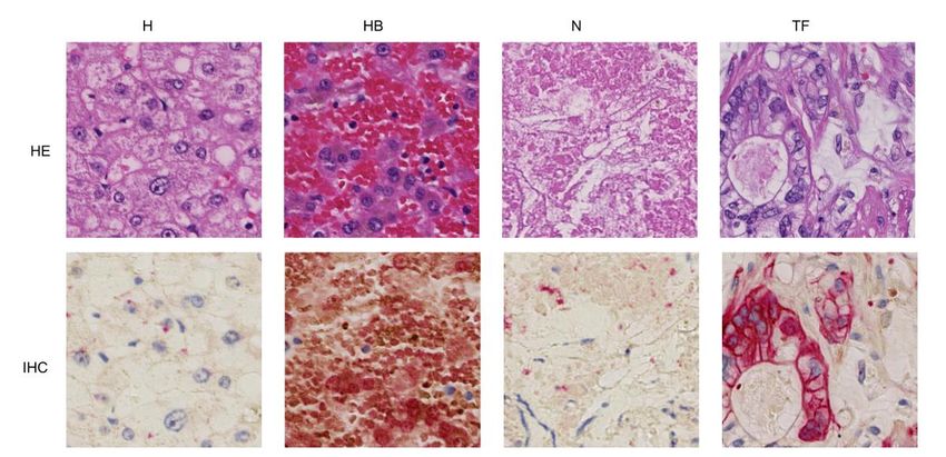
4      Zhaoyang Xu? , Carlos Fernández Moro† ,Béla Bozóky† , Qianni Zhang?

common features of images with different stainings, the most relevant problem
is the unpaired image-to-image translation that will transfer the original image
with style from the reference image.
    Therefore in this study, we tackle the task of virtual staining tissue samples
by focusing on producing virtual IHC images from original H&E images as a
start. With the proposed novel cCGAN model, the challenging objective of un-
paired image-to-image translation for multi-class virtual staining is addressed,
with additional patch-wise labels. In particular, to make sure the structural de-
tails of the original image remains unchanged in the virtual staining process, a
photorealism and structure similarity loss is introduced to regulate the transla-
tion process. The overall aims are to improve the effectiveness of pathological
examination and enable a more precise tissue segmentation and labelling for fur-
ther computer aided applications, eventually based on the virtual IHC images.

1.2   Related Work
Generative Adversarial Networks Generative Adversarial Networks (GANs)
[2], as an important branch of deep learning, is gaining more and more attention
from the researchers around the world. A significant amount of investigations
have been made to explore the potential of GAN in natural images related tasks
like image synthesizing, image super-resolution, and style transfer [3, 4].The gen-
erative model is a very promising approach for histology image processing as well.
The fundamental idea of a generative adversarial network is to train the gener-
ative model G that can generate fake images though learning the real images,
to fool the discriminator D. Once the discriminator D can not tell the fake ones
from the real, it means that the network has learned to model the distribution
of the input data appropriately. Modelling the patterns in the histology images
is a particularly complicated task for generative models. Although there are un-
derlying regulations that control the growth pattern of the cells, the regulations
are limited and unknown to human experts. Tissue morphologies can be treated
as orderless texture patterns. In this case, the generative adversarial networks
are often used as a data augmentation method that helps generate more tissue
regions. This is one of the most intuitive uses of generative models in histology
image processing.
Conditional GAN / Paired Image to Image Translation Based on the
original GAN networks, conditional GAN[5] has been proposed to take into ac-
count certain constraints that can help to improve the truthfulness of images,
in additional to the imaginary information. The conditions encode the labels of
the generated images which contain category information. Furthermore, pix2pix
GAN, as a paired image-to-image translation method, provides segmentation
masks as conditions to generate images on a pixel level [6]. From the image
translation point of view, the pix2pix model translates the images from abstract
representation to real images while keeping the semantic meaning.
Unpaired Image Translation Among the above mentioned paired image-to-
image translational models, an obvious drawback is the requirement for cor-
responding masks on the input images. Thus, a few unpaired image transla-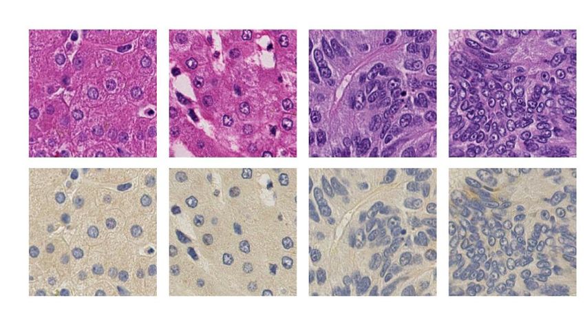
Title Suppressed Due to Excessive Length        5

tion models are proposed including SimGAN [7], CoGAN [8] and CycleGAN[1].
Though the images are unpaired and are from different domains, they shared
similar semantic and structural features, which form the basis for unpaired im-
age translation. For example, in CycleGAN, the model tried to translate a horse
to a zebra and an apple to an orange. The objects before and after the trans-
lation have to demonstrate similar semantic structures to make the translation
meaningful.
Style Transfer Instead of re-arranging the contents in the images, the style
transfer approach attempts to replace the low-level representations from another
domain regardless of whether the input and reference images share the same
semantic information or not[9]. However, the transferred images may change
significantly compared to the original, in terms of both content semantics and
structure. Hence, deep photo style transfer tries to keep the structural informa-
tion as much as possible while changing the semantic meaning by manipulating
the colour space [10].

    Among the application domains for image translation, histology image anal-
ysis poses unique challenges. How to improve the visualization of histopathology
images is at the core of the challenge due to its crucial impact on the efficiency
of pathology examination and diagnostic accuracy. Research on virtual staining
has been conducted for many years and applied on many different types of im-
ages. The recent advances in GAN based approaches with their superior abilities
open new roads in this direction.
Low Level Virtual Staining Early virtual staining research mainly focuses
on the low visual level. For example, in the work of Sasajima et al., a real-time
endoscopy system is developed to visualize the endocytoscopic images in a H&E
staining style [11]. In Bautista and Yagi’s work, they present a linear spectral
transformation method for ”digital staining of histopathology multispectral im-
ages” [12] . Other works like in [13, 14], employ a simpler mapping method to
virtually stain the fluorescence images to H&E. Some other research attempt to
modify the hardware to achieve the virtual staining results. Tao et al. present a
non-linear microscope to assist the diagnosis of breast cancer [15]. Recent work
has demonstrated that with the help of deep learning, the different tissue com-
ponents can be separated semantically. This means that the staining of differ-
ent tissue components can be separately re-colourized regardless of the original
staining technique. Bayramoglu et al. utilise a Conditional Generative Adver-
sarial Networks (cGAN) to virtually stain unstained specimens [16]. Rivenson
et. al[17] also employ the GAN model to virtually stain the fluorescence images
to H&E images.
Semantic Virtual Staining Generally speaking, semantic virtual staining can
be considered as a deeper process of raw images which is usually related to image
segmentation or classification tasks. For multi-class tissue type classification and
segmentation, once the results are mapped back to the original images, the new
results can be a form of virtual staining. The research by Litjens et al. overlapped
a heatmap of the likelihood of cancerous regions on the original image, which is
also a kind of virtual staining [18] . Recently, a team from Google successfully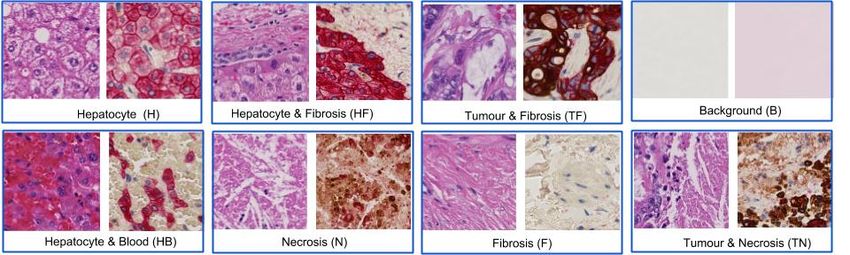
6         Zhaoyang Xu? , Carlos Fernández Moro† ,Béla Bozóky† , Qianni Zhang?

build an augment reality microscope which has built-in ”virtual staining func-
tion” through real-time image analysis using deep learning [19]. Trahearn et al.
[20] propose a hyper stain inspector for image alignment and cancer detection as
well. Another innovative way of ”virtual staining” method is proposed in [21].
The authors represent different tissue components in the image with colourful
plates of different size, facilitating diagnosis.

1.3     Our contribution
In this study, we explore the potential of unpaired image-to-image translation
as ”virtual staining” for histopathology image analysis. We propose a condi-
tional CycleGAN which could perform multi-class virtual staining with addi-
tional patch-wise labelling. Furthermore, to make sure the virtual staining does
not change the structural details of the original image, we introduce photorealism
and structure similarity losses to regulate the translation process.

2      Method
The overall objective of the proposed network is to learn a multi-class mapping
between two domains X and Y . In fact, the transformation takes place between
their sub-domains X t and Y t ,t ∈ [1, C], where C denotes the number of pre-
defined classes. For two unpaired samples x and y , their distributions of the
training dataset are denoted as x ∼ p(x|c) and y ∼ p(y|c) where c represents the
category condition. As illustrated in Fig. 2, there are two conditional mappings
in our model, the encoder network Enc maps the input image from domain X
to Y while the decoder network Dec maps back from Y to X.

           Fig. 2: Mapping between two domains and their sub-domains

    Our method extends the original CycleGAN model proposed in [1] for the
histology image tasks, by introducing the following two improvements:
    – Patch-wise label information is employed to enforce the model to learn mu-
      tual representation with conditional query. This is achieved by appending
      a few classifier layers to the original generative network for the sub-domain
      consistency.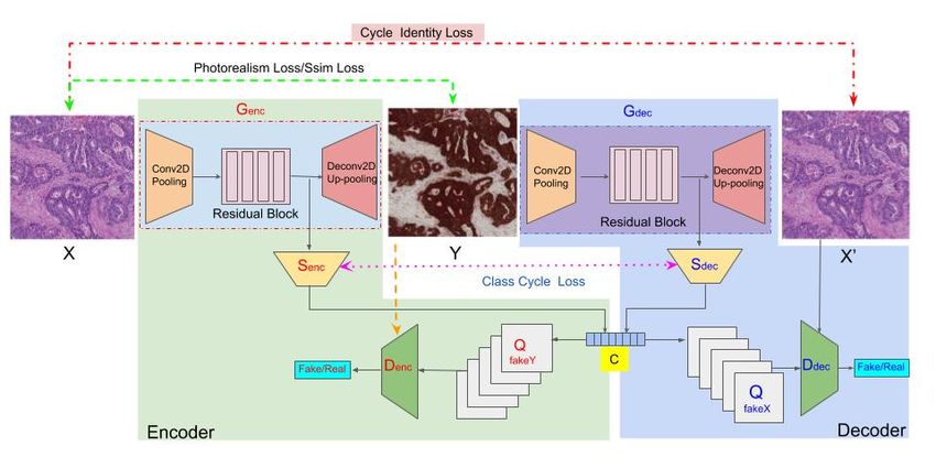
Title Suppressed Due to Excessive Length        7

 – Two constraints are introduced to regularize changes to the structural de-
   tails: the photorealism loss and the structural similarity loss.

                Fig. 3: Overall structure of the proposed network

As shown in Fig. 3, the whole framework consists of two big modules- an encoder
network (in green zone) and a decoder network(in blue zone), both of which
have the same subnetwork structure, a generator G, a discriminator D and a
classifier S. In the encoder network, they are denoted as Genc , Denc and Senc .
Likewise, in the decoder network, they are Gdec , Ddec and Sdec . The generator
is in charge of translating the image between domains, while the discriminator
provides feedback on True or False for the generated image. At the same time,
the classifiers will provide extra information about the image’s sub-domain. To
train the proposed model for decent performance on the task, we facilitate the
following losses during the training phase.

2.1   Conditional Adversarial Loss

To include the category information for guiding the translation process, we apply
conditions on the inputs for the discriminator Denc , Ddec . For the mapping from
X to Y , the conditional adversarial loss inspired by the loss in[2] within the class
c and the generator Genc is defined as :

                   LcGAN (Genc , Denc ) = Ey∼p(y|c) [logDenc (y)]
                                                                                 (1)
                            +Ex∼p(x|c) [log(1 − Denc (Genc (x)))]

Likely, for the mapping from Y to X, the same loss is calculated with the dis-
criminator Ddec and generator Gdec .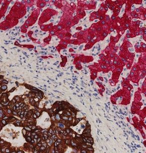
8      Zhaoyang Xu? , Carlos Fernández Moro† ,Béla Bozóky† , Qianni Zhang?

2.2   Deterministic Loss for Tissue Classification Networks

The tissue subtype is a kind of crucial information for the histopathology image
translation. Without appropriate class inference, it will lead to a failed trans-
lation. In our network, the classifiers S infer the category information indepen-
dently of the generator but share the same base network structure. Practically,
by adding extra category information to the network, we attempt to map the
distribution of the data category information during the transformation, and
include the categories information in the output as well. The networks employ
the Softmax Entropy Loss ` to regularize the category information with regards
to their class information. The loss of the classifiers is defined as :

      Lclass (Senc , Sdec ) = Ex∼p(x|c) [`(Senc (x), c)] + Ey∼p(y|c) [`(Sdec (x), c)]   (2)

2.3   Cycle Loss and Classification Cycle loss

The core idea of cycle-GAN is to use the encoder and decoder process as a cycle
                                               0
to make X ≈ X 0 after one cycle X → Y → X . By minimizing the difference
                    0
between X and X , the network will be able to learn the shared features. The
original cycle loss defined as [1] :

               Lcyc (Genc , Gdec ) = Ex∼p(x|c) [||Gdec (Genc (x)) − x||1 ]
                                                                                        (3)
                                    +Ey∼p(y|c) [||Genc (Gdec (y)) − y||1 ]

During the cycle process, the outputs of the classifiers S should be identical as
well. Thus the classification cycle loss is defined with L1 loss:

            Lclcyc (Senc , Sdec ) = E(x,y)∼p(x,y|c) [||Senc (x) − Sdec (y)||1 ]         (4)

The cycle consistency of the classification information is of signification impor-
tance to final results.

2.4   Identity Loss , Photorealism Loss and Structural Similarity Loss

The identity loss proposed by Taigmen et al. [22] is introduced in the original
cycle GAN paper to preserve the color composition especially when the input
image is very close to the output image domain[1] . The definition of identity
loss for the proposed network is demonstrated as follow:

                   Lid (Genc , Gdec ) = Ex∼p(x|c) [||(Genc (y) − x||1 ]
                                                                                        (5)
                                        +Ey∼p(y|c) [||Gdec (y) − y||1 ]

However, in virtual staining, identity loss is not strong enough to keep the
structural information unchanged, because the mapping is on the same domain.
Hence, we introduced two other loss functions, photorealism loss [10] and struc-
tural similarity loss (SSIM)[23], to enhance the original cycle-GAN network.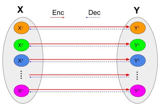
Title Suppressed Due to Excessive Length        9

These two losses serve the same purpose which is to preserve the texture struc-
ture of the input image. Photorealism loss uses Matting Laplacian transform to
measure the structural differences and is defined as :

                                    3
                                    X                     3
                                                          X
                     P ho(x, y) =         ykT [Mx ]yk +         xTk [My ]xk         (6)
                                    k=1                   k=1

where k denotes the RGB channels while Mx /My is the transformed matrix
regarding to the input images x/y. For more details, please refer to [10].

              Lpho (Genc , Gdec ) = P ho(Genc (x), x) + P ho(Gdec (y), y)           (7)

    SSIM has been used for assessing the image quality in many related studies
[24] .We introduce it to our model to regulate the structural changes between
the input and output images. For each pixel in the image, the SSIM is defined
as:
                               2µx µy + Q1      2σxy + Q2
                Ssim(x, y) = 2              + 2                            (8)
                              µx + µ2y + Q1   σx + σy2 + Q2

where µx , µy are the mean of a fixed window centered as the pixel, σx , σy are the
standard derivations. Q1 , Q2 are the regularization term for division stabiliza-
tion. Hence, the loss function of SSIM of the whole network can be formulated
as:

      Lsim (Genc , Gdec ) = (1 − Ssim(Genc (x), x)) + (1 − Ssim(Gdec (y), y))       (9)

2.5     Our Approach

Our full objective can be achieved by a weighted linear combination of:

           L(G, D, S, x, y, c) = LcGAN (Genc , Denc ) + LcGAN (Gdec , Ddec )
                                    +λLcyc (Genc , Gdec ) + δLid (Genc , Gdec )
                                                                                   (10)
                                +γLclass (Senc , Sdec ) + γLclcyc (Senc , Sdec )
                               +αLssim (Genc , Gdec ) + βLpho (Genc , Gdec )

The parameters λ, γ, δ, α, β in the loss function regulate the importance of dif-
ferent losses to the overall objective. By solving the following equation,

                      G, D, S = arg min maxL(G, D, S, x, y, c)                     (11)
                                      G,S     D

optimal models can be found for the generators Genc , Gdec , the decoders Denc , Ddec
and the classifiers Senc , Sdec .
10 Zhaoyang Xu? , Carlos Fernández Moro† ,Béla Bozóky† , Qianni Zhang?
Fig. 4: Example of the training datasets
3 Dataset and Implementation
3.1 Dataset preparation
Our dataset consists of whole slide images from resected colorectal liver metas-
tases (CRLM) operated at **(hospital name)**. The study was approved by the
**(ethical approval number)**. Matched H&E and IHC stained slides (2-plex
CK19/CK18) were scanned at 40X with Hamamatsu NanoZoomer slide scanner.
On H&E stain, all cell types show varying tones of dark blue and pink. CK19
stains in brown colorectal cancer cells using DAB chromogen, while CK18 stains
in red the surrounding and benign liver cells (hepatocytes) with AP chromogen.
Immune cells, stromal cells and extracellular matrix (fibrosis) are negative for
both cytokeratine (CK) stainings. Immunohistochemical sections are also coun-
terstained with hematoxylin (H ) to visualize the background context of cells and
tissue components on the slide.
As histological sections are 4 µm thick, there is always some degree of inter-
slide variation between the matched slides. The differences may not be apparent
at low magnification levels, for example 10×, but become evident at higher
magnification levels, e.g. 40×. In this study, the dataset are cropped from 20×
magnification level with a patch size of 256 × 256 pixels.
According to the color properties of the IHC stained image, for the training
dataset we divide the training patches into 8 different categories. As demon-
strated in Fig.4, they are Hepatocyte (H), Fibrosis (F), Necrosis (N), Tumour &
Fibrosis (TF), Hepatocyte & Fibrosis (HF), Hepatocyte & Blood (HB), Tumour
& Necrosis (TN) and Background (BG). The testing dataset are cropped from
aligned WSIs with a patch size of 256 × 256 pixels. The number of patches that
are used for training are listed in Table 1.
3.2 Implementation Details
Network Architecture This section describes the implementation details in-
cluding the network structure and the parameters set-up. The generator networks
have the same architecture as proposed in [25]. Two convolutional layers at the
begining for downsampling and two deconvolutional layers for upsampling. InTitle Suppressed Due to Excessive Length 11
the middle, there are 9 resnet blocks [26]. Both the discriminators and classifiers
are composed in a fully convolutional fashion. For the discriminators, there are
5 layers inside, while for the classifiers, the number of convolutional layers are 8.
Training Details During the training, the history of generated images is used
to reduce model oscillation. However, to fit with the conditional generative net-
work, the image query process is applied on condition as well. For the parameters
λ, γ, δ, α, β in the loss functions Eq.10, λ is set to a fixed value of 10, γ is set to
0.5 and the other three (δ, α, β) are set to different values to compare the perfor-
mances. The rest of the parameters are identical with the original CycleGAN.
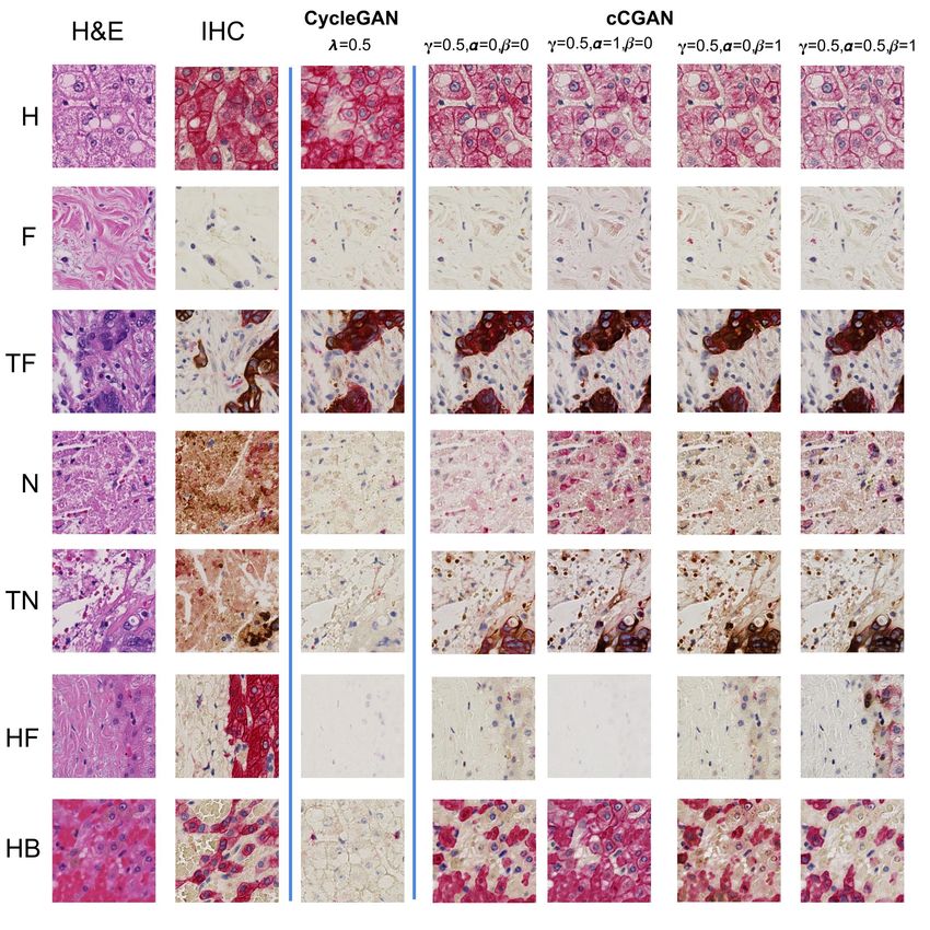
Fig. 5: Experiment results with different parameters settings12 Zhaoyang Xu? , Carlos Fernández Moro† ,Béla Bozóky† , Qianni Zhang?
4 Results and Evaluation
Evaluating the results of the generated images for tasks like style transfer, image
re-colorization is well-known challenge for a long time. One of the wide adopted
methods is to use Amazon Mechanical Turk (AMT) perceptual study that asks
human observers to differentiate the fake from the true ones. However, in our
study, we need to employ well-trained histopathologist experts to distinguish
them. Meanwhile, we will ask computer vision researchers to assess on the image
by a visual quality error score.
For the computer vision researchers, all the patches (real and fake) from
different classes are mixed together and the observers need to make decisions
based on the image quality. While, for professional assessment, both of the orig-
inal H&E stained and virtually stained patches will be provided. The experts
will give a staining score that indicates if the patches have been properly virtual
stained. Two pathology experts and ten computer vision researchers are invited
to perform the evaluation. 240 patches are generated from the evaluation dataset
(30 patches per class). The results of experts are listed in Table 1. The results
of different settings are demonstrated in Fig. 5. In this figure, for each H&E
stained patch, the patch from the same location on the roughly aligned IHC
images are used as references. Comparing to the results by CycleGAN, the re-
sults of proposed method greatly improve the results especially in the class with
mixed components and limited training examples such as TN, HF, HB. From the
experiments that we can observe the γ = 0.5, α = 0.5 and β = 1 output the best
results. Hence, we employ the results from this model for further evaluations.
Further results are listed in Table 1.
Table 1: Quantitative Evaluation Results
H TF N F HF TN HB BG Overall
No. Annotated patches (H&E) 1828 950 538 798 212 48 394 2016 3392
No. Annotated patches(IHC) 1440 738 438 1184 210 52 88 1252 2711
No. Test patches (H& E) 30 30 30 30 30 30 30 30 240
Vision experts on CycleGAN* 3% 26% 1% 4% 3% 0% 18% 0% 7%
Vision experts on cCGAN* 0% 10% 4% 0% 0% 0% 0% 0% 2%
Pathologists on CycleGAN* 34% 71% 22% 11% 83% 95% 97% 0% 52%
Pathologists on cCGAN* 23% 39% 23% 4% 76% 86% 99% 0% 44%
* The number indicates the visual quality error score and staining error score. The
larger the number indicate a worse performance.
The results from the non-experts view demonstrated that the proposed method
is better to preserve the image contents than the original cycleGAN. The intro-
duction of the structural losses to the model greatly suppresses the ”imaginary”
ability, especially when encountering new features. The class TF has the highest
fake rate, which has a strong relationship with its complex features. The poor
performance in the classes HB, HF, and TN is due to the insufficient and unbal-Title Suppressed Due to Excessive Length 13
anced training data in different classes. By inspecting the failed transformation
cases, we can find that mapping to the wrong domain is the main cause for the
failure. Some examples are illustrated in Fig. 6. The patch from H is mapped to
F, while for the HB, N, and TN, the staining style is N, F and H respectively.
Fig. 6: Unsuccessful mapping examples
5 Conclusions and Future Work
The results indicate that the proposed virtual staining method is capable of
virtually staining the H&E images into IHC style image based on the underlying
mutual features in the two kinds of images. In other words, IHC stained WSI
are distinguishable enough for guiding the cross-domain translation.
With the help of virtual staining, the laborious and expensive experts’ manual
annotation can be minimized. In fact, the presented method for computerized
virtual staining entails great potential for a wide range of clinical diagnostic and
AI related applications. A few of these are listed below:
1. Fast and low-cost generation of IHC staining for improved tumour diagnosis.
Currently, pathologists devote a great amount of time and efforts in the error-
prone eyeball work of searching for tumour cells within the low-contrast H&E
slides. Virtual IHC of tumour cells, by enabling high-contrast visualization,
may contribute to greatly reducing visual screening time and improving the
accuracy of cancer detection.
2. Virtual multiplex IHC staining for spatial mapping and objective quantita-
tion of intertwined cell types. As a generalization of GAN networks can be
trained to classify and virtually stain multiple cell types of interest, provided
multiplex immunostainings as training. For example, the spatial analysis and
quantitation of tumour and immune cells is a crucial assessment in the cur-
rent cancer immunotherapy. IHC stained cells can be then readily quantified
using available digital pathology image analysis tools.14 Zhaoyang Xu? , Carlos Fernández Moro† ,Béla Bozóky† , Qianni Zhang?
3. WSI registration for colocation analysis in multiple IHC stainings. Registra-
tion of multiple serial sections is another challenge of increasing interest in
the diagnosis and biomedical research. In clinical practice, the pathologists
employ multiple IHC stainings (bio-markers) to detect relevant features in
cells and tissues within the WSIs. As it is often not feasible to align the sam-
ples during lab processing, image registration is the only option to accomplish
this task, but very difficult to achieve. By exploiting virtual staining’s high
accurate matching of image pairs, multiple aligned IHC stainings can be
combined into a new staining style achieving increasing levels of multiplex
immunostaining far beyond current laboratory techniques.
4. Automatic generation or augmentation of highly accurate training datasets.
As, once learned, virtual staining can be exploited bi-directionally, H&E
staining can also be faithfully reproduced from an IHC stained slide as
demonstrated in Fig. 7. The original, high-contrast IHC staining can then
be easily segmented and used as mask to automatically extract annotations
from the virtual H&E staining with a pixel-level accuracy, reducing the need
for manual annotation and overcoming the intrinsic limitation of inter-slide
variation in serial (4 µm thick) tissue slides. This may enable also to improve
the accuracy of patch-based training data, especially in areas with a mixture
of cell and tissue types.
5. Color deconvolution and nuclei detection. Virtual staining can also be used
to decouple the information of the H&E stained image to obtain only the H
channel of the image. By using H&E, H-only, and E-only stained WSIs as
training data, the model will learn to ”unmix” the two color components in
the H&E images. The H/E-only images can then be easily normalized. This
may facilitate nuclei detection tasks, especially in cell dense and cancerous
areas. Some preliminary results are demonstrated in Fig. 8.
Fig. 7: IHC to H&E examples Fig. 8: Color deconvolution examples
One of the limitations of the current study is the low number of samples
for training and the significant imbalance on the amount of annotated patches
among different classes. In future work, we attempt to minimize or discarding
the labelling information that is used as input in order to pursue unsupervised
image translation. To that end, we plan to model the staining variance within
staining methods by introducing other controllable parameters.Title Suppressed Due to Excessive Length 15
References
1. Zhu, J.Y., Park, T., Isola, P., Efros, A.A.: Unpaired image-to-image translation
using cycle-consistent adversarial networks, (IEEE) 2242–2251
2. Goodfellow, I., Pouget-Abadie, J., Mirza, M., Xu, B., Warde-Farley, D., Ozair, S.,
Courville, A., Bengio, Y.: Generative adversarial nets. In Ghahramani, Z., Welling,
M., Cortes, C., Lawrence, N.D., Weinberger, K.Q., eds.: Advances in Neural Infor-
mation Processing Systems 27. (Curran Associates, Inc.) 2672–2680
3. Wang, T.C., Liu, M.Y., Zhu, J.Y., Tao, A., Kautz, J., Catanzaro, B.: (High-
resolution image synthesis and semantic manipulation with conditional GANs)
4. Zhang, R., Isola, P., Efros, A.A.: Colorful image colorization. In: Computer Vision
ECCV 2016. Lecture Notes in Computer Science. (Springer, Cham) 649–666
5. Mirza, M., Osindero, S.: (Conditional generative adversarial nets)
6. Isola, P., Zhu, J.Y., Zhou, T., Efros, A.A.: Image-to-image translation with con-
ditional adversarial networks, (IEEE) 5967–5976
7. Shrivastava, A., Pfister, T., Tuzel, O., Susskind, J., Wang, W., Webb, R.: Learn-
ing from simulated and unsupervised images through adversarial training. In: The
IEEE Conference on Computer Vision and Pattern Recognition (CVPR). Vol-
ume 3. (2017) 6
8. Liu, M.Y., Tuzel, O.: Coupled generative adversarial networks. In: Advances in
neural information processing systems. (2016) 469–477
9. Gatys, L.A., Ecker, A.S., Bethge, M.: Image style transfer using convolutional
neural networks. In: Computer Vision and Pattern Recognition (CVPR), 2016
IEEE Conference on, IEEE (2016) 2414–2423
10. Luan, F., Paris, S., Shechtman, E., Bala, K.: Deep photo style transfer, (IEEE)
6997–7005
11. Sasajima, K., Kudo, S.e., Inoue, H., Takeuchi, T., Kashida, H., Hidaka, E.,
Kawachi, H., Sakashita, M., Tanaka, J., Shiokawa, A.: Real-time in vivo virtual
histology of colorectal lesions when using the endocytoscopy system. (63) 1010–
1017
12. Bautista, P.A., Yagi, Y.: Digital simulation of staining in histopathology multi-
spectral images: enhancement and linear transformation of spectral transmittance.
(17) 056013
13. Elfer, K.N., Sholl, A.B., Wang, M., Tulman, D.B., Mandava, S.H., Lee, B.R.,
Brown, J.Q.: DRAQ5 and eosin (d&e) as an analog to hematoxylin and eosin
for rapid fluorescence histology of fresh tissues. (11) e0165530
14. Giacomelli, M.G., Husvogt, L., Vardeh, H., Faulkner-Jones, B.E., Hornegger, J.,
Connolly, J.L., Fujimoto, J.G.: Virtual hematoxylin and eosin transillumination
microscopy using epi-fluorescence imaging. (11) e0159337
15. Tao, Y.K., Shen, D., Sheikine, Y., Ahsen, O.O., Wang, H.H., Schmolze, D.B., John-
son, N.B., Brooker, J.S., Cable, A.E., Connolly, J.L., Fujimoto, J.G.: Assessment
of breast pathologies using nonlinear microscopy. (111) 15304–15309
16. Bayramoglu, N., Kaakinen, M., Eklund, L., Heikkila, J.: Towards virtual h&e stain-
ing of hyperspectral lung histology images using conditional generative adversarial
networks, (IEEE) 64–71
17. Rivenson, Y., Wang, H., Wei, Z., Zhang, Y., Gunaydin, H., Ozcan, A.: (Deep
learning-based virtual histology staining using auto-fluorescence of label-free tis-
sue)
18. Litjens, G., Snchez, C.I., Timofeeva, N., Hermsen, M., Nagtegaal, I., Kovacs, I.,
Kaa, C.H.v.d., Bult, P., Ginneken, B.v., Laak, J.v.d.: Deep learning as a tool for
increased accuracy and efficiency of histopathological diagnosis. (6) srep2628616 Zhaoyang Xu? , Carlos Fernández Moro† ,Béla Bozóky† , Qianni Zhang?
19. PoHsuan, C., Krishna, G., Robert, M., Yun, L., Kunal, N., Timo, K., Greg S.,
C., Jason D., H., Martin C., S.: (An augmented reality microscope for cancer
detection)
20. Trahearn, N., Epstein, D., Cree, I., Snead, D., Rajpoot, N.: Hyper-stain inspector:
A framework for robust registration and localised co-expression analysis of multiple
whole-slide images of serial histology sections. (7) 5641
21. Tosun, A.B., Nguyen, L., Ong, N., Navolotskaia, O., Carter, G., Fine, J.L., Taylor,
D.L., Chennubhotla, S.C.: Histological detection of high-risk benign breast lesions
from whole slide images. In: Medical Image Computing and Computer-Assisted
Intervention MICCAI 2017. Lecture Notes in Computer Science, (Springer, Cham)
144–152
22. Taigman, Y., Polyak, A., Wolf, L.: (Unsupervised cross-domain image generation)
23. Wang, Z., Bovik, A.C., Sheikh, H.R., Simoncelli, E.P.: Image quality assessment:
from error visibility to structural similarity. IEEE transactions on image processing
13 (2004) 600–612
24. Wang, Z., Simoncelli, E.P., Bovik, A.C.: Multiscale structural similarity for image
quality assessment. In: Signals, Systems and Computers, 2004. Conference Record
of the Thirty-Seventh Asilomar Conference on. Volume 2., Ieee (2003) 1398–1402
25. Johnson, J., Alahi, A., Fei-Fei, L.: (Perceptual losses for real-time style transfer
and super-resolution)
26. He, K., Zhang, X., Ren, S., Sun, J.: Deep residual learning for image recognition,
(IEEE) 770–778You can also read